Health-Related Quality of Life in Patients with Bone Tumor around the Knee after Resection Arthrodesis
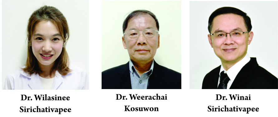
Vol 5 | Issue 1 | Jan-April 2019 | page: 17-20 | Wilasinee Sirichativapee, Weerachai Kosuwon, Winai Sirichativapee.
Authors: Wilasinee Sirichativapee [1], Weerachai Kosuwon [1], Winai Sirichativapee [1].
[1] Department of Orthopaedics, Faculty of Medicine, Khon Kaen University, Khon Kaen, Thailand.
Address of Correspondence
Dr. Winai Sirichativapee,
Department of Orthopaedics, Srinagarind Hospital, 123 Khon Kaen University, Nai Mueang Sub-District, Mueang District, Khon Kaen Province – 40002, Thailand.
E-mail: winaisiri@yahoo.com
Abstract
Background: This study aimed to compare the health-related quality of life (HRQoL) of patient with bone tumor around the knee after resection arthrodesis.
Methods: Patients between 15 and 70 years of age who underwent resection arthrodesis in Srinagarind Hospital >1 year were recruited. Patients were interviewed using a short form-36 questionnaire (social functioning-36 [SF-36] Ver2.0 Thai version) regarding their daily life problems.
Results: Eighteen patients with the mean age of 36.6 years (15–63 years) were included (15 females) in the study. Histological diagnoses were giant cell tumor 10 cases, osteosarcoma seven cases, and low-grade chondrosarcoma one case. Site of lesions was distal femur 15 cases and proximal tibia 3 cases. According to HRQoL, patients have good quality of life (score SF-36 >70) in all domains: Mean score: Physical functioning 75.55 ± 21.88, role physical 71.18 ± 22.70, bodily pain 85.41 ± 22.51, vitality 77.43 ± 16.76, general health 74.44 ± 19.16, SF 83.05 ± 26.40, role emotional 80.09 ± 22.89, and mental health 77.77 ± 21.29. Complications post-operative are broken implants (3 cases, 16.7%) and infections (4 cases, 22.2%).
Conclusion: In patients with bone tumor around the knee after resection, arthrodesis has a good quality of life in all domains in SF-36 version 2.0 questionnaire including function, pain, and mentality.
Keywords: Limb salvage, Arthrodesis, Quality of life, social functioning-36 version 2.0, Osteosarcoma, Giant cell tumor.
References
1. Tarnawska-Pierścińska M, Hołody Ł, Braziewicz J, Królicki L. Bone metastases diagnosis possibilities in studies with the use of 18F-NaF and 18F-FDG. Nucl Med Rev Cent East Eur 2011;14:105-8.
2. Sampath SC, Sampath SC, Mosci C, Lutz AM, Willmann JK, Mittra ES, et al. Detection of osseous metastasis by 18F-NaF/18F-FDG PET/CT versus CT alone. Clin Nucl Med 2015;40:e173-7.
3. Harisankar CN, Agrawal K, Bhattacharya A, Mittal BR. F-18 fluoro-deoxy-glucose and F-18 sodium fluoride cocktail PET/CT scan in patients with breast cancer having equivocal bone SPECT/CT. Indian J Nucl Med 2014;29:81-6.
4. Roop MJ, Singh B, Singh H, Watts A, Kohli PS, Mittal BR, et al. Incremental value of cocktail 18F-FDG and 18F-NaF PET/CT over 18F-FDG PET/CT alone for characterization of skeletal metastasesin breast cancer. Clin Nucl Med 2017;42:335-40.
5. Chan HP, Hu C, Yu CC, Huang TC, Peng NJ. Added value of using a cocktail of F-18 sodium fluoride and F-18 fluorodeoxyglucose in positron emission tomography/computed tomography for detecting bony metastasis: A case report. Medicine (Baltimore) 2015;94:e687.
6. Iagaru A, Mittra E, Mosci C, Dick DW, Sathekge M, Prakash V, et al. Combined 18F-fluoride and 18F-FDG PET/CT scanning for evaluation of malignancy: Results of an international multicenter trial. J Nucl Med 2013;54:176-83.
7. Gradishar WJ, Anderson BO, Balassanian R, Blair SL, Burstein HJ, Cyr A, et al. NCCN Clinical Practice Guidelines in Oncology Breast Cancer Version 2; 2016. Available from: https://www.nccn.org/professionals/physician_gls/pdf/breast.pdf. [Last accessed on 2016 Oct 19].
8. Yoon SH, Kim KS, Kang SY, Song HS, Jo KS, Choi BH, et al. Usefulness of (18)F-fluoride PET/CT in breast cancer patients with osteosclerotic bone metastases. Nucl Med Mol Imaging 2013;47:27-35.
9. Israel O, Goldberg A, Nachtigal A, Militianu D, Bar-Shalom R, Keidar Z, et al. FDG-PET and CT patterns of bone metastases and their relationship to previously administered anti-cancer therapy. Eur J Nucl Med Mol Imaging 2006;33:1280-4.
10. Lapa P, Saraiva T, Silva R, Marques M, Costa G, Lima JP. Superiority of 18F-Fna PET/CT for detecting bone metastases in comparison with other diagnostic ımaging modalities. Acta Med Port 2017;30:53-60.
11. Araz M, Aras G, Küçük ÖN. The role of 18F-NaF PET/CT in metastatic bone disease. J Bone Oncol 2015;4:92-7.
12. Schirrmeister H, Glatting G, Hetzel J, Nüssle K, Arslandemir C, Buck AK. Prospective evaluation of the clinical value of planar bone scans, SPECT, and (18)F-labeled NaF PET in newly diagnosed lung cancer. J Nucl Med 2001;42:1800-4.
13. Piccardo A, Puntoni M, Morbelli S, Massollo M, Bongioanni F, Paparo F, et al. 18F-FDG PET/CT is a prognostic biomarker in patients affected by bone metastases from breast cancer in comparison with 18F-naF PET/CT. Nuklearmedizin 2015;54:163-72.
14. Iagaru A, Young P, Mittra E, Dick DW, Herfkens R, Gambhir SS. Pilot prospective evaluation of 99mTc-MDP scintigraphy, 18F NaF PET/CT, 18F FDG PET/CT and whole-body MRI for detection of skeletal metastases. Clin Nucl Med 2013;38:e290-6.
15. Hillner BE, Siegel BA, Hanna L, Duan F, Quinn B, Shields AF. 18F-fluoride PET used for treatment monitoring of systemic cancer therapy: Results from the national oncologic PET registry. J Nucl Med 2015;56:222-8.
16. Iagaru A, Mittra E, Dick DW, Gambhir SS. Prospective evaluation of (99m)Tc MDP scintigraphy, (18)F NaF PET/CT, and (18)F FDG PET/CTfor detection of skeletal metastases. Mol Imaging Biol 2012;14:252-9.
| How to Cite this article: Koç Z P, Kara P Ö, Sezer E, Erçolak V.Diagnostic Comparison of F-18 Sodium FluorideNaF, Bone Scintigraphy, and F-18 Fluorodeoxyglucose Positron Emission Tomography/Computed Tomography in the Detection of Bone Metastasis. Journal of Bone and Soft Tissue Tumors Jan-Apr 2019;5(1): 17-20. |
