Tag Archive for: Osteosarcoma

Vertebral Osteosarcoma – A Report of Five Cases

Case Report | Volume 7 | Issue 3 | JBST September – December 2021 | Page 13-15 | Chitturi Ramya, Jayasree Kattoor, Narayanan Geetha, Muraleedharan Venugopal. DOI:0.13107/jbst.2021.v07i03.59

Author: Chitturi Ramya [1], Jayasree Kattoor [2], Narayanan Geetha [3], Muraleedharan Venugopal [4]

[1] Department of Pathology, NRI Medical College, Chinakakani, Andhra Pradesh, India.

[2] Department of Pathology, Regional Cancer Centre, Trivandrum, Kerala, India.

[3] Department of Medical Oncology, Regional Cancer Centre, Trivandrum, Kerala, India.

[4] Department of Radiology, Regional Cancer Centre, Trivandrum, Kerala, India.

Address of Correspondence
Dr. Jayasree Kattoor,
Department of Pathology, Regional Cancer Centre, Trivandrum, Kerala, India.
E-mail: jayasreeramdas@gmail.com


Abstract

Background: Vertebral osteosarcoma is rare, accounting for 3%–5% of all osteosarcomas. It tends to occur in a slightly older age group. The prognosis is poor compared to osteosarcoma of extremities.
Case Report: We present five cases of vertebral osteosarcomas. The patient’s age ranged from 16 years to 54 years. There were four females and one male. They presented with pain, swelling, or weakness of limbs. Destructive lesions, mixed lytic and sclerotic lesions involving vertebral bodies, and/or pedicles were seen on imaging. A definitive diagnosis could not be made in two cases. On histopathological examination, all the cases turned out to be osteosarcomas.
Conclusion: Osteosarcoma of vertebrae has high rate of recurrence, metastasis, and mortality. Differentiation of vertebral osteosarcoma from other common bony lesions involving the spine is of utmost importance as the treatment is entirely different. Combination therapies including surgery, radiation, and chemotherapy achieve adequate short-term survival rates for vertebral osteosarcoma.

Keywords: Osteosarcoma, Spine, Vertebra.


References:

1. Zils K, Bielack S, Wilhelm M, Werner M, Schwarz R, Windhager R, et al. Osteosarcoma of the mobile spine. Ann Oncol 2013;24:2190-5.
2. World Health Organization Classification of Tumours Editorial Board. Soft Tissue and Bone Tumours. WHO Classification of Tumours Series. 5th ed., Vol. 3. Lyon France: International Agency for Research on Cancer; 2020. p. 404-5.
3. Ozaki T, Flege S, Liljenqvist U, Hillmann A, Delling G, Salzer-Kuntschik M, et al. Osteosarcoma of the spine: Experience of the cooperative osteosarcoma study group. Cancer 2002;94:1069-77.
4. Green R, Saifuddin A, Cannon S. Pictorial review: Imaging of primary osteosarcoma of the spine. Clin Radiol 1996;51:325-9.
5. Ilaslan H, Sundaram M, Unni KK, Shives TC. Primary vertebral osteosarcoma: Imaging findings. Radiology 2004;230:697-702.
6. Egea-Gamez RM, Ponz-Lueza V, Cendrero-Torrado A, Martínez-Gonzalez C, Certucha-Barragan JA, Gonzalez-Díaz R. Spinal osteosarcoma in the paediatric age group: Case series and literature review. Rev Esp Cir Ortop Traumatol 2019;63:122-31.
7. Yalniz E, Özcan M, Copuroglu C, Memisoglu S, Yalçin O. Osteosarcoma of the lumbar vertebra: Case report and a review of the literature. Arch Orthop Trauma Surg 2009;129:1701.
8. Sansur CA, Pouratian N, Dumont AS, Schiff D, Shaffrey CI, Shaffrey ME. Part II: Spinal-cord neoplasms-primary tumours of the bony spine and adjacent soft tissues. Lancet Oncol 2007;8:137-47.
9. Murphey MD, Andrews CL, Flemming DJ, Temple HT, Smith WS, Smirniotopoulos JG, et al. From the archives of the AFIP. Primary tumors of the spine: Radiologic pathologic correlation. Radiographics 1996;16:1131-58.
10. Deyrup AT. Skeletal metastases. Surg Pathol 2012;5:287-300.
11. Sundaresan N, Rosen G, Huvos AG, Krol G. Combined treatment of osteosarcoma of the spine. Neurosurgery 1988;23:714-9.
12. Kokubo Y, Uchida K, Kobayashi S, Yayama T, Sato R, Kakuyama M, et al. Primary osteosarcoma of the thoracic spine: Report of an unusual elderly patient with autopsy findings. Spinal Cord 2005;43:508-11.


How to Cite this article: Ramya C, Kattoor J, Geetha N, Venugopal M | Vertebral Osteosarcoma – A Report of Five Cases. | Journal of Bone and Soft Tissue Tumors | Sep-Dec 2021; 7(3): 13-15.

[Full Text HTML] [Full Text PDF] [XML]


Effect of Combination Chemotherapy and Radiotherapy in the Management of a Pathological Fracture in High-grade Osteosarcoma with Limb Salvage Procedure – A Case Report

Original Article | Volume 6 | Issue 3 | JBST September – December 2020 | Page 9-12 | V.R.Ganesan, T.C.Prem Kumar, Sanjay.C, Roopesh Kumar.S DOI: 10.13107/jbst.2020.v06i03.32

Author: V.R.Ganesan[1], T.C.Prem Kumar[1], Sanjay.C[1], Roopesh Kumar.S[1]

[1]Department of Orthopaedic Surgery & Traumatology,
Madurai Medical College & GRH, Madurai, Tamilnadu. India.

Address of Correspondence
Dr. V R Ganesan,
Department of Orthopaedic Surgery & Traumatology,
Madurai Medical College & GRH , Madurai, Tamilnadu. India.
E-mail: sphospital@hotmail.com


Introduction: Osteosarcoma (OS), the most common primary bone tumor, is known to be relatively a radioresistant tumor. Pathological fracture in OS denotes its aggressive biological response and so it was considered a contraindication to limb salvage in earlier days. Radiotherapy has its role only in cases that are inoperable or have poor prognostic factors. In recent years, there have been major advances in the management of pathological fractures in high-grade OS (HGOS). This case report is about the effect of combination chemotherapy and radiotherapy in the management of pathological fracture in HGOS with limb salvage procedure.
Case Report: A 19-year-old male with pain and swelling in his right lower third of thigh and inability to walk for 3 months was diagnosed as a case of OS right distal femur with a pathological fracture. Open biopsy was done which confirmed the diagnosis as HGOS, staged Enneking IIb. He was treated with a combination of chemotherapy, external beam radiotherapy, tumor resection, and modular resection prosthesis.
Results: There were no immediate, early, and late complications. At the end of 1½ years, his functional recovery is good and he has reached more than 70% of the right knee functions. He has no signs of recurrence at present. He has got a better quality of life and functional activity with the prosthesis compared to what an amputated limb can produce.
Conclusion: Pathological fracture in OS is not a contraindication to limb salvage. Radiotherapy can be used in combination with chemotherapy and limb salvage surgery in HGOS with a pathological fracture. This combination treatment helps in increasing the chances of limb sparing surgery with good local control and tumor necrosis rate. The new knowledge that radiotherapy can be effective when used with chemotherapy has shown good result in our case.
Keywords: Osteosarcoma, pathological fracture, external beam radiotherapy, limb salvage, custom modular prosthesis.


Reference:
1. Abudu A, Sferopoulos NK, Tillman RM, Carter SR, Grimer RJ. The surgical treatment and outcome of pathological fractures in localized osteosarcoma. J Bone Joint Surg Br 1996;78:694-8.
2. Ferguson PC, McLaughlin CE, Griffin AM, Bell RS, Deheshi BM, Wunder JS. Clinical and functional outcomes of patients with a pathologic fracture in high-grade osteosarcoma. J Surg Oncol 2010;102:120-4.
3. Scully SP, Ghert MA, Zurakowski D, Thompson RC, Gebhardt MC. Pathologic fracture in osteosarcoma: Prognostic importance and treatment implications. J Bone Joint Surg Am 2002;84:49-57.
4. Glasser DB, Lane JM, Huvos AG, Marcove RC, Rosen G. Survival, prognosis and therapeutic response in osteogenic sarcoma: The Memorial Hospital experience. Cancer 1992;69:698-708.
5. Finn HA, Simon MA. Limb-salvage surgery in the treatment of osteosarcoma in skeletally immature individuals. Clin Orthop Relat Res 1991;262:108-18.
6. Mankin HJ, Mankin CJ, Simon MA. The hazards of the biopsy, revisited: Members of the Musculoskeletal Tumor Society. J Bone Joint Surg 1996;78:656-63.
7. Bacci G, Ferrari S, Longhi A, Donati D, Manfrini M, Giacomini S, et al. Nonmetastatic osteosarcoma of the extremity with pathologic fracture at presentation: Local and systemic control by amputation or limb salvage after preoperative chemotherapy. Acta Orthop Scand 2003;74:449 54.
8. Kim MS, Lee SY, Lee TR, Cho WH, Song WS, Cho SH, et al. Prognostic effect of pathologic fracture in localized osteosarcoma: A cohort/case controlled study at a single institute. J Surg Oncol 2009;100:233 9.
9. Jaffe N, Spears R, Eftekhari F, Robertson R, Cangir A, Takaue Y, et al. Pathologic fracture in osteosarcoma. Impact of chemotherapy on primary tumor and survival. Cancer 1987;59:701-9.
10. Longhi A, Errani C, De Paolis M, Mercuri M, Bacci G. Primary bone osteosarcoma in the pediatric age: State of the art. Cancer Treat Rev 2006;32:423-36.
11. Jeon DG, Lee SY, Kim JW. Bone primary sarcomas undergone unplanned intralesional procedures the possibility of limb salvage and their oncologic results. J Surg Oncol 2006;94:592-8.
12. Xie J, Diener M, Sorg R, Wu EQ, Namjoshi M. Cost-effectiveness of denosumab compared with zoledronic acid in patients with breast cancer and bone metastases. Clin Breast Cancer 2012;12:247-58.
13. Ebeid W, Amin S, Abdelmegid A. Limb salvage management of pathologic fractures of primary malignant bone tumors. Cancer Control 2005;12:57-61.
14. Natarajan MV, Govardhan RH, Williams S, Raja GT. Limb salvage surgery for pathological fractures in osteosarcoma. Int Orthop 2000;24:170-2.
15. Cui Q, Li DF, Liu C, Guo J, Liu SB, Liu YS, et al. Two case-reports of the limb salvage treatment of osteosarcoma consolidated with obvious pathological fractures. Pathol Oncol Res 2011;17:973-9.
16. Godley K, Watts AC, Robb JE. Pathological femoral fracture caused by primary bone tumour: A population-based study. Scott Med J 2011;56:5-9.
17. Chandrasekar CR, Grimer RJ, Carter SR, Tillman RM, Abudu AT, Jeys LM. Outcome of pathologic fractures of the proximal femur in nonosteogenic primary bone sarcoma. Eur J Surg Oncol 2011;37:532-6.
18. Yin K, Liao Q, Zhong D, Ding J, Niu B, Long Q, et al. Metaanalysis of limb salvage versusamputation for treating high grade and localized osteosarcoma in patients with pathological fracture. Exp Ther Med 2012;4:889-94.
19. Scully SP, Temple HT, O’Keefe RJ, Mankin HJ, Gebhardt M. The surgical treatment of patients with osteosarcoma who sustain a pathological fracture. Clin Orthop 1996;324:227-32.
20. Machak GN, Tkachev SI, Solovyev YN, Sinyukov PA, Ivanov SM, Kochergina NV, et al. Neoadjuvant chemotherapy and local radiotherapy for high-grade osteosarcoma of the extremities. Mayo Clin Proc 2003;78:147-55.
21. Caceres E, Zaharia M, Valdivia S, Hilsenbeck S. Tejada F. Local control of osteogenic sarcoma by radiation and chemotherapy. Int J Radiat Oncol Biol Phys 1984;10:35-9.
22. Dincbas FO, Koca S, Mandel NM, Hiz M, Dervişoğlu S, Seçmezacar H, et al. The role of preoperative radiotherapy in non-metastatic high-grade osteosarcoma of the extremities for limb-sparing surgery. Int J Radiat Oncol Biol Phys 2005;62:820-8.


How to Cite this article: Ganesan VR, Kumar TCP, Sanjay C, Kumar SR| Effect of Combination Chemotherapy and Radiotherapy in the Management of a Pathological Fracture in High-grade Osteosarcoma with Limb Salvage Procedure – A Case Report | Journal of Bone and Soft Tissue Tumors | September-December 2020; 6(3): 9-12.

[Full Text HTML] [Full Text PDF] [XML]


Osteosarcoma of the Rib: A Case Report

Original Article | Volume 6 | Issue 2 | JBST May-August 2020 | Page 17-20 | Kanu Priya Bhatia, Sameer Rastogi, Ekta Dhamija, Adarsh Barwad, Nishant Bhatia, Jyoutishman Saika. DOI: 10.13107/jbst.2020.v06i02.26

Author: Kanu Priya Bhatia[1], Sameer Rastogi[1], Ekta Dhamija[2], Adarsh Barwad[3], Nishant Bhatia[4], Jyoutishman Saika[5]

[1]Department of Medical Oncology, Dr. B.R Ambedkar Institute Rotary Cancer Hospital, All India Institute of Medical Sciences, New Delhi, India,
[2]Department of Radiodiagnosis, Dr. B.R Ambedkar Institute Rotary Cancer Hospital, All India Institute of Medical Sciences, New Delhi, India,
[3]Department of Pathology, All India Institute of Medical Sciences, New Delhi, India,
[4]Department of Orthopaedics, Maulana Azad Medical College and Associated Lok Nayak Hospital, New Delhi, India,
[5]Department of Surgical Oncology, All India Institute of Medical Sciences, New Delhi, India.

Address of Correspondence
Dr. Kanu Priya Bhatia,
Department of Medical Oncology, Dr. B.R Ambedkar Institute Rotary Cancer Hospital, All India Institute of Medical Sciences, New Delhi, India.
E-mail: bhatiakanu13@gmail.com


Abstract

Introduction: Osteosarcoma (OS) is the most common primary malignancy of bone in children and adolescents. OS has a predilection for the metaphyseal region of the long bones. The most common site of involvement is the distal femur followed by proximal tibia, proximal humerus, middle and proximal femur, and other bones. Primary OS of chest wall is a very rare entity.
Case Report: We, here, describe a case of a 14-year-old boy who presented to our center with a chest wall swelling and pleural effusion, which was subsequently diagnosed as chest wall OS originating from the rib. We treated him with neoadjuvant chemotherapy (Ifosfamide, Adriamycin, and Carboplatin). He showed an excellent transient response to the chemotherapy, after which he underwent an en bloc resection of the tumor. He was then started on adjuvant chemotherapy, but unfortunately, he relapsed soon after the last cycle and later on succumbed to the disease.
Conclusion: Chest wall OS is an infrequent malignancy. Pathological diagnosis is difficult and requires a high index of suspicion. Data regarding the prognostic factors are scarce, and no concrete guidelines are available for the management of such patients.
Keywords: Chest wall, osteosarcoma, rib.


Reference:
1. Moghazy K, Al-Jehani Y, El-Baz A, El-Ghoneimy Y. Incidental finding of a large chest wall osteosarcoma–a case report. Gulf J Oncolog 2007;1:93-7.
2. Sabatier R, Bouvier C, de Pinieux G, Sarran A, Brenot-Rossi I, Pedeutour F, et al. Low-grade extraskeletal osteosarcoma of the chest wall: Case report and review of literature. BMC Cancer 2010;10:645.
3. Qian J, Zhang XY, Gu P, Shao JC, Han BH, Wang HM. Primary thoracic extraskeletal osteosarcoma: A case report and literature review. J Thorac Dis 2017;9:E1088-95.
4. Bathurst N, Sanerkin N. Osteoclast-rich osteosarcoma. Br J Radiol 1986;59:667-73.
5. Chow LT. Giant cell rich osteosarcoma revisited-diagnostic criteria and histopathologic patterns, Ki67, CDK4, and MDM2 expression, changes in response to bisphosphonate and denosumab treatment. Virchows Arch 2016;468:741-55.
6. Daw N, Neel M, Rao B, Billups C, Wu J, Jenkins J, et al. Frontline treatment of localized osteosarcoma without methotrexate. Cancer 2011;117:2770-8.
7. Baez JC, Lee EY, Restrepo R, Eisenberg RL. Chest wall lesions in children. Am J Roentgenol 2013;200:W402-19.
8. Burt M, Fulton M, Wessner-Dunlap S, Karpeh M, Huvos AG, Bains MS, et al. Primary bony and cartilaginous sarcomas of chest wall: Results of therapy. Ann Thorac Surg 1992;54:226-32.
9. Ikeda H, Takeo M, Kayata H, Mikami R, Nakamoto Y, Yamamoto M. A case of rapidly growing osteosarcoma of the rib. Ann Thorac Cardiovasc Surg 2014;20:521-4.
10. Inchara Y, Crasta J, Ananthamurthy A, Mohanty S. An unusual case of primary osteosarcoma of the rib in an adult. Indian J Med Paediatr Oncol 2010;31:18.
11. Rad MP, Masoum SF, Layegh P, Rad MS. Primary osteosarcoma of the sternum: A case report and review of the literature. Arch Bone Joint Surg 2014;2:272-5.
12. Lim W, Sarji SA, Yik Y, Ramanujam T. Osteosarcoma of the rib. Biomed Imaging Interv J 2008;4:e7.
13. Masoud S. Scapula osteosarcoma. Biomed J Sci Tech Res 2017;1:739-42.
14. Bielack SS, Kempf-Bielack B, Delling G, Exner GU, Flege S, Helmke K, et al. Prognostic factors in high-grade osteosarcoma of the extremities or trunk: An analysis of 1,702 patients treated on neoadjuvant cooperative osteosarcoma study group protocols. J Clin Oncol 2002;20:776-90.


How to Cite this article: Bhatia KP, Rastogi S, Dhamija E, Barwad A, Bhatia N, Saika J | Osteosarcoma of the Rib: A Case Report | Journal of Bone and Soft Tissue Tumors | May-August 2020; 6(2): 17-20.

[Full Text HTML] [Full Text PDF] [XML]


Health-Related Quality of Life in Patients with Bone Tumor around the Knee after Resection Arthrodesis

Vol 5 | Issue 1 | Jan-April 2019 | page: 17-20 | Wilasinee Sirichativapee, Weerachai Kosuwon, Winai Sirichativapee.


Authors: Wilasinee Sirichativapee [1], Weerachai Kosuwon [1], Winai Sirichativapee [1].

[1] Department of Orthopaedics, Faculty of Medicine, Khon Kaen University, Khon Kaen, Thailand.

Address of Correspondence
Dr. Winai Sirichativapee,
Department of Orthopaedics, Srinagarind Hospital, 123 Khon Kaen University, Nai Mueang Sub-District, Mueang District, Khon Kaen Province – 40002, Thailand.
E-mail: winaisiri@yahoo.com


Abstract

Background: This study aimed to compare the health-related quality of life (HRQoL) of patient with bone tumor around the knee after resection arthrodesis.
Methods: Patients between 15 and 70 years of age who underwent resection arthrodesis in Srinagarind Hospital >1 year were recruited. Patients were interviewed using a short form-36 questionnaire (social functioning-36 [SF-36] Ver2.0 Thai version) regarding their daily life problems.
Results: Eighteen patients with the mean age of 36.6 years (15–63 years) were included (15 females) in the study. Histological diagnoses were giant cell tumor 10 cases, osteosarcoma seven cases, and low-grade chondrosarcoma one case. Site of lesions was distal femur 15 cases and proximal tibia 3 cases. According to HRQoL, patients have good quality of life (score SF-36 >70) in all domains: Mean score: Physical functioning 75.55 ± 21.88, role physical 71.18 ± 22.70, bodily pain 85.41 ± 22.51, vitality 77.43 ± 16.76, general health 74.44 ± 19.16, SF 83.05 ± 26.40, role emotional 80.09 ± 22.89, and mental health 77.77 ± 21.29. Complications post-operative are broken implants (3 cases, 16.7%) and infections (4 cases, 22.2%).
Conclusion: In patients with bone tumor around the knee after resection, arthrodesis has a good quality of life in all domains in SF-36 version 2.0 questionnaire including function, pain, and mentality.
Keywords: Limb salvage, Arthrodesis, Quality of life, social functioning-36 version 2.0, Osteosarcoma, Giant cell tumor.


References

1. Tarnawska-Pierścińska M, Hołody Ł, Braziewicz J, Królicki L. Bone metastases diagnosis possibilities in studies with the use of 18F-NaF and 18F-FDG. Nucl Med Rev Cent East Eur 2011;14:105-8.
2. Sampath SC, Sampath SC, Mosci C, Lutz AM, Willmann JK, Mittra ES, et al. Detection of osseous metastasis by 18F-NaF/18F-FDG PET/CT versus CT alone. Clin Nucl Med 2015;40:e173-7.
3. Harisankar CN, Agrawal K, Bhattacharya A, Mittal BR. F-18 fluoro-deoxy-glucose and F-18 sodium fluoride cocktail PET/CT scan in patients with breast cancer having equivocal bone SPECT/CT. Indian J Nucl Med 2014;29:81-6.
4. Roop MJ, Singh B, Singh H, Watts A, Kohli PS, Mittal BR, et al. Incremental value of cocktail 18F-FDG and 18F-NaF PET/CT over 18F-FDG PET/CT alone for characterization of skeletal metastasesin breast cancer. Clin Nucl Med 2017;42:335-40.
5. Chan HP, Hu C, Yu CC, Huang TC, Peng NJ. Added value of using a cocktail of F-18 sodium fluoride and F-18 fluorodeoxyglucose in positron emission tomography/computed tomography for detecting bony metastasis: A case report. Medicine (Baltimore) 2015;94:e687.
6. Iagaru A, Mittra E, Mosci C, Dick DW, Sathekge M, Prakash V, et al. Combined 18F-fluoride and 18F-FDG PET/CT scanning for evaluation of malignancy: Results of an international multicenter trial. J Nucl Med 2013;54:176-83.
7. Gradishar WJ, Anderson BO, Balassanian R, Blair SL, Burstein HJ, Cyr A, et al. NCCN Clinical Practice Guidelines in Oncology Breast Cancer Version 2; 2016. Available from: https://www.nccn.org/professionals/physician_gls/pdf/breast.pdf. [Last accessed on 2016 Oct 19].
8. Yoon SH, Kim KS, Kang SY, Song HS, Jo KS, Choi BH, et al. Usefulness of (18)F-fluoride PET/CT in breast cancer patients with osteosclerotic bone metastases. Nucl Med Mol Imaging 2013;47:27-35.
9. Israel O, Goldberg A, Nachtigal A, Militianu D, Bar-Shalom R, Keidar Z, et al. FDG-PET and CT patterns of bone metastases and their relationship to previously administered anti-cancer therapy. Eur J Nucl Med Mol Imaging 2006;33:1280-4.
10. Lapa P, Saraiva T, Silva R, Marques M, Costa G, Lima JP. Superiority of 18F-Fna PET/CT for detecting bone metastases in comparison with other diagnostic ımaging modalities. Acta Med Port 2017;30:53-60.
11. Araz M, Aras G, Küçük ÖN. The role of 18F-NaF PET/CT in metastatic bone disease. J Bone Oncol 2015;4:92-7.
12. Schirrmeister H, Glatting G, Hetzel J, Nüssle K, Arslandemir C, Buck AK. Prospective evaluation of the clinical value of planar bone scans, SPECT, and (18)F-labeled NaF PET in newly diagnosed lung cancer. J Nucl Med 2001;42:1800-4.
13. Piccardo A, Puntoni M, Morbelli S, Massollo M, Bongioanni F, Paparo F, et al. 18F-FDG PET/CT is a prognostic biomarker in patients affected by bone metastases from breast cancer in comparison with 18F-naF PET/CT. Nuklearmedizin 2015;54:163-72.
14. Iagaru A, Young P, Mittra E, Dick DW, Herfkens R, Gambhir SS. Pilot prospective evaluation of 99mTc-MDP scintigraphy, 18F NaF PET/CT, 18F FDG PET/CT and whole-body MRI for detection of skeletal metastases. Clin Nucl Med 2013;38:e290-6.
15. Hillner BE, Siegel BA, Hanna L, Duan F, Quinn B, Shields AF. 18F-fluoride PET used for treatment monitoring of systemic cancer therapy: Results from the national oncologic PET registry. J Nucl Med 2015;56:222-8.
16. Iagaru A, Mittra E, Dick DW, Gambhir SS. Prospective evaluation of (99m)Tc MDP scintigraphy, (18)F NaF PET/CT, and (18)F FDG PET/CTfor detection of skeletal metastases. Mol Imaging Biol 2012;14:252-9.


How to Cite this article: Koç Z P, Kara P Ö, Sezer E, Erçolak V.Diagnostic Comparison of F-18 Sodium FluorideNaF, Bone Scintigraphy, and F-18 Fluorodeoxyglucose Positron Emission Tomography/Computed Tomography in the Detection of Bone Metastasis. Journal of Bone and Soft Tissue Tumors Jan-Apr 2019;5(1): 17-20.


  (Abstract    Full Text HTML)   (Download PDF)


Is Limb Salvage Surgery a Contra Indication in Pathological Fractures Secondary to Osteosarcoma? Do We Know The Answer?

Volume 2 | Issue 2 | May-Aug 2016 | Page 10-12 | Zeeshan Khan, Shakir Hussain, Simon Carter.


Authors: Zeeshan Khan [1], Shakir Hussain [1], Simon Carter[1].

1Orthopedic Oncology Services, Department of Surgical Oncology, Tata Memorial Hospital, Mumbai.

Address of Correspondence
Dr. Ashish Gulia
Associate Professor, Orthopedic oncology, Department of Surgical Oncology, Tata Memorial Hospital, Mumbai.
Email: aashishgulia@gmail.com


Abstract

 Osteosarcoma is the commonest primary bone tumour with a bimodal age distribution. The survivorship of patients with osteosarcoma has improved with advances in chemotherapy making limb salvage surgery the commonest surgical procedure. Pathological fractures associated with osteosarcoma, however are rare and suggests the aggressiveness of the tumour. These patients are considered as a special group due to the variable outcomes reported in the literature due to some special characteristics, prompting the discussion between limb salvage surgery versus ablative surgery.
This article reviews the reasons why this group of patients are considered challenging and also the various outcomes reported in the literature.
Keywords: Pathological fracture, osteosarcoma, outcomes


Introduction

Primary bone and soft tissue sarcomas are rare tumours. Osteosarcoma is the commonest primary bone tumour with a bimodal age distribution and with a reported incidence of 2-3 per million population per year [1, 2]. With advances in chemotherapy, the survivorship of patients with osteosarcoma has improved significantly with various studies revealing similar results with limb salvage surgery when compared with amputation [5]. Contra indications to limb salvage surgery may include involvement of the neurovascular bundle, joint involvement, progression of disease whilst on treatment, patient choice, infection and a pathological fracture (Fig. 1).
A pathological fractureassociated with osteosarcoma at presentation or during treatment is even rarer with a reported incidence of 5-10% [3, 4]. A pathological fracture can be the mode of presentation for osteosarcomas in certain cases whereas it can occur during treatment in others. This is generally considered to be an aggressive biological behaviour of the disease which in turn, historically, has been considered as a poor prognostic factor in the outcome of this special group of patients [6].

Why is this group of patients special?
This select group of patients poses a challenge to the treating orthopaedic surgeon about the modality of surgical procedure. There has been a debate over the years whether these patients should have ablative or limb salvage surgery and if there is a difference in the outcome of both with varying results reported by different authors [8].

What makes these patients special is the associated hematoma with the pathological fracture which is considered to have tumour cells which spreads locally in the tissues [7]. The extent of spread also depends on the anatomic location of fracture and whether it is intra or extra capsular. The disruption of local microvasculature is also considered to be a risk factor for development of metastasis [7]. Understandably, extra articular resection for intra articular extension of tumours is a more challenging procedure particularly when limb salvage surgery is attempted with the reported outcomes of extra articular resections in limb salvage surgery considered to be compromised as well [9]. The local contamination of soft tissues with the tumour cells is also considered to be a risk factor for local recurrence. This prompted the thought that early and aggressive surgery in the form of ablative surgery will halt the progression and spread of disease any further. The presence of a pathological fracture in osteosarcoma, therefore, has been considered as a poor prognostic factor by some authors but not by all [8, 10, 11].

Initial treatment& work up
Perhaps the most important step in the management of this select group of patients is the early recognition of the aggressiveness of the lesion and prompt referral to a specialist unit. Failure of recognition of these fractures as being pathological can lead to inappropriate treatment and potentially worse outcomes (Fig. 2) [14]. The rest of the management in a multidisciplinary team setting involves a detailed history, examination of the involved limb and joints for any effusion, local and systemic staging, biopsy and neoadjuvant chemotherapy after confirmation of diagnosis of osteosarcoma.
It is also important to note that one of the most challenging issues with this group of patients is pain management and immobilisation during the pre-operative period whilst they wait for surgery and have neoadjuvant chemotherapy. 

Immobilisation
Immobilisation for pain relief can be challenging as this depends on the location of the fracture and may involve a plaster cast, simple sling, skin traction or in some cases external fixators [15]. Significant attention should be paid to the placement of the schanz pins if an external fixator is used due to the risk of tumour spread into non-involved compartments and risk of infection which would compromise limb sparing surgery.

Prognostic factors
A pathological fracture is independently considered a poor prognostic factor in osteosarcoma but was not considered one in cases of chondrosarcoma and Ewing’s sarcoma [8, 10]. A poor response to chemotherapy and local recurrence are also considered to be poor prognostic factors for survivorship [8, 10-13]. It is however, important to note that the efficacy of chemotherapy and healing of fracturesin these special cases are considered as supportive factors for limb salvage surgery [20].

Fracture consolidation
It has been noted that these fractures heal whilst patients are on chemotherapy and in most of the cases these patients have had significant post chemotherapy necrosis(Fig. 3) [8]. On the contrary some fractures might happen whilst patients are on chemo which depicts the aggressive nature of the disease.

What is the verdict?
Limb salvage surgery should be attempted, if possible, in these patients after neoadjuvant treatment but if clear surgical margins cannot be obtained during surgery or limb salvage will result in a poor functioning limb, then ablative surgery should be considered, particularly in the paediatric population where they can adapt to prosthetics earlier than adults [16]. It is however, also important to note that after wide resection of tumour, limb salvage is still a viable option with reconstruction performed with either arthrodesis or rotationplasty where appropriate.
Scully et al, suggested that a pathological was a poor prognostic factor but it is important to note that this study was performed over a 30 year period where some patients in their series had not received any chemortherapy and there have been advances in this field over the study time period [8]. Similarly Finn et al, suggested early amputation due to the risk of local and distant tumour spread [14]. In another study, the 5 year survival in patients with pathological fractures secondary to osteosarcoma was lower than those without a fracture [18]. On the contrary Bacci et al, and Abudu et al, showed that there was no difference in the survivorship of these patients when they were treated with neoadjuvant chemotherapy [7, 11]. In a recent meta-analysis comparing limb salvage with ablative surgery for pathological fractures in high grade osteosarcomas, no significant difference between local recurrence and 5 year survival was noted [19]. Adjuvant radiotherapy in these patients has not been shown to reduce the risk of local recurrence and in fact might increase the risk of these patients undergoing further surgical procedures compromising there outcomes [7].

Future direction
All the studies performed on this select group of patients are retrospective and contain a small number of patients over a prolonged period of time. The results are further effected by variables including the heterogeneity of the patient and fracture characteristics and also the advances in chemotherapy over a period of time. Improvements in surgical techniques have also resulted in improved outcomes. Most of these variables are un avoidable due to the rarity of these cases but in order to come to a definite conclusion, a multi central randomised trial will eradicate all these bias and should guide treatment.


References

1. 1.Bielack S, Carrle D, Jost L. ESMO guidelines working group osteosarcoma: ESMO clinical recommendations for diagnosis, treatment and follow up. Annals of Oncology.2008; 19, supplement 2: 94-96.
2. Widhe B, Widhe T. Initial symptoms and clinical features in osteosarcoma and Ewing sarcoma. J Bone Joint Surg Am. 2000; 82:667-74.
3. Jaffe N, Spears R, Eftekhari F, Robertson R, Cangir A, Takaue Y, Carrasco H, Wallace S, Ayala A,Raymond K, et al. Pathologic fracture in osteosarcoma. Impact of chemotherapy on primarytumorand survival. Cancer. 1987; 59:701-09.
4. Mulder JO, Schutte HE, Kroon HM, Taconis WK. Radiologic atlas of bone tumors. Amsterdam:Elsevier Science. 1993. Intraosseous osteosarcoma: conventional type: 51-5.
5. Simon MA. Current concepts review. Limb salvage in osteosarcoma. J Bone Joint Surg Am.1988; 70:307-10.
6. Coley BL, Pool JL. Factors influencing the prognosis in osteogenic sarcoma. Ann Surg. 1940; 112:1114-28.
7. Abudu A, Sferopoulos NK, Tillman RM, Carter SR, Grimer RJ. The surgical treatment and outcome of pathological fractures in localised osteosarcoma. J Bone Joint Surg Br. 1996; 78:694-8.
8. Scully SP1, Ghert MA, Zurakowski D, Thompson RC, Gebhardt MC. Pathologic fracture in osteosarcoma: prognostic importance and treatment implications. J Bone Joint Surg Am. 2002 Jan;84-A (1):49-57.
9. Hardes J1, Henrichs MP, Gosheger G, Gebert C, Höll S, Dieckmann R, Hauschild G, Streitbürger A. Endoprosthetic replacement after extra-articular resection of bone and soft-tissue tumours around the knee. Bone Joint J. 2013 Oct; 95-B (10):1425-31.
10. Bramer JAM, Abudu AA, Grimer RJ, Carter SR, Tillman RM. Do pathological fractures influence survival and local recurrence rate in bony sarcomas?. Eur J Cancer. 2007 Sep; 43(13):1944-51.
11. Bacci G1, Ferrari S, Longhi A, Donati D, Manfrini M, Giacomini S, Briccoli A, Forni C, Galletti S.Nonmetastatic osteosarcoma of the extremity with pathologic fracture at presentation: local andsystemic control by amputation or limb salvage after preoperative chemotherapy. ActaOrthop Scand. 2003 Aug; 74(4):449-54.
12. Meyers PA, Heller G, Healey J, Huvos A, Lane J, Marcove R, ApplewhiteA, Vlamis V, Rosen G. Chemotherapy for nonmetastatic osteogenic sarcoma: the Memorial Sloan-Kettering experience. J ClinOncol. 1992; 10:5-15.
13. Glasser DB, Lane JM, Huvos AG, Marcove RC, Rosen G. Survival, prognosis, and therapeutic response in osteogenic sarcoma. The Memorial Hospital experience. Cancer. 1992; 69:698-708.
14. Mankin HJ, Mankin CJ, Simon MA. The hazards of the biopsy, revisited: Members of the Musculoskeletal Tumor Society. J Bone Joint Surg [Am] 1996; 78-A: 656–663.
15. Chandrasekar CR, Grimer RJ, Carter SR, et al. Pathological fracture of the proximal femur in osteosarcoma: need for early radical surgery? ISRN Oncol 2012; 2012:512389.
16. Hosalkar HS, Dormans JP. Limb sparing surgery for pediatric musculoskeletal tumors. Pediatr Blood Cancer 2004; 42:295–310.
17. Finn HA, Simon MA. Limb-salvage surgery in the treatment of osteosarcoma in skeletally immature individuals. ClinOrthopRelat Res 1991; 262:108–118.
18. Ferguson PC, McLaughlin CE, Griffin AM, et al. Clinical and functional outcomesof patients with a pathologic fracture in high-grade osteosarcoma. J SurgOncol2010; 102:120–124.
19. Yin K, Liao Q, Zhong D, Ding J, Niu B, Long Q, Ding D. Meta-analysis of limb salvage versusamputation for treating high-grade and localized osteosarcoma in patients with pathological fracture. ExpTher Med. 2012 Nov; 4(5):889-894.
20. Scully SP, Temple HT, O’Keefe RJ, et al. The surgical treatment of patients with osteosarcoma who sustain a pathological fracture. ClinOrthop. 1996; 324:227-232.


How to Cite this article: Khan Z, Hussain S, Carter S. Is Limb Salvage Surgery a Contra Indication in Pathological Fractures Secondary to Osteosarcoma? Do We Know The Answer? Journal of Bone and Soft Tissue Tumors May- Aug 2016;2(2):10-12 .


                                              (Abstract    Full Text HTML)      (Download PDF)


Radiological Review of Extremity Osteosarcoma

 Volume 2 | Issue 1 | Jan-Apr 2016 | Page 13-18  Amit Janu, Nikshita Jain, Shashikant Juvekar, Ashish Gulia.


Author: Amit Janu[1], Nikshita Jain[1], Shashikant Juvekar[1], Ashish Gulia[2].

[1]Dept of Radiodiagnosis, Tata Memorial Hospital, Parel, Mumbai. India.
[2]Orthopedic Oncology, Dept. of Surgical Oncology, Tata Memorial Hospital, Parel, Mumbai, India.

Address of Correspondence
Dr Ashish Gulia, MS (Ortho), Mch (Surgical Oncology)
Associate Professor, Orthopedic Oncology, Dept. of Surgical Oncology, Tata Memorial Hospital,
Mumbai – 400012, India.
Email: aashishgulia@gmail.com


Abstract

The paradigm shift in the overall outcomes of osteosarcoma is multi factorial. Be it introduction of chemotherapy, introduction of better
imaging or advances in the technology to produce better prosthesis, the crux lies in the correct and timely diagnosis. Radiology along with
clinical evaluation and histopathological confirmation is an essential part of the three tier system which leads to an accurate diagnosis
which forms the platform to initiate an ideal treatment to have desired oncological and functional outcomes. Plain radiography has been
the age old diagnostic tool which is still as important as was in yester century. Inclusion of high end cross sectional imaging likeCT and
MRI has not only helped in early and price diagnosis but has also proved to be a boon to operative surgeons to stage the disease locally and
plan complex limb salvage strategies. In the present article we have discussed the radiological features of various sub types of osteosarcoma
to help a clinician to assess these complex varieties of lesions.
Keywords: osteosarcoma, radiological assessment.


Introduction
Osteosarcoma is the most common primary nonhematologic bone malignancy and is the most common primary bone malignancy in children. There are various subtypes of osteosarcoma, each with distinct clinical and imaging characteristics and variable survival and an incidence of 0.2 to 0.3 per 100,000/year. Osteosarcomas can be classified as intramedullary (high grade, telangiectatic, low grade, small cell, osteosarcomatosis, gnathic), juxtacortical (parosteal, periosteal, intracortical, high-grade surface), or secondary lesions [1]. Though majority of cases are of conventional high grade intramedullary osteosarcoma accounting for 75%- 90%, it is important to differentiate them from other low grade and (low grade central and parosteal osteosarcoma) and intermediate grade osteosarcoma (Periosteal osteosarcoma). An understanding of the systematic imaging approach helps to more accurately diagnose these lesions and direct effective treatment. This article provides an organized approach to analyzing and subtyping of osteosarcoma based on radiographs and guiding the referring physician if any further imaging is warranted as there is frequently overlap with other benign and malignant entities, creating substantial diagnostic challenges. For accurate diagnosis, it is important to be aware of radiographic and cross-sectional imaging features that allow differentiation of each subtype of osteogenic sarcoma from its mimics. Osteosarcoma is a malignant tumor that is characterized by production of osteoid matrix (immature bone) and variable amounts of cartilage matrix and fibrous tissue [2]. Each subtype of the osteosarcoma exhibit distinct imaging features mimicking different benign and malignant entities, however with critical evaluation of specific features a correct diagnosis can be made. Furthermore, important prognostic information, as well therapeutic options can be evaluated based on imaging.

Conventional osteosarcoma
Conventional intramedullary osteosarcoma is the most common subtype of osteosarcoma, accounting for 75% of all cases. Conventional osteosarcoma is a high-grade neoplasm produces osteoid matrix by the tumour cells centrally within the bone and eventually involves the entire width of the bone. It is often described as amorphous, fluffy, cloud-like, solid, cotton like or ivory like on the plain radiographs. It appears as homogenously increased density within bone and in soft tissues. Approximately 90 % of the osteosarcomas show some degree of osteoid matrix on radiographs [3]. Histologically osteosarcoma is pleomorphic and can produce variable amounts of cartilage, fibrous tissue, or other components. Some osteosarcomas produce more than one type of matrix and depending on the dominant cell type, they can be further subdivided into osteoblastic (50%–80%), fibroblastic-fibrohistiocytic (7%–25%), chondroblastic (5%–25%), telangiectatic (2.5%–12%), or small cell (1%) (4). Most cases of conventional osteosarcoma are seen in second and third decades of life, peaking when patients are aged 10 to 15 years, while they are unusual in patients younger than 6 years or older than 60 years [5]. The imaging characteristics of the various subtypes of osteosarcoma are summarized in Table 1.

Table 1
Conventional osteosarcoma most frequently affects long bones (70%–80%), particularly near the knee, in the femur, tibia, and humerus. This lesion originates in the metaphysis, with extension to the epiphysis (seen in up to 80% of MR imaging studies) (Fig. 1C), however initial manifestation in epiphysis alone is extremely rare [6]. Patients with conventional osteosarcoma may present with pathologic fractures. Skip lesions also occur in about 5% of patients. The intraosseous and extraosseous extent of tumor seen in cross sectional imaging should be measured and documented which is vital in preoperative assessment and staging of osteosarcoma. Joint involvement is seen in 19% to 24% of cases and is diagnosed when hyaline cartilage is penetrated and synovial involvement is rarely seen [7]. Radiographic findings are characteristic, osteosarcoma tends to destroy cortex without expanding osseous contours, reflects its aggressive nature with osteoid matrix having a pattern of fluffy opacities, with aggressive periosteal reaction (laminated, hair-on-end, sunburst, or Codman triangle) and with a soft tissue mass in 80-90 % of the cases (Fig.1). Occasionally, the lesions are purely lytic (fibroblastic) or sclerotic (osteoblastic), but most common pattern seen is mixed lytic and sclerotic [8]. MR imaging is the examination of choice for local staging and for planning biopsies or surgery because of superior contrast resolution and multiplanar imaging. The entire involved bone should be scanned to evaluate for skip metastases. The lytic areas appears low signal on T1-weighted images and high signal on T2-weighted images, whereas the mineralized matrix appears low signal on both T1- weighted (see Fig.1C) and T2-weighted images. The T1- weighted images gives vital information regarding the anatomical extent of the marrow involvement, invasion into epiphysis and skip lesions. Treatment includes chemotherapy followed by wide surgical resection and limb salvage or amputation. Local recurrence is high if there has been a pathologic fracture. Staging work-up should include a non contrast chest CT and a whole-body bone scan. Approximately 15% to 20 % of patients present with radiographic metastases. Most common site for metastasis are lungs followed by other bones. All high-grade osteosarcoma are treated with a multimodality management. The standard sequence include a multiagent chemotherapy (Doxorubicin, cisplatin, high-dose methotrexate, etoposide and ifosphamide) followed by wide surgical excision of the primary tumor which is followed by adjuvant chemotherapy. Addition of chemotherapy has dramatically improved the overall outcomes of extremity osteosarcomas from a mere 20% to 60–70%.

Figure 1, 2

Telangiectatic osteosarcoma
Telangiectatic osteosarcoma accounts for 2 %–7% of all osteosarcomas cases and most commonly occurs in the 1st and 2nd decades of life. Telangiectatic osteosarcomas are located in the metaphysis of long bones and show asymmetric expansion, geographic lysis of bone, with an aggressive growth pattern (ill defined margins) with cortical destruction, minimal sclerosis and soft tissue mass [9, 10]. On pathological analysis, it shows dilated cavities filled with blood and septa and a small solid mass or a rim that contains high-grade osteosarcomatous cells. Commonly it appears low attenuation mass on computed tomography (CT), low signal on T1-weighted MR imaging, and high signal on T2-weighted MR imaging with fluid-fluid levels are seen in up to 90% of these lesions (Fig. 2). The imaging and pathologic features of these lesions may be confused with those of aneurysmal bone cysts, giant cell tumor, metastases and chondroblastic conventional osteosarcoma [11, 12]. The presence of thick, nodular, solid tissue within or around the cystic spaces, best seen on contrast-enhanced MR imaging along with aggressive pattern of growth and presence of matrix mineralization, is also helpful in making the diagnosis. Matrix mineralization in these lesions may be subtle on radiographs and it is better seen on CT. Imaging also helps to guide biopsy of the viable tumour areas. It is of utmost importance that these tumors must not be confused with other differentials and a biopsy should be performed before embarking on any form of definitive surgical procedure. The staging work up and management of telangiectatic osteosarcoma is similar to that of a conventional osteosarcoma.

Small-cell osteosarcoma
Small cell osteosarcoma is a distinct but rare subtype of conventional osteosarcoma which represents approximately 1- 4% of osteosarcoma cases. It most often affects patients in the 2nd and 3rd decades of life. The pathologic characteristics may be mistaken for Ewing sarcoma or primitive neuro- ectodermal tumor because its cells are small and have round and hyperchromatic nuclei, but cells of small cell osteosarcoma lack uniformity and consistently produce osteoid [13]. These lesions are most commonly seen in the metaphysis like conventional osteosarcoma, but they can be seen purely in the diaphysis in 15% of cases [14, 15]. Small-cell osteosarcoma is an intramedullary, permeative lytic lesion that is associated with cortical destruction, aggressive periosteal reaction, and soft tissue mass (Fig. 3) [16]. Differential diagnosis includes Ewing sarcoma, lymphoma, and conventional osteosarcoma. Although osteoid matrix is typically seen, purely lytic lesions may occur in up to 40% of cases. The prognosis is poor than that of conventional osteosarcoma. Some centres modify the chemotherapy like that of Ewing sarcoma due to presence of round cells but no standard concensus exist [14]. Over all staging and management of these tumors is also similar to other high grade osteosarcoma.

Low-grade central osteosarcoma
Low-grade central osteosarcoma is uncommon variant of conventional osteosarcoma also referred to as well differentiated or sclerosing osteosarcoma [20]. The mean age of presentation is slightly older and occurs in 3rd or 4th decade of life [21]. The radiologic and pathologic findings simulates those of fibrous dysplasia and benign fibro-osseous lesions often resulting in erroneous radiographic and histologic diagnosis. The presence of aggressive features like cortical destruction, permeative pattern or a soft tissue mass is helpful in differentiation of low-grade central osteosarcoma from benign fibro-osseous lesions which are better seen on CT and MRI. These cases are usually staged with a chest radiograph only. Surgery forms the main cornerstone of the management. Chemotherapy is not warranted and these are treated with wide surgical excision. The outcomes are usually excellent with wide excision, however intra-lesional resection and curettage can result in high local recurrences and transformation of initial lesion into high grade sarcoma as well [22].

Juxtacortical osteosarcoma
Juxtacortical or surface osteosarcoma refers to originating from the surface of bone and accounts for 4% to 10% of all osteosarcomas. It is usually associated with the periosteum and cortex with variable medullary canal involvement. These lesions are further divided into parosteal, periosteal, high grade surface, and intracortical osteosarcomas because of different radiological and histological findings.

Figure 3, 4

Parosteal osteosarcoma
Parosteal osteosarcoma is the most common type of juxtacortical osteosarcoma originates from the outer layer of the periosteum, accounting for 65% of juxtacortical osteosarcomas and typically manifesting in the third and fourth decades [23]. The lesion is slightly more commonly seen in women. The tumor usually occurs in the metaphysis of long bones and posterior aspect of the distal femur is the most frequent site. Pathologically, it is usually a low grade tumour with extensive osteoid matrix and minimal fibroblastic stroma with occasional areas of cartilage are seen. At radiography, the classic appearance is a lobulated, cauliflower like, juxtacortical centrally dense ossific mass, separated by radiolucent cleavage plane with adjacent normal cortex in its early stages (approximately 30% of cases at radiographs and in 65% of cases at MRI) [24, 25]. This cleavage plane refers to the periosteum interposed between the normal cortex and the tumor mass. Cortical thickening with a relative lack of aggressive periosteal reaction may also be seen (Fig.4). The ossified matrix is predominantly low in signal intensity on both T1- and T2-weighted images (Fig.4), while unmineralized soft-tissue mass larger than 1 cm3 is predominantly high in T2 signal intensity. High signal intensity indicates high grade tumour [24]. Medullary cavity invasion may be seen in 8% to 59% of cases on MR imaging, and although the prognosis of these patients of these patients is controversial, knowledge of this invasion helps in complete surgical resection. Prognosis in patients with parosteal osteosarcoma is excellent, with a 10-year survival rate of 80%. High-grade foci warrant adjuvant chemotherapy. The main differential diagnosis includes myositis ossificans, osteochondroma and periosteal chondroma. Apart from trauma history, the gradual ossification of the lesion from the periphery toward the center of the mass and no attachment to the cortex is a characteristic radiographic finding of myositis ossificans [26]. Osteochondroma have corticomedullary continuity between the tumor and the underlying medullary canal which lacks in parosteal osteosarcoma [27].

Periosteal osteosarcoma
Periosteal osteosarcoma is the second most common type of juxtacortical osteosarcoma originates from the inner germinative layer of periosteum, accounts for 25% of juxtacortical osteosarcomas and usually presents in second and third decades with slight male preponderance [28, 29]. Pathologically, it is predominantly cartilaginous with small areas of osteoid, intermediate cytologic grade distinctly lower than that of conventional osteosarcoma but higher than that of parosteal osteosarcoma. Periosteal osteosarcoma characteristically occurs in diaphysis or metadiaphysis and usually involves 50% of the osseous circumference (Fig.5). Common radiographic findings include a broad-based mass on the surface of the bone, with cortical erosions, cortical thickening and periosteal reaction. Though medullary extension can occur, it is still rare and reactive marrow changes can occur in 50% of the cases [28, 29]. Periosteal reaction is seen as perpendicular low signal intensity areas on all MR sequences arising from the inner cortex to the outer margin of the tumor (Fig.5). Pathologically tumours are chondroblastic and they usually appear low attenuation on CT and high signal on T2-weighted images. Perpendicular periosteal reaction is seen as rays of low signal intensity on all MR imaging sequences. Prognosis of patients is better than conventional osteosarcoma but worse than parosteal osteosarcoma. Treatment consists of wide local excision with limb salvage. Perisoteal osteosarcomas are intermediate grade tumors and are staged like high grade osteosarcomas. A wide surgical resection is mandatory but role of chemotherapy is controversial.

Figure 5

High-grade surface osteosarcoma
High-grade surface osteosarcoma is least common type of osteosarcoma and accounts for 10% of juxtacortical osteosarcomas. It usually manifests in second and third decade of life. Pathologically, it is high grade like conventional osteosarcoma. Radiologically, it affects the diaphysis or metadiaphysis of the long bones, involves the entire circumference of the bone and may invade the medullary cavity [30, 31]. These tumors are staged and treated like other high grade osteosarcoma.

Intracortical osteosarcoma
Intracortical osteosarcoma is a rare type of osteosarcoma that arises from the cortex and is most commonly seen in second decade. Radiologically, they affect diaphysis long bones and have a geographic lytic area with variable amounts of mineralized osteoid. The lesions may also have smooth margins and variable cortical thickening with common differential diagnosis for this condition includes osteoid osteoma or osteoblastoma. Medullary invasion is rare.

Secondary osteosarcoma
Although conventional osteosarcoma and secondary osteosarcoma are histologically indistinguishable, diagnosis is made on the basis of typical radiographic appearances in the pre existing lesions such as MFH or paget disease (Fig.7) or secondary to radiation. The prognosis for these patients is usually poor.

Osteosarcomatosis (multifocal osteosarcoma)
Osteosarcomatosis is a condition characterized by multiple intraosseous osteosarcomas believed to represent rapidly progressive multicentric metastatic disease. It accounts for 3% to 4% of all osteosarcomas. Most of these patients have a multiple radiographic lesions and pulmonary metastatic disease. Mean survival for these patients is less than 1 year

Figure 6, 7


Conclusion

Osteosarcoma is the most common primary bony malignancy in children’s. The radiologic appearances vary over a wide spectrum and may be mimicked by various benign and malignant lesions still each subtype have often characteristic radiographic features and are suggestive of the specific diagnosis most of the time. Perhaps more important, additional cross sectional imaging modalities specifically MR imaging, provide vital information for planning biopsies or preoperative staging in surgical management. Recognition of these imaging features is an important guide for the accurate diagnosis which helps our clinical colleagues regarding the often difficult and complex multimodality treatment of patients with osteosarcoma to improve the clinical outcome.


References

1.White LM, Kandel R. Osteoid-producing tumours of bone. Semin Musculoskelet Radiol 2000;4(1):25–43.
2.Klein MJ, Siegal GP. Osteosarcoma: anatomic and histologic variants. Am J Clin Pathol 2006;125(4): 555–581.
3.Murphy M.D., Robbin M.R., McRae G.A., et al: The many faces of osteosarcoma. Radiographics 1997; 17: pp. 1205-1231.
4.Mirra JM: Bone Tumors: Clinical, Radiologic and Pathologic Correlations. Philadelphia, Lea & Febiger 1989. 248-438.
5.Huvos AG: Osteogenic sarcoma. In: Bone Tumors: Diagnosis, Treatment and Prognosis. Philadelphia: WB Saunders 1991; 85-156.
6.Ghandur-Mnaymneh L, Mnaymneh WA, Puls S.The incidence and mechanism of transphyseal spread of osteosarcoma of long bones. Clin Orthop 1983; 177:210-215.
7.Schima W, Amann G, Stiglbauer R, et al. Preoperative staging of osteosarcoma: efficacy of MR imaging in detecting joint involvement. AJR 1994; 163:1171-1175.
8.Dahlin DC, Coventy MB. Osteogenic sarcoma: A study of six hundred cases. J. Bone and Joint surgery 1967; 49:101-110.
9. Weiss A, Khoury JD, Hoffer FA, et al. Telangiectatic osteosarcoma: the St. Jude Children’s Research Hospital’s experience. Cancer 2007;109(8):1627– 1637.
10. Murphey MD, wan Jaovisidha S, Temple HT, Gan- non FH, Jelinek JS, Malawer MM. Telangiectatic osteosarcoma: radiologic-pathologic comparison. Radiology 2003;229(2):545–553.
11.Murphey MD, Nomikos GC, Flemming DJ, Gan- non FH, Temple HT, Kransdorf MJ. Imaging of gi- ant cell tumor and giant cell reparative granuloma of bone: radiologic-pathologic correlation. Radio- Graphics 2001;21(5):1283–1309.
12.Kransdorf MJ, Sweet DE. Aneurysmal bone cyst: concept, controversy, clinical presentation, and im- aging. AJR Am J Roentgenol 1995;164(3):573–580.
13.Sim FH, Unni KK, Beabout JW, Dahlin DC. Osteosarcoma with small cells simulating Ewing’s tumor. J Bone Joint Surg Am 1979;61(2):207–215.
14.Nakajima H, Sim FH, Bond JR, Unni KK. Small cell osteosarcoma of bone: review of 72 cases. Can- cer 1997;79(11):2095–2106.
15.Ayala AG, Ro JY, Papadopoulos NK, Raymond AK, Edeiken J. Small cell osteosarcoma. Cancer Treat Res 1993;62:139–149.
16.Edeiken J, Raymond AK, Ayala AG, Benjamin RS, Murray JA, Carrasco HC. Small-cell osteosarcoma. Skeletal Radiol 1987;16(8):621–628.
17.Finklestein DUB. Osteosarcoma of the jaw bones. Radiol Clin North Am 1970; 8:425-433.
18.Garrington GE, Scofield HH, CornynJ, Hooker SP. Osteosarcoma of the jaws: analysis of 56 cases. Cancer 1967; 20:377-391.
19.Lee YY, Van Tassel P, Nauert C, Raymond AK, EdeikenJ. Craniofaciab osteosarcomas: plain film. CT, and MR findings in 46 cases. AJR 1988; 150: 1397- 1402.
20.Unni KK, Dahlin DC, McLeod RA, Pritchard DJ. Intraosseous well-differentiated osteosarcoma. Cancer 1977;40(3):1337–1347.
21.Andresen KJ, Sundaram M, Unni KK, Sim FH. Imaging features of low-grade central osteosarcoma of the long bones and pelvis. Skeletal Radiol 2004;33 (7):373–379.
22.Kurt AM, Unni KK, McLeod RA, Pritchard DJ. Low-grade intraosseous osteosarcoma. Cancer 1990;65(6):1418–1428.
23.Antonescu CR, Huvos AG. Low-grade osteogenic sarcoma arising in medullary and surface osseous locations. Am J Clin Pathol 2000;114(suppl):S90– S103.
24.Jelinek JS, Murphey MD, Kransdorf MJ, Shmookler BM, Malawer MM, Hur RC. Parosteal osteosarcoma: value of MR imaging and CT in the prediction of histologic grade. Radiology 1996;201(3): 837–842.
25.Campanacci M, Picci P, Gherlinzoni F, Guerra A, Bertoni F, Neff JR. Parosteal osteosarcoma. J Bone Joint Surg Br 1984;66(3):313–321.
26.Kransdorf MJ, Meis JM, Jelinek JS. Myositis ossificans: MR appearance with radiologic-pathologic correlation. AJR Am J Roentgenol 1991;157(6): 1243–1248.
27.Lin J, Yao L, Mirra JM, Bahk WJ. Osteochondromalike parosteal osteosarcoma: a report of six cases of a new entity. AJR Am J Roentgenol 1998;170(6): 1571–1577.
28. Murphey MD, Jelinek JS, Temple HT, Flemming DJ, Gannon FH. Imaging of periosteal osteosarcoma: radiologic-pathologic comparison. Radiology 2004;233(1):129–138. 19.
29.Revell MP, Deshmukh N, Grimer RJ, Carter SR, Tillman RM. Periosteal osteosarcoma: a review of 17 cases with mean follow-up of 52 months. Sarcoma 2002;6(4):123–130.
30.Staals EL, Bacchini P, Bertoni F. High-grade surface osteosarcoma: a review of 25 cases from the Rizzoli Institute. Cancer 2008;112(7):1592–1599.
31.Okada K, Unni KK, Swee RG, Sim FH. High grade surface osteosarcoma: a clinicopathologic study of 46 cases. Cancer 1999;85(5):1044–1054.


How to Cite this article:.Janu A, Jain N, Juvekar S, Gulia A. Radiological Review of Extremity Osteosarcoma. Journal of  Bone and Soft Tissue Tumors Jan-Apr 2016;2(1): 13-18.

Photos


(Abstract    Full Text HTML)      (Download PDF)


Emerging role of PET/CT in osteosarcoma

Volume 2 | Issue 1 | Jan-Apr 2016 | Page 19-21 |Nilendu C Purandare1, Venkatesh Rangarajan1


Nilendu C Purandare[1], Venkatesh Rangarajan[1]

Department of Nuclear Medicine and Molecular Imaging, Tata Memorial Hospital, Parel, Mumbai 400012

Address of Correspondence
Dr.Nilendu C Purandare
Department of Nuclear Medicine and Molecular Imaging, Tata Memorial Hospital, Parel, Mumbai 400012
Email: nilpurandare@gmail.com


Abstract

The role of PET/CT in malignant tumors has grown exponentially in the past few years. Newer methods have been investigated and older methods have been refined. This article is a brief review of its current and potential utility in osteosarcoma.
Keywords: PET/CT radioisotope scans, osteosarcoma.


Introduction:
FDG PET/CT has been used to evaluate various malignancies including those of the musculoskeletal system. It is an imaging technique that provides information about the metabolic changes associated with cancer. PET imaging uses molecules that are labelled with radio nuclides. In clinical PET practice the principal radio-isotope used is the positron emitting 18 F-FDG which is a glucose analogue labelled with radionuclide 18 F. FDG is injected intravenously and is transported from the plasma to the cells by glucose transporters (GLUT 1 & GLUT 4). It then undergoes phosphorylation within the cell by the enzyme hexokinase and is converted to FDG-6-phosphate. FDG-6-phosphate does not get further metabolised and gets trapped in the cell. Cancer cells demonstrate increased anaerobic glycolysis (Warburg effect) which is believed to be due to upregulation of glucose transporters and hexokinase and reduced levels of glucose-6-phosphatase thus limiting further metabolism of the tracer in cancer cells [1]. Thus FDG PET provides unique functional information by taking advantage of the propensity of malignant tumors to demonstrate increased glucose utilization (metabolism) as compared with normal tissue. A semiquantitative measure of the metabolic activity (corrected for the amount of radiotracer injected per kilogram of body weight and for blood glucose level) is known as the standard uptake value (SUV) . Although the degree to which both benign and malignant tumors accumulate FDG can be quite variable, malignant tumors tend to have higher SUV values. In general significant difference is noted in the SUV of benign and malignant primary bone tumors. It should be noted that the interpretation of absolute SUV values can be misleading. No cutoff value of SUV can be established in clinical practice to reliably differentiate benign from malignant primary bone tumors. Osteosarcomas (OGS) and Ewings sarcomas tend to be FDG avid whereas chondrosarcomas and soft tissue sarcomas showing a wide range of uptake depending on the histology and grade of the tumor. Since OGS is FDG concentrating tumor, FDG PET can be used for staging and restaging. FDG PET can also be used to monitor response to neo-adjuvant chemotherapy and as a prognostic and predictive marker.

Use of FDG PET in staging:
Effective treatment stratification in patients with musculoskeletal malignancies requires accurate assessment of the extent of primary tumor and evaluation for the presence of metastatic disease. MRI is the modality of choice in assessing the local extent of the primary lesion for surgical planning and PET plays a limited role. However PET may allow the noninvasive estimation of the histologic grade of tumors. The SUV of bone and soft tissue sarcomas has been used as a prognostic marker to predict patient outcomes. It has been shown that the baseline SUVmax is an independent predictor of overall survival [2]. Also FDG accumulation within a large heterogeneous tumor allows identification of the areas with the highest biologic activity. This can allow targeted biopsy from the most metabolically viable portion of the tumor, which can help in ascertaining the accurate histological grade [3]. Since OGS metastasizes to multiple skeletal sites an accurate whole body bone imaging modality is important for accurate staging. Bone scanning using 99m Tc- methylene diphosphonate(MDP) has been for several years the work horse for detection of skeletal metastases in several cancers including OGS. MDP bone scan seems to be well suited to detect skeletal involvement because of the osteoblastic nature of skeletal metastases in OGS which can be detected by a conventional MDP bone scintigraphy with a high degree of sensitivity. However MDP bone scanning can suffer from limited spatial resolution due to planar imaging. In addition the high tracer uptake in the region of the growth plates can also conceal metastatic lesions. Another limitation of bone scanning is the high number of false positives and indeterminate findings which need further confirmation with anatomical imaging. Recent studies have shown a better sensitivity of FDG PET/CT than MDP bone scan primarily due to its greater ability to detect metastases in the region of the growth plate which are often masked on bone scans [4]. Addition of CT information in an integrated PET/CT scan also reduce the number of false positives leading to better accuracy compared to bone scintigraphy. Volker et al in their study in paediatric sarcomas (Ewings, OGS & RMS) showed a higher sensitivity of FDG PET (88%) over conventional imaging (37%) for skeletal metastases from Ewings sarcoma [5]. The sensitivity however was not much different (90% for PET Vs 81% for conventional imaging) for skeletal metastases in OGS. PET was superior to conventional imaging in the correct detection of lymph node involvement (sensitivity, 95% v 25% respectively). With the availability of integrated PET/CT machines, pulmonary metastases can be diagnosed using the CT component of the PET/CT study with same accuracy as that of a chest CT obviating the need for a separate CT examination of the chest. Thus an integrated FDG PET/CT can serve as a single stop shop modality for metastatic work up of OGS patients (figure 1). Various metabolic parameters can be obtained from the FDG PET/CT study that provide valuable prognostic information. Metabolic tumor volume (MTV) and total lesion glycolysis (TLG) are indicators of tumor metabolism which are independent prognostic markers and can predict metastases and survival in OGS [6].

Figures 1, 2

Role of FDG PET as a surrogate marker in assessing response to neoadjuvant therapy:
Since functional and biochemical changes in a tumor in response to therapy occur much earlier than morphologic changes, FDG PET has proven to be very useful in looking at tumor viability before the changes are evident on standard morphological imaging techniques. Various studies have found a strong correlation between the degree of tumor necrosis on histology following neo-adjucvant chemotherapy and reduction in FDG concentration in Osteosarcomas [7,8]. In patients classified as having a good response to chemotherapy there is significant reduction in tumor FDG concentration (figure 2). Thus PET using FDG can be potentially used as a non-invasive surrogate to predict response as well for prognostication [9]. A multiparameter analysis technique based on kinetic 18F-FDG data (dynamic PET) of a baseline study and after 2 cycles is helpful for the very early prediction of chemosensitivity in patients with soft-tissue sarcomas receiving neoadjuvant chemotherapy. This can have potential implications on management in the form of whether to change or intensify chemotherapy or to decide whether to salvage or ampute the limb in chemo non-responsive tumors.

Role of FDG PET in detection of disease recurrence:
Conventional imaging modalities such as CT and MR imaging are limited in their ability to differentiate treatment related changes from recurrent tumor. Distortion of normal anatomy, symmetry and tissue planes following surgery or radiation therapy makes detection of tumor recurrence difficult. Also degradation of image quality due to artifiacts produced by metallic prosthesis limits evaluation of local tumor recurrence. The whole body imaging capabilities of FDG PET allow detection of local as well as distant tumor recurrence. FDG PET/CT is useful in detecting recurrence at the primary site and is often complementary to other imaging modalities [9]. It is fairly accurate in detecting sites of distant failure as well. Its potential benefits and limitations compared to conventional imaging modalities will have be studied in larger homogenous patient groups.

18F Sodium fluoride (NaF) PET scan:
18F NaF is a bone seeking radiotracer which was introduced way back in 1962 for skeletal imaging [10]. The availability of integrated PET/CT systems has led to a renewed interest in the use of 18F-NaF for imaging skeletal metastases (Fig. 3). PET/CT imaging allows high-resolution functional imaging of the skeleton with greater sensitivity than that of planar scintigraphy/MDP bone scans. Integrated PET and CT system allows the interpretation of 18FNaF in conjunction with CT images. This enables better morphologic characterization and improved differentiation between benign and malignant lesions reducing the number if false positives and the indeterminate lesions. Studies have shown better accuracy and lesions detectability for 18F NaF PET scans as compared to MDP bone scans in several cancers.(11,12). 18F NaF PET/CT scan can image skeletal as well as lung metastases in a single examination and can be used for metastatic work up of OGS patients. Comparison of the diagnostic accuracy and cost effectiveness of FDG PET/CT and NaF PET/CT in OGS patients has not been investigated in detail and studies addressing the same need to be carried out.

Figure 3


References

1. Warburg O, Wind F, Negelein E. The metabolism of tumors. J Gen Physiol 1927;8:519-30.
2. Brenner W, Bohuslavizki KH, Eary JF. PET imaging of osteosarcoma. J Nucl Med 2003;44:930–942.
3. Eary JF, Conrad EU, Bruckner JD, et al. Quantitative[F-18] fluorodeoxyglucose positron emission tomography in pretreatment and grading of sarcoma. Clin Cancer Res 1998;4:1215–1220.
4. Byun BH, Kong CB, Lim I et al. Comparison of (18)F-FDG PET/CT and (99 m)Tc-MDP bone scintigraphy for detection of bone metastasis in osteosarcoma. Skeletal Radiol. 2013;42(12):1673-81.
5.Volker T, Denecke T, Steffen I et al. Positron Emission Tomography for Staging of Pediatric Sarcoma Patients: Results of a Prospective Multicenter Trial. J Clin Oncol. 2007; 25:5435-41
6.Byun BH, Kong CB, Park J, Seo Y, Lim I, Choi CW, Cho WH, Jeon DG, Koh JS, Lee
SY, Lim SM. Initial metabolic tumor volume measured by 18F-FDG PET/CT can predict
the outcome of osteosarcoma of the extremities. J Nucl Med. 2013 Oct;54:1725-32.
6.Hawkins DS, Schuetze SM, Butrynski JE, et al. [18F] Fluorodeoxyglucose positron emission tomography predicts outcome for Ewing sarcoma family of tumors. J Clin Oncol 2005;23:8828–8834.
7.Im HJ, Kim TS, Park SY, et al. Prediction of tumour necrosis fractions using metabolic and volumetric18F-FDG PET/CT indices, after one course and at the completion of neoadjuvant chemotherapy, in children and young adults with osteosarcoma. Eur J Nucl Med Mol Imaging. 2012;39:39–49.
8. Cheon GJ, Kim MS, Lee JA, et al. Prediction model of chemotherapy response in osteosarcoma by 18F-FDG PET and MR imaging. J Nucl Med. 2009;50:1435–1440.
9.Franzius C, Daldrup-Link HE, Wagner-Bohn A, et al. FDG-PET for detection of recurrences from malignant primary bone tumors: comparison with conventional imaging. Ann Oncol 2002;13:157–16.
10. Blau M, Nagler W, Bender MA. Fluorine-18: a new isotope for bone scanning. J Nucl Med 1962;3:332–334
11. Schirrmeister H, Guhlmann A, Kotzerke J, et al. Early detection and accurate description of extent of metastatic bone disease in breast cancer with fluoride ion and positron emission tomography. J Clin Oncol 1999;17:2381–2389.
12. Schirrmeister H, Guhlmann A, Elsner K, et al. Sensitivity in detecting osseous lesions depends on anatomic localization: planar bone scintigraphy versus 18F PET. J Nucl Med 1999;40:1623–1629.


How to Cite this article:Purandare NC, Rangarajan V. EPurandare NC, Rangarajan V. Emerging role of PET/CT in osteosarcoma. Journal of  Bone and Soft Tissue Tumors Jan-Apr 2016;2(1):19-21 .

Photo


(Abstract    Full Text HTML)      (Download PDF)


How important are surgical margins in Osteosarcoma?

 Volume 2 | Issue 1 | Jan-Apr 2016 | Page 22-26 |Thomas P Cloake, Lee M Jeys.


Authors: Thomas P Cloake[1], Lee M Jeys[2].

[1]The Royal Orthopaedic Hospital, Bristol Road South, Birmingham, B31 2AP, UK.
[2]School of Health and Life Sciences, Aston University, Aston Triangle, Birmingham, B4 7ET, UK.

Address of Correspondence
Professor Lee M. Jeys
Professor of Health and Life Sciences
Aston University, Aston Triangle, Birmingham, B4 7ET, UK.
E-mail: lee.jeys@nhs.net


Abstract

Surgical resection combined with chemotherapy is the mainstay of treatment of osteosarcoma. Traditionally, surgical margins were based upon tumour grade and classified into marginal, wide or radical resection. The definition of these margins, however, remains subjective and recent research has questioned the need for wide or radical margins. Advances in surgical technique and the use of neo-adjuvant chemotherapy have led to an improvement in outcome. By reducing tumour burden, chemotherapy has provided surgeons with the option of limb salvage surgery rather than radical resection. Surgical margins and response to chemotherapy are now considered the two most important predictors of outcome in osteosarcoma. This review focuses on surgical margins with respect to limb salvage surgery and discusses the importance of response to chemotherapy.
Keywords: osteogenic sarcoma, osteosarcoma, surgical margins, chemotherapy, limb salvage surgery.


Introduction
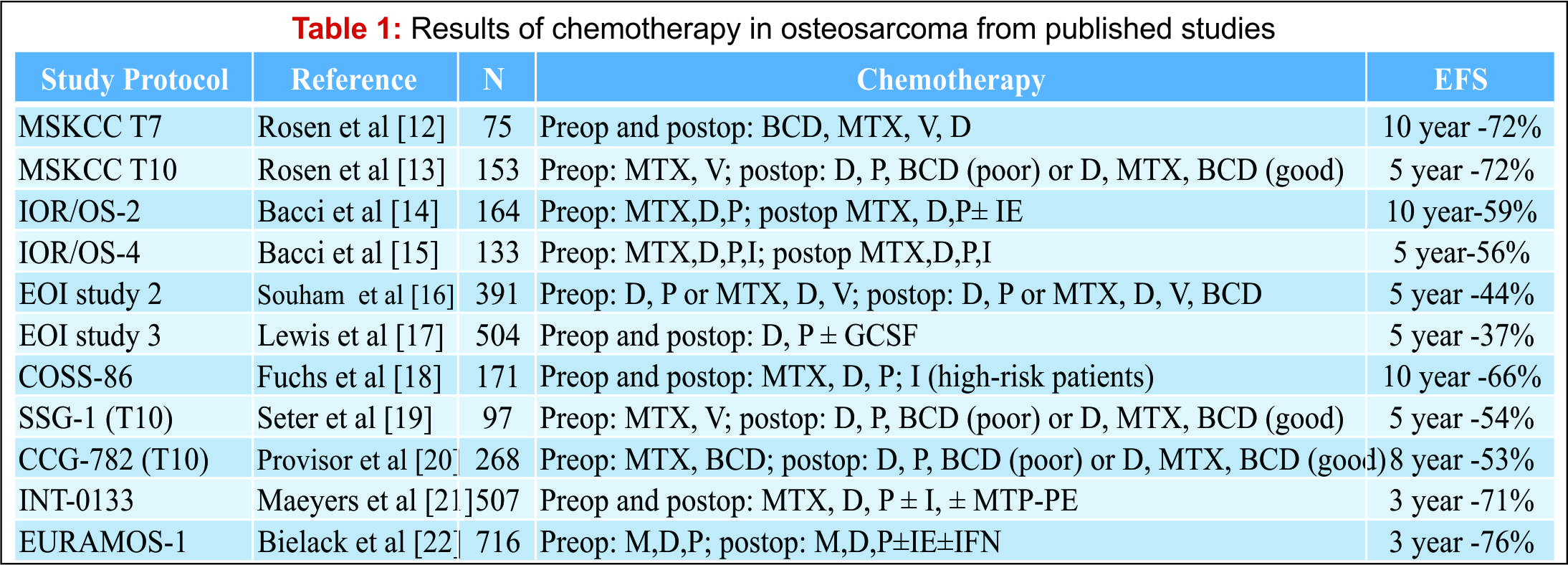
Osteosarcoma is a high grade, primary tumour of bone in which the tumour cells produce osteoid [1]. It is the most common primary bone tumour, with an annual incidence rate of 5.0 per million [2]. Osteosarcoma is predominantly a disease of the young with a peak incidence in the second decade and displays a male predominance which is most pronounced at a younger age [3]. The treatment of osteosarcoma is challenging. The use of neo-adjuvant chemotherapy regimes combined with surgical resection has led to an improvement in outcome. Nevertheless, despite recent advances in surgical technique and chemotherapy agents, the survival rate has plateaued over the last 30 years [4]. There has been much research into prognostic factors that may help predict outcome in osteosarcoma, a number of these have been identified (see Table 1). Authors have suggested the most important, independent risk factors are the response to adjuvant chemotherapy and resection margins [5-7]. This review considers the impact of resection margins with a focus on limb salvage surgery and discusses the significance of response to chemotherapy.

Table 1

Resection margins
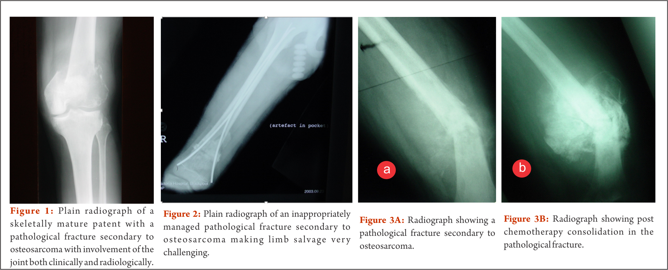
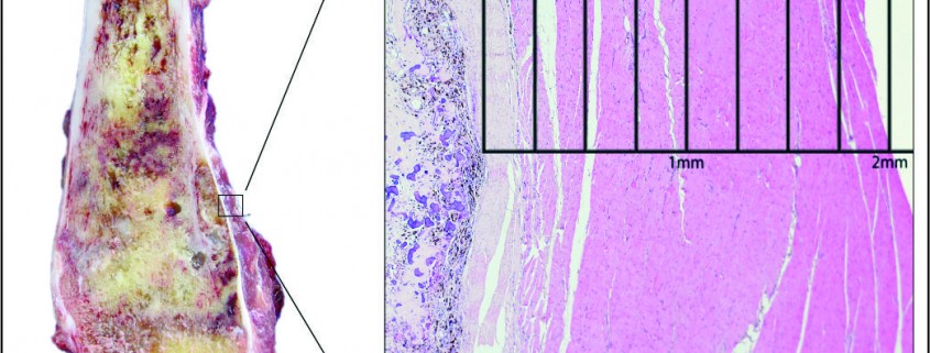
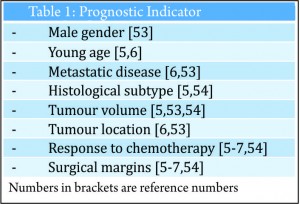
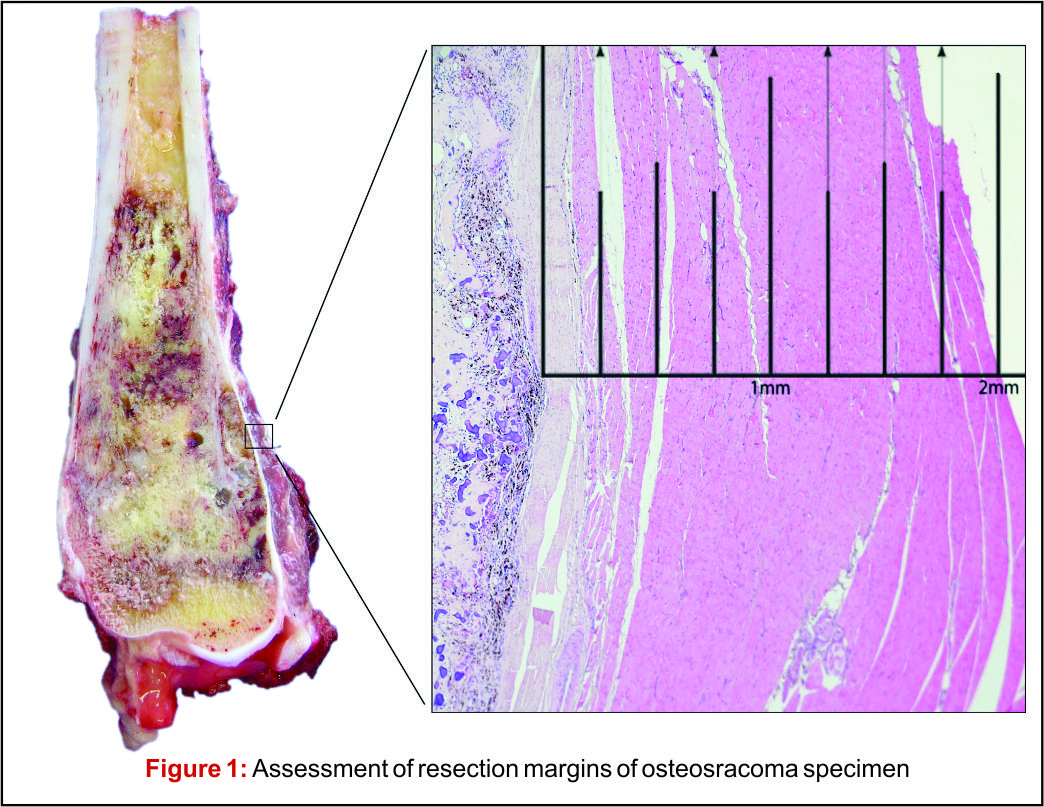
There has been much debate around the margin of clearance required for surgical treatment of osteosarcoma.  Enneking et al. were the first group to formally stage osteosarcoma into three distinct grades according to biologic aggressiveness, tumour site and distant metastases [8].  The authors suggested this system be used in surgical planning and inform the use of marginal, wide or radical resection margins. Nonetheless the definition of marginal or wide resection remains subjective and may vary between surgeons or units and has never been objectively defined (Fig 1).  Kawaguchi et al. developed this concept by giving distinct numerical values for desired resection margin according to the grade to tumour suggesting a 2cm margin was required for low-grade tumours and a 3cm margin was needed for high-grade neoplasms such as osteosarcoma [9].More contemporary studies have failed to reach a consensus on a numerical value for an adequate resection margin. Li et al. reported there was no difference in local recurrence when wide (>5mm) margins and close (<5mm) margins were used [10]. Bispo et al. failed to detect a difference in local recurrence using a margin of 2mm [11]. Betrand et al. found surgical margin to be the only independent risk factor for local recurrence and suggested a margin of 1mm may be adequate [12].  These papers suggest resection does not require a strict numerical margin, however efforts should be made to ensure no margins are intralesional. However, international consensus is in equipoise regarding margins, and this has made interpreting research articles very difficult. Even within units, tumour clear margins and ‘wide’ margins have become interchangeable when in reality they may be completely different and may lead to inappropriate treatment for patients. In the oncological world, the concept of patient specific treatment or ‘personalised medicine’ is gaining popularity and what is correct for one patient, may not be suitable for another patient, even with the same tumour type.

Figure 1

Limb salvage surgery

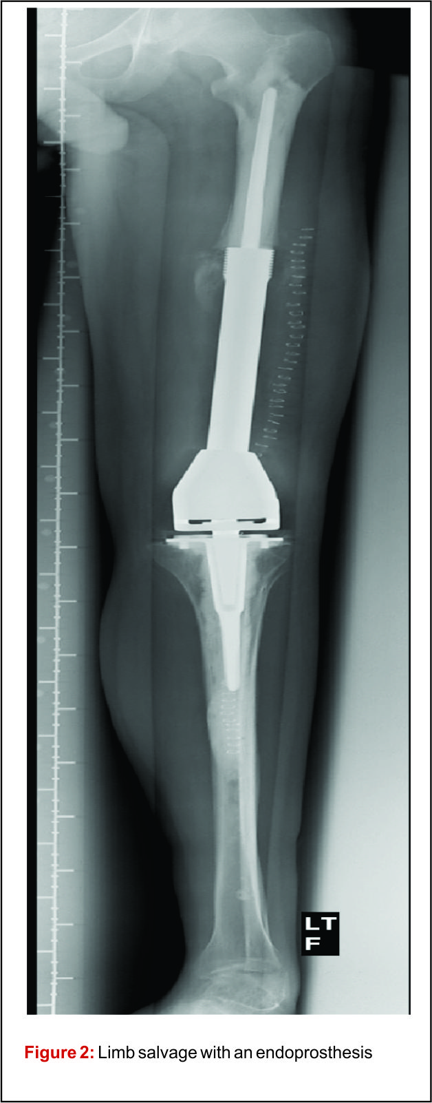
Prior to the advent of effective chemotherapy, the surgical treatment for osteosarcoma involved early radical amputation or disarticulation of the affected limb. Whilst ensuring complete removal of the tumour, performing this radical surgery on young patients caused loss of function and permanent disability, without improving patient survival. Limb salvage surgery (LSS) aims to resect the tumour, whilst maintaining function of the preserved limb, all with minimal risk to the patient (Fig 2).


Figure 2 Figure 3


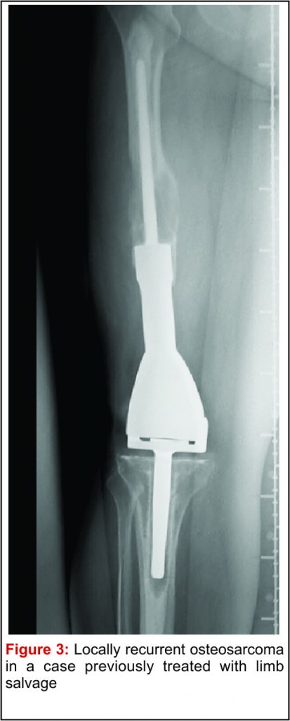
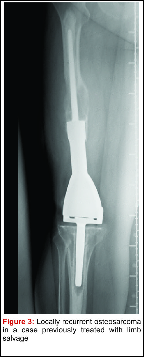
The emergence of efficacious chemotherapy regimes, which acted to reduce tumour burden and reduce metastatic spread, and enhanced imaging techniques such as CT and MRI have led to the increased use of LSS [13-15]. By definition, the use of LSS requires preservation of limb neurovascular structures and narrower surgical margins when compared to amputation. Preservation of tissue during tumour resection has led to the inevitable decrease in resection margins, which potentially risks causing an increase in local recurrence (Fig 3). There are conflicting reports on the rate of local recurrence in LSS with some studies reporting an increase [15-17] and others a decrease [18], when compared to amputation. Considering local recurrence is associated with poor outcome, much work has been done to examine the impact of LSS on survival. Simon et al. were one of the first groups to investigate outcomes following LSS in a multi-centre retrospective review of 227 patients. They reported LSS had a comparable survival rate with amputation at 5 years follow up [19] and provided the impetus for increased uptake of LSS amongst surgeons. A large study by Bacci et al. retrospectively compared the outcome in patients who underwent LSS to amputation. The authors report that whilst LSS was associated with reduced resection margins, local recurrence and 5-year disease free survival were comparable to amputation [20]. These results are confirmed by a number of other groups, with each describing a survival rate equal to or better than that of amputation [15,17,18,21-27].It is important to consider, however, these studies are limited by their retrospective nature. Without robust methods of randomisation, treatment decisions have been based on individual patient and tumour characteristics, local practice and patient choice, leaving them open to the influence of selection bias. Postoperative quality of life is an important outcome measure in osteosarcoma. As patients with osteosarcoma are young and can expect a prolonged period of survival following treatment, the demands put upon a salvaged limb or prosthesis can be great. It is essential, therefore to ensure there is minimal risk of technical failure, the limb provides adequate function for the individual patient and has an acceptable cosmesis for both the patient and their care givers. Measurement of quality of life in children is difficult and there have been relatively few studies assessing this outcome measure. Using objective quality of life scores, LSS and amputation groups report reduced quality of life compared to population norms [29,30]. A meta-analysis comparing quality of life in patients who underwent LSS and amputation found there was no significant difference between the 2 groups. Taking into consideration all the above evidence LSS remains a safe and effective management option and when used in combination with adjuvant chemotherapy offers a good survival outcome.

Chemotherapy/chemonecrosis

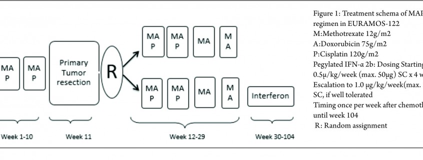
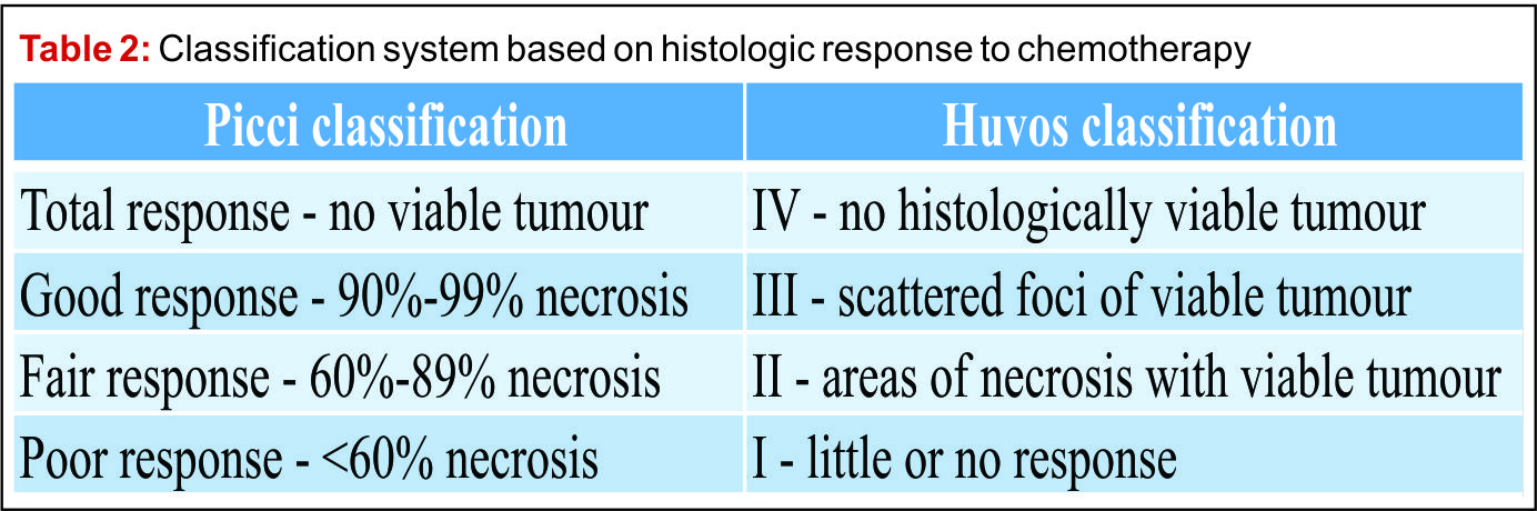
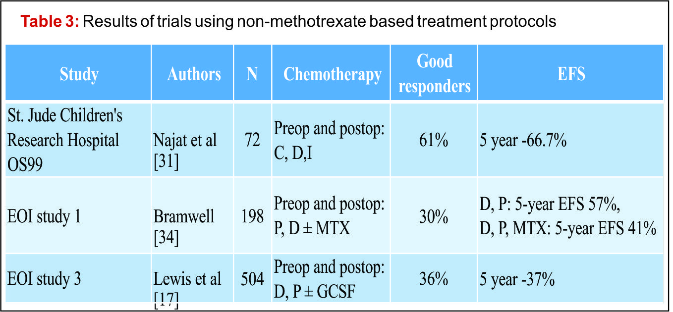
The introduction of chemotherapy regimes alongside surgical resection has led to a dramatic improvement in survival. The use of chemotherapy in the treatment of osteosarcoma began in the 1970s with the use of doxorubicin and high dose methotrexate regimens [31]. Administration of chemotherapy agents before surgical resection as neo-adjuvant therapy enhanced survival from10 – 20% to 70% [32].Current modern chemotherapy regimes are based on combination therapy using methotrexate, adriamycin/doxirubicin and cisplatin. Poor response to chemotherapy has been identified as an important independent risk factor for poor prognosis. Histological evaluation of surgical resection specimens permits the classification of response to chemotherapy as good (>90% tumour necrosis) and poor (<90% tumour necrosis). Patients who display poor response are consistently reported to have worse outcome [33,34]. A number of strategies have been employed to improve results in poor responders. Evidence suggests modification of chemotherapy regime may improve results. Several groups have showed intensification of pre-operative chemotherapy enhances tumour response [35-37] and may improve survival [38-40]. This benefit however, is limited and intensification of chemotherapy beyond a certain level does not improve outcome [36,41-43]. The use of high dose, intensive treatment to induce a good response early in the disease process has also been shown not to convey overall survival benefit [38,42-44]. Further work is therefore required to optimize tumour response and improve outcome in patients with poor chemotherapy response. A recent, large, multi-national study EURAMOS-1 investigated the effect of adding the additional agents, ifosfamide and etoposide, to salvage poor response to chemotherapy, as well as evaluating the addition of pegylated interferon for good responding tumours [45]. The published initial results suggest that the addition of interferon for good responding tumours appears beneficial, however, it was poorly tolerated and frequently refused by patients. Current practice involves assessing tumour response using resection specimens following surgery, after the completion of neo-adjuvant chemotherapy, to advise further treatment[45]. Considering tumour response to chemotherapy is such a significant prognostic factor, measuring response early in the disease process may inform further management choices. Non-invasive imaging techniques such as CT [46], MRI [47-49] and F-FDG PET [50,51] have all be used to investigate response to neo-adjuvant chemotherapy. A combination of F-FDG PET and CT (F-FDG PET-CT) scanning is widely used for the detection of many cancers. Meta-analysis of the current evidence for its use in osteosarcoma has shown F-FDG PET-CT to be a valuable modality to assess chemotherapy-induced necrosis [52]. Newer techniques for evaluating response to chemotherapy prior to surgery, such as functional MRI (fMRI) are also promising and may inform surgeon’s decisions in planning surgical margins.
Patients with poor response to chemotherapy present a complex management challenge. There have been few studies presenting evidence to guide the surgical management of these patients. Bacci et al. suggested that amputation should be considered in the setting of poor response to chemotherapy due to its significant correlation with local recurrence rates [20]. Recent work in Birmingham investigated the influence of resection margins on survival in patients with poor response to chemotherapy [28]. The authors showed there was no survival benefit gained from amputation when compared to LSS with close margins, irrespective of the risk of developing local recurrence [28]. These data demonstrate resection with preservation of the limb to be a safe surgical option even in patients with poor chemonecrosis.

Predicting outcome
The current classification systems used to grade osteosarcoma, pioneered by Enneking, incorporate tumour characteristics including the presence of metastases to guide surgical management and predict prognosis [8]. However, despite the widely accepted importance of response to chemotherapy in prognosis, the current classification fails to reflect this.  In a recent presentation at International Society of Limb Salvage (ISOLS 2015), Jeys et al introduced The Birmingham Classification, which uses numerically defined tumour margins and response to chemotherapy to predict both local recurrence and survival. In this series, chemotherapy response was reported to show a significant effect on the rate of local recurrence and overall survival. It was also reported that a margin of 2mm was a statistically significant cut off value for predicting local recurrence.  Furthermore, combining resection margins (greater or lesser then 2mm) with response to chemotherapy (good, >90% or poor, <90%) was more effective in predicting local recurrence and survival than other staging systems.  This classification, however, requires further validation on a multi-centre basis.


Conclusion

Osteosarcoma continues to present a number to treatment challenges. Although surgical resection margins are an important predictor of outcome, limb salvage surgery with close margins has been shown to be a safe and effective surgical option.  Response to chemotherapy is an important independent predictor of survival.  A distinct group of poor responders exist, who despite modification to chemotherapy regimes and complete surgical excision of the tumour continue to have a poor outcome.  Current classification systems have so far failed to reflect important prognostic indicators, the Birmingham Classification represents a new, robust system for classifying osteosarcoma and predicting outcome.


References

1. Fletcher C, Bridge J, Hogendoorn P, Mertens F. WHO Classification of Tumours of Soft Tissue and Bone. Fourth Edition ed.; 2013. p. 247.
2. Ottaviani G, Jaffe N. The epidemiology of osteosarcoma. Cancer Treat Res 2009;152:3-13.
3. Dorfman HD, Czerniak B. Bone cancers. Cancer 1995 Jan 1;75(1 Suppl):203-210.
4. Allison DC, Carney SC, Ahlmann ER, Hendifar A, Chawla S, Fedenko A, et al. A meta-analysis of osteosarcoma outcomes in the modern medical era. Sarcoma 2012;2012:704872.
5. Bacci G, Longhi A, Versari M, Mercuri M, Briccoli A, Picci P. Prognostic factors for osteosarcoma of the extremity treated with neoadjuvant chemotherapy: 15-year experience in 789 patients treated at a single institution. Cancer 2006 Mar 1;106(5):1154-1161.
6. Bielack SS, Kempf-Bielack B, Delling G, Exner GU, Flege S, Helmke K, et al. Prognostic factors in high-grade osteosarcoma of the extremities or trunk: an analysis of 1,702 patients treated on neoadjuvant cooperative osteosarcoma study group protocols. J Clin Oncol 2002 Feb 1;20(3):776-790.
7. Picci P, Sangiorgi L, Rougraff BT, Neff JR, Casadei R, Campanacci M. Relationship of chemotherapy-induced necrosis and surgical margins to local recurrence in osteosarcoma. J Clin Oncol 1994 Dec;12(12):2699-2705.
8. Enneking WF, Spanier SS, Goodman MA. A system for the surgical staging of musculoskeletal sarcoma. Clin Orthop Relat Res 1980 Nov-Dec;(153)(153):106-120.
9. Kawaguchi N, Ahmed AR, Matsumoto S, Manabe J, Matsushita Y. The concept of curative margin in surgery for bone and soft tissue sarcoma. Clin Orthop Relat Res 2004 Feb;(419)(419):165-172.
10. Li X, Moretti VM, Ashana AO, Lackman RD. Impact of close surgical margin on local recurrence and survival in osteosarcoma. Int Orthop 2012 Jan;36(1):131-137.
11. Bispo Junior RZ, Camargo OP. Prognostic factors in the survival of patients diagnosed with primary non-metastatic osteosarcoma with a poor response to neoadjuvant chemotherapy. Clinics (Sao Paulo) 2009;64(12):1177-1186.
12. Bertrand TE, Cruz A, Binitie O, Cheong D, Letson GD. Do Surgical Margins Affect Local Recurrence and Survival in Extremity, Nonmetastatic, High-grade Osteosarcoma? Clin Orthop Relat Res 2015 May 27.
13. Ayerza MA, Farfalli GL, Aponte-Tinao L, Muscolo DL. Does increased rate of limb-sparing surgery affect survival in osteosarcoma? Clin Orthop Relat Res 2010 Nov;468(11):2854-2859.
14. Kotz R, Dominkus M, Zettl T, Ritschl P, Windhager R, Gadner H, et al. Advances in bone tumour treatment in 30 years with respect to survival and limb salvage. A single institution experience. Int Orthop 2002;26(4):197-202.
15. Tsuchiya H, Tomita K. Prognosis of osteosarcoma treated by limb-salvage surgery: the ten-year intergroup study in Japan. Jpn J Clin Oncol 1992 Oct;22(5):347-353.
16. Brosjo O. Surgical procedure and local recurrence in 223 patients treated 1982-1997 according to two osteosarcoma chemotherapy protocols. The Scandinavian Sarcoma Group experience. Acta Orthop Scand Suppl 1999 Jun;285:58-61.
17. Rougraff BT, Simon MA, Kneisl JS, Greenberg DB, Mankin HJ. Limb salvage compared with amputation for osteosarcoma of the distal end of the femur. A long-term oncological, functional, and quality-of-life study. J Bone Joint Surg Am 1994 May;76(5):649-656.
18. Sluga M, Windhager R, Lang S, Heinzl H, Bielack S, Kotz R. Local and systemic control after ablative and limb sparing surgery in patients with osteosarcoma. Clin Orthop Relat Res 1999 Jan;(358)(358):120-127.
19. Simon MA, Aschliman MA, Thomas N, Mankin HJ. Limb-salvage treatment versus amputation for osteosarcoma of the distal end of the femur. 1986. J Bone Joint Surg Am 2005 Dec;87(12):2822.
20. Bacci G, Ferrari S, Lari S, Mercuri M, Donati D, Longhi A, et al. Osteosarcoma of the limb. Amputation or limb salvage in patients treated by neoadjuvant chemotherapy. J Bone Joint Surg Br 2002 Jan;84(1):88-92.
21. Mavrogenis AF, Abati CN, Romagnoli C, Ruggieri P. Similar survival but better function for patients after limb salvage versus amputation for distal tibia osteosarcoma. Clin Orthop Relat Res 2012 Jun;470(6):1735-1748.
22. Lindner NJ, Ramm O, Hillmann A, Roedl R, Gosheger G, Brinkschmidt C, et al. Limb salvage and outcome of osteosarcoma. The University of Muenster experience. Clin Orthop Relat Res 1999 Jan;(358)(358):83-89.
23. Bacci G, Ruggieri P, Bertoni F, Ferrari S, Longhi A, Biagini R, et al. Local and systemic control for osteosarcoma of the extremity treated with neoadjuvant chemotherapy and limb salvage surgery: the Rizzoli experience. Oncol Rep 2000 Sep-Oct;7(5):1129-1133.
24. Eilber FR, Mirra JJ, Grant TT, Weisenburger T, Morton DL. Is amputation necessary for sarcomas? A seven-year experience with limb salvage. Ann Surg 1980;192(4):431-438.
25. Jiang F, Shi Y, Li GJ, Zhou F. A meta-analysis of limb-salvage versus amputation in the treatment of patients with Ennekingdouble daggerU pathologic fracture osteosarcoma. Indian J Cancer 2015 Feb;51 Suppl 2:e21-4.
26. Yin K, Liao Q, Zhong D, Ding J, Niu B, Long Q, et al. Meta-analysis of limb salvage versus amputation for treating high-grade and localized osteosarcoma in patients with pathological fracture. Exp Ther Med 2012 Nov;4(5):889-894.
27. Schrager J, Patzer RE, Mink PJ, Ward KC, Goodman M. Survival outcomes of pediatric osteosarcoma and Ewing’s sarcoma: a comparison of surgery type within the SEER database, 1988-2007. J Registry Manag 2011 Autumn;38(3):153-161.
28. Reddy KI, Wafa H, Gaston CL, Grimer RJ, Abudu AT, Jeys LM, et al. Does amputation offer any survival benefit over limb salvage in osteosarcoma patients with poor chemonecrosis and close margins? Bone Joint J 2015 Jan;97-B(1):115-120.
29. Bekkering WP, Vliet Vlieland TP, Koopman HM, Schaap GR, Schreuder HW, Beishuizen A, et al. Quality of life in young patients after bone tumor surgery around the knee joint and comparison with healthy controls. Pediatr Blood Cancer 2010 May;54(5):738-745.
30. Eiser C, Darlington AS, Stride CB, Grimer R. Quality of life implications as a consequence of surgery: limb salvage, primary and secondary amputation. Sarcoma 2001;5(4):189-195.
31. Rosen G, Murphy ML, Huvos AG, Gutierrez M, Marcove RC. Chemotherapy, en bloc resection, and prosthetic bone replacement in the treatment of osteogenic sarcoma. Cancer 1976 Jan;37(1):1-11.
32. Anninga JK, Gelderblom H, Fiocco M, Kroep JR, Taminiau AH, Hogendoorn PC, et al. Chemotherapeutic adjuvant treatment for osteosarcoma: where do we stand? Eur J Cancer 2011 Nov;47(16):2431-2445.
33. Winkler K, Beron G, Delling G, Heise U, Kabisch H, Purfurst C, et al. Neoadjuvant chemotherapy of osteosarcoma: results of a randomized cooperative trial (COSS-82) with salvage chemotherapy based on histological tumor response. J Clin Oncol 1988 Feb;6(2):329-337.
34. Goorin AM, Schwartzentruber DJ, Devidas M, Gebhardt MC, Ayala AG, Harris MB, et al. Presurgical chemotherapy compared with immediate surgery and adjuvant chemotherapy for nonmetastatic osteosarcoma: Pediatric Oncology Group Study POG-8651. J Clin Oncol 2003 Apr 15;21(8):1574-1580.
35. Fuchs N, Bielack SS, Epler D, Bieling P, Delling G, Korholz D, et al. Long-term results of the co-operative German-Austrian-Swiss osteosarcoma study group’s protocol COSS-86 of intensive multidrug chemotherapy and surgery for osteosarcoma of the limbs. Ann Oncol 1998 Aug;9(8):893-899
36. Smeland S, Muller C, Alvegard TA, Wiklund T, Wiebe T, Bjork O, et al. Scandinavian Sarcoma Group Osteosarcoma Study SSG VIII: prognostic factors for outcome and the role of replacement salvage chemotherapy for poor histological responders. Eur J Cancer 2003 Mar;39(4):488-494.
37. Winkler K, Bielack SS, Delling G, Jurgens H, Kotz R, Salzer-Kuntschik M. Treatment of osteosarcoma: experience of the Cooperative Osteosarcoma Study Group (COSS). Cancer Treat Res 1993;62:269-277.
38. Fuchs N, Bielack SS, Epler D, Bieling P, Delling G, Korholz D, et al. Long-term results of the co-operative German-Austrian-Swiss osteosarcoma study group’s protocol COSS-86 of intensive multidrug chemotherapy and surgery for osteosarcoma of the limbs. Ann Oncol 1998 Aug;9(8):893-899
39. Ferrari S, Mercuri M, Picci P, Bertoni F, Brach del Prever A, Tienghi A, et al. Nonmetastatic osteosarcoma of the extremity: results of a neoadjuvant chemotherapy protocol (IOR/OS-3) with high-dose methotrexate, intraarterial or intravenous cisplatin, doxorubicin, and salvage chemotherapy based on histologic tumor response. Tumori 1999 Nov-Dec;85(6):458-464.
40. Bacci G, Ferrari S, Bertoni F, Ruggieri P, Picci P, Longhi A, et al. Long-term outcome for patients with nonmetastatic osteosarcoma of the extremity treated at the istituto ortopedico rizzoli according to the istituto ortopedico rizzoli/osteosarcoma-2 protocol: an updated report.J Clin Oncol 2000 Dec 15;18(24):4016-4027.
41. Ferrari S, Smeland S, Mercuri M, Bertoni F, Longhi A, Ruggieri P, et al. Neoadjuvant chemotherapy with high-dose Ifosfamide, high-dose methotrexate, cisplatin, and doxorubicin for patients with localized osteosarcoma of the extremity: a joint study by the Italian and ScandinavianSarcoma Groups. J Clin Oncol 2005 Dec 1;23(34):8845-8852.
42. Lewis IJ, Nooij MA, Whelan J, Sydes MR, Grimer R, Hogendoorn PC, et al. Improvement in histologic response but not survival in osteosarcoma patients treated with intensified chemotherapy: a randomized phase III trial of the European Osteosarcoma Intergroup. J Natl Cancer Inst 2007Jan 17;99(2):112-128.
43. Meyers PA, Gorlick R, Heller G, Casper E, Lane J, Huvos AG, et al. Intensification of preoperative chemotherapy for osteogenic sarcoma: results of the Memorial Sloan-Kettering (T12) protocol. J Clin Oncol 1998 Jul;16(7):2452-2458.
44. Eselgrim M, Grunert H, Kuhne T, Zoubek A, Kevric M, Burger H, et al. Dose intensity of chemotherapy for osteosarcoma and outcome in the Cooperative Osteosarcoma Study Group (COSS) trials. Pediatr Blood Cancer 2006 Jul;47(1):42-50.
45. Whelan JS, Bielack SS, Marina N, Smeland S, Jovic G, Hook JM, et al. EURAMOS-1, an international randomised study for osteosarcoma: results from pre-randomisation treatment. Ann Oncol 2015 Feb;26(2):407-414.
46. Wellings RM, Davies AM, Pynsent PB, Carter SR, Grimer RJ. The value of computed tomographic measurements in osteosarcoma as a predictor of response to adjuvant chemotherapy. Clin Radiol 1994 Jan;49(1):19-23.
47. Bajpai J, Gamnagatti S, Kumar R, Sreenivas V, Sharma MC, Khan SA, et al. Role of MRI in osteosarcoma for evaluation and prediction of chemotherapy response: correlation with histological necrosis. Pediatr Radiol 2011 Apr;41(4):441-450.
48. Uhl M, Saueressig U, Koehler G, Kontny U, Niemeyer C, Reichardt W, et al. Evaluation of tumour necrosis during chemotherapy with diffusion-weighted MR imaging: preliminary results in osteosarcomas. Pediatr Radiol 2006 Dec;36(12):1306-1311.
49. Hayashida Y, Yakushiji T, Awai K, Katahira K, Nakayama Y, Shimomura O, et al. Monitoring therapeutic responses of primary bone tumors by diffusion-weighted image: Initial results. Eur Radiol 2006 Dec;16(12):2637-2643.
50. Im HJ, Kim TS, Park SY, Min HS, Kim JH, Kang HG, et al. Prediction of tumour necrosis fractions using metabolic and volumetric 18F-FDG PET/CT indices, after one course and at the completion of neoadjuvant chemotherapy, in children and young adults with osteosarcoma. Eur J Nucl MedMol Imaging 2012 Jan;39(1):39-49.
51. Cheon GJ, Kim MS, Lee JA, Lee SY, Cho WH, Song WS, et al. Prediction model of chemotherapy response in osteosarcoma by 18F-FDG PET and MRI. J Nucl Med 2009 Sep;50(9):1435-1440.
52. Hongtao L, Hui Z, Bingshun W, Xiaojin W, Zhiyu W, Shuier Z, et al. 18F-FDG positron emission tomography for the assessment of histological response to neoadjuvant chemotherapy in osteosarcomas: a meta-analysis. Surg Oncol 2012 Dec;21(4):e165-70.
53. Duchman KR, Gao Y, Miller BJ. Prognostic factors for survival in patients with high-grade osteosarcoma using the Surveillance, Epidemiology, and End Results (SEER) Program database. Cancer Epidemiol 2015 Aug;39(4):593-599.
54. Ferrari S, Bertoni F, Mercuri M, Picci P, Giacomini S, Longhi A, et al. Predictive factors of disease-free survival for non-metastatic osteosarcoma of the extremity: an analysis of 300 patients treated at the Rizzoli Institute. Ann Oncol 2001 Aug;12(8):1145-1150.


How to Cite this article: Cloake T, Jeys L.How important are surgical margins in Osteosarcoma? . Journal of  Bone and Soft Tissue Tumors Jan-Apr 2016;2(1):22-26.

Dr. Thomas P Cloake

Dr. Thomas P Cloake

Prof Lee M Jeys

Prof Lee M Jeys


 (Abstract    Full Text HTML)      (Download PDF)


Chemotherapy in Osteosarcoma: Current Strategies

Volume 2 | Issue 1 | Jan-Apr 2016 | Page 27-32 | Sandeep Jain, Gauri Kapoor.


Authers: Sandeep Jain[1], Gauri Kapoor[1]

[1]Department of Pediatric Hematology and Oncology, Rajiv Gandhi Cancer Institute & Research Centre, Delhi.

Address of Correspondence
Dr. Gauri Kapoor MD, PhD
Director Department of Pediatric Hematology and Oncology,
Rajiv Gandhi Cancer Institute & Research Centre, Delhi.
Email: kapoor.gauri@gmail.com


Abstract

Incorporation of chemotherapy to multi-modality management of high grade osteosarcoma has led to remarkable improvement in survival rates. Its use in the neoadjuvant setting is now accepted as standard of care and has the added advantage of providing important information on histologic response. Survival rates for non-metastatic disease are nearly 70%. Outcome of patients with poor histological response and those with metastatic and recurrent disease continues to be unsatisfactory and an ongoing challenge. Therefore, there is a need to develop novel agents and biologically driven strategies to target these disease subgroups. The current review focusses on evolution of chemotherapy, controversies in its use and current standard of care for osteosarcoma.
Keywords: Osteosarcoma, chemotherapy, neoadjuvant chemotherapy.


Introduction
Osteosarcoma is the most common primary malignant bone tumor in children and adolescents accounting for 4% of all pediatric malignancies. Approximately 20% of children present with metastatic disease at diagnosis and it remains, unquestionably the most important factor affecting long term survival. Prior to 1970, the prognosis of patients with osteosarcoma was dismal, with a 10–20 % overall survival despite being treated with radical surgeries [1-3]. Outcome of patients with osteosarcoma has improved in the past three decades with the addition of effective systemic polychemotherapy and advances in surgical resection. These have led to improvements in overall survival of patients with localized disease to the tune of 70% [4-5]. Various well coordinated systemic trials by different co-operative groups in North America and Europe have identified high-dose methotrexate (HD-MTX), cisplatin, doxorubicin, ifosfamide and etoposide as active cytotoxic agents and combinations of these drugs make up the cornerstone of treatment. Chemotherapy not only takes care of micrometastatic disease at diagnosis but also facilitates limb salvage surgery. The choice of regimen and optimal schedule of chemotherapy is somewhat controversial. In this review we focus on evolution of chemotherapy, controversies in chemotherapy use and current standard of care.

Evolution of chemotherapy
Before the introduction of chemotherapy, the outcome of patients with osteosarcoma was only 15-20%, despite adequate local control. Most patients succumbed to metastatic lung disease. These findings led to the conclusion that patients with osteosarcoma have microscopic metastatic disease at the time of diagnosis and this prompted investigators to identify active agents to target it. Initial studies to demonstrate chemosensitivity of osteosarcoma were done in the early 1970s by Sutow et al [6]. He developed a regimen called “Conpadri”which included cyclophosphamide, Oncovin (vincristine), doxorubicin (adriamycin), and L-phenylalaninemustard. Later on with the inclusion of HD-MTX, the acronym was changed to “Compadri’ [6-7]. These regimes were the first rational attempt at confirming the role of adjuvant combination chemotherapy using drugs with non-overlapping toxicities in osteosarcoma. Compadri I–III yielded a 41% 18-month disease-free survival [8]. These results suggested that addition of chemotherapy improved survival in patients with osteosarcoma. However, in the absence of randomized trials, it was not clear, to what extent improvement in surgical techniques and radiological studies contributed to achieving these results. These observations were further supported by the first randomized trial from Mayo clinic wherein patients were randomized to receive adjuvant vincristine and HD-MTX versus surgery alone[9]. This trial did not show any difference between the two arms. All these concerns were put to rest by two subsequent randomized controlled trials from North America that clearly established the survival benefit of adjuvant chemotherapy. In both these trials patients receiving no adjuvant treatment had a 2 year event free survival of just 20% compared to 66% and 55% in patients who received adjuvant chemotherapy [10-11]. These trials also established adriamycin, cisplatin, HD-MTX and alkylating drugs like ifosfamide and etoposide as active agents in the treatment of osteosarcoma. The various trials showing benefit of chemotherapy in osteosarcoma are listed in Table 1[12-22].

Table 1

Role of neoadjuvant chemotherapy
The concept of neoadjuvant chemotherapy (NACT) was first introduced at Memorial Sloan-Kettering Cancer Center (MSKCC) in their T-10 protocol [23]. Preoperative chemotherapy was administered in an effort to increase the number of patients who could undergo limb salvage as the surgeons needed time to order the prosthetic devices. Administration of NACT also had the theoretical advantage of treating presumed microscopic metastatic disease. The outcome of the T-10 trial was similar to that of the Multi Institutional Osteosarcoma Study (MIOS), with a 65% survival rate at 5 years. Importantly, the results of this trial laid the foundation for the subsequent important association between histologic necrosis and prognosis. However, there were concerns regarding the impact of delayed surgery among patients with chemo-resistant disease as well as the probability of development of resistant clone in those with high volume disease. To answer this concern Pediatric Oncology Group conducted a randomized clinical trial (POG 8651) between 1986 and 1993, comparing NACT with adjuvant chemotherapy. This trial compared immediate surgery followed by postoperative adjuvant chemotherapy with 10 weeks of the NACT (same drugs) followed by surgery in 100 patients under the age of 30 years with non-metastatic,high grade osteosarcoma. Chemotherapy consisted of alternating courses of HD-MTX with leucovorin rescue, cisplatin, doxorubicin, and bleomycin, cyclophosphamide,dactinomycin (BCD). The five-year relapse-free survival rates were similar between the two groups, 65% versus 61% for adjuvant and neoadjuvant arms respectively. There was also no difference in the number of patients who underwent limb salvage procedures (55% and 50 % for immediate and delayed surgery, respectively) [24]. On the basis of these results, the use of preoperative chemotherapy has become standard of care, given its advantages, as it allows sufficient time for surgical planning, potentially facilitates tumor removal, and permits evaluation of response to therapy. Several investigators in single and multi-institutional studies in the United States and across Europe, support this general strategy [13,14,16,18].

Histological response to chemotherapy
Most trials reveal that patients with greater than 90% necrosis following NACT have significantly better event free survival (EFS) compared to those with less than 90% necrosis. Several grading systems have been developed for assessing the effect of preoperative chemotherapy on the tumor. The two most commonly used classification systems are the Picci and Huvos classifications[Table 2]. The Institute of Rizzoli (IOR) reviewed data on localized extremity osteosarcoma in more than 1000 patients over the 19-year period from 1983 to 2002 [25]. Fifty-nine percent of all patients had good response to chemotherapy (Picci), and had a 5-year survival of 76%, compared to 56% for poor responders. The Cooperative Osteosarcoma Study group (COSS) database analyzed 1,700 patients between 1980 and 1998 that included all sites, ages, and presence or absence of metastases [26]. The data revealed that 55.6% of patients had good response to therapy. The 5-year survival rate for good and poor responders was 77.8% and 55.5% respectively. The European Osteosarcoma Intergroup (EOI) analyzed data of two consecutive studies between 1983 and 1986 and 1986 and 1991 [27]. A total of 570 patients were analyzed in the report. This analysis is notable for several differences compared to the COSS and IOR analyses. Only 28% of patients had a good histologic response, whereas 72% of patients had a poor histologic response. Their 5-year survival rate was 75% and 45% respectively. Interestingly, many of the patients included in the analysis did not receive HD-MTX as they were randomized to receive either doxorubicin and cisplatin or more intensive therapy including doxorubicin and HD-MTX. This data clearly established that histological response to chemotherapy is an important prognostic factor.

Table 2

Intensification of neoadjuvant and adjuvant chemotherapy
As it became clear that the degree of histological necrosis after pre-operative chemotherapy predicts survival, efforts were directed to intensify chemotherapy so as to achieve maximum therapeutic response. This strategy of preoperative chemotherapy intensification has been tested in COSS-86 and MSKCC T-12 study [14,28]. Although this strategy resulted in increased proportion of good responders achieving >90% necrosis, it did not translate into improved overall survival (OS) or EFS rates. Till date only INT-0133 study has shown benefit of NACT intensification [19]. The next group of trials focused to alter or intensify chemotherapy for patients with sub-optimal response to preoperative chemotherapy. In the early 1980s at Memorial Sloan-Kettering Cancer Center, poor responders had cisplatin substituted for HD-MTX in addition to continuing BCD (bleomycin, cyclophosphamide, and dactinomycin) and doxorubicin [13]. Survival of patients with intensified adjuvant treatment was similar to others. Several other reports have also failed to demonstrate benefit of intensification of therapy for poor responders[20,29]. Thus, till date it has not been possible to improve the outcome of poor responders by altering postoperative chemotherapy. An explanation for this may be that the NACT response is a surrogate measure of chemo-sensitivity of tumor and an inherently biologic unresponsive tumor is not modifiable by currently available therapies.

Table 3

Role of intra-arterial chemotherapy
The intra-arterial route was introduced in an attempt to enhance the efficacy of drugs by increasing the local concentration of chemotherapy. Alkylating agents like ifosfamide and cyclophosphamide could not be used as they required phosphorylation in liver for activation. Doxorubicin was not a suitable agent as it is associated with skin and subcutaneous necrosis. MTX achieved high tumoricidal concentrations intra-arterially but similar concentrations could also be attained via the intravenous route. Intra-arterial cisplatin was therefore, selected and found to be highly effective. Response rates with the intra-arterial route were better when compared to the intravenous route[30]. It has been used extensively at the MD Anderson Cancer Center in the TIOS pediatric trials. It was highly effective in patients with pathological fractures and neurovascular involvement. Unfortunately, intra-arterial route is labor intensive and requires general anaesthesia or conscious sedation in a radiological suite. It also requires intensive monitoring of the distal arterial vascular status during and after the infusion. Moreover, similar results could be achieved with multiple courses of combination chemotherapy administered by the intravenous route over a more prolonged period. Therefore, intra-arterial route is generally not preferred.

High Dose Methotrexate
High dose methotrexate is one of the oldest drugs used in the treatment of osteosarcoma. It is generally administered over 4-6 hours and requires aggressive hydration, leucovorin rescue, serum level monitoring and adequate infrastructure to safeguard delivery and manage toxicity. Moreover, it adds substantially to the overall cost of treatment. In addition, there are no randomized studies to compare the efficacy of higher versus intermediate doses of HD-MTX plus doxorubicin and cisplatin versus doxorubicin/cisplatin alone. Furthermore, investigators at St. Jude Children’s Research Hospital have demonstrated good outcomes with five-year EFS and OS of 66% and 75% respectively with non-methotrexate-containing chemotherapy regimen consisting of carboplatin, ifosfamide and doxorubicin [31]. All of this has led to considerable controversy regarding the optimum role of HD-MTX. Methotrexate is the only active agent that has been subjected to a comparative trial of efficacy with another active agent i.e. cisplatin. Compared to 5-20% survival of historical controls in pre-chemotherapy era, HD-MTX increased survival to 40% – 60% as a single agent. When combined with other active agents like cisplatin and doxorubicin the long term survival of 65% – 75% was reported [12-15]. Many studies have shown a favorable correlation between peak serum levels and outcome [19,32-33]. Therefore, optimum doses and administration schedule is crucial to derive optimum benefit from HD-MTX therapy. Chemotherapy regimes devoid of HD-MTX were considered, among the “major poor prognostic factors” in the treatment of osteosarcoma by Graf et al. [33]. Despite the absence of randomized trials evaluating osteosarcoma treatment with and without HD-MTX, it is generally acknowledged that methotrexate is a standard component of almost all contemporary osteosarcoma protocols in children and adolescents.

Current standard of care for patients with osteosarcoma
It is well established that chemotherapy is an integral component of osteosarcoma treatment and is essential in addition to local surgery in order to achieve a reasonable expectation of cure. Therefore, optimum treatment for osteosarcoma demands a multidisciplinary strategy. The treatment generally consists of three stages: initial cytoreduction with chemotherapy to eradicate micro metastatic disease and facilitate effective local control measures with wide negative margins; and consolidation therapy for eradication of occult residual disease to reduce the likelihood of tumor recurrence. Importantly, NACT not only helps to achieve optimal cytoreduction in facilitating limb salvage procedures but also provides a chance to assess the histologic response to chemotherapy. Most treatment protocols include cisplatin, doxorubicin and HD-MTX with or without ifosfamide plus etoposide(IE). In the recently concluded EURAMOS study, all patients received NACT: 2 blocks of MAP (methotrexate, doxorubicin and cisplatin) chemotherapy for 10 weeks followed by surgery (wide excision). Surgical excision of tumor with oncologically safe margins was the best means of local control. Post surgery, poor responders were randomized to receive MAP for 28 weeks with or without IE. All good responders continued on MAP for 28 weeks and then were randomized to no further therapy and maintenance therapy with pegylated interferon. This is the largest international trial in the history of osteosarcoma treatment and its results show that intensification of adjuvant chemotherapy by addition of IE in poor responders did not improve survival. Furthermore, in good responders addition of pegylated interferon maintenance was not useful [22]. Schema of this treatment is shown in [Figure 1] and most of the study groups endorse this strategy as current standard of care.

Figure 1

Non-methotrexate based chemotherapy for countries with limited resources
There is paucity of published data on osteosarcoma from India. Historically, the role of high dose methotrexate in the treatment of osteosarcoma has always been debatable. From the practical perspective, it requires rigorous pharmacokinetic monitoring and often the infrastructure required for monitoring is not available in many centers with limited resources. Therefore, most of the centers in India use cispaltin, doxorubicin and ifosfamide based chemotherapy. Pathak et al have reported relapse-free survival was 72% nonmetastatic osteogenic sarcoma of the extremities using cisplatin and doxorubicin as adjuvant therapy [35]. Recently, results from a single center study from India have revealed 2yr progression free survival of 70 % for patients with non-metastatic osteosarcoma [36]. In light of these results use of non methotrexate based therapy in resource constraint setting seems justified. In addition, it is desirable to focus on developing infrastructure to provide limb salvage procedures and direct resources to develop indigenous affordable prosthesis.

Treatment of relapsed osteosarcoma
Treatment of relapsed osteosarcoma has not been tested in randomized clinical trials, and thus, there is no single standard approach. Prognosis of patients with relapse depends on duration of off therapy and site of relapse. In a large database of 565 osteosarcoma patients who relapsed after being treated with one of three different NACT protocols within the European Osteosarcoma Intergroup, five year survival post relapse in those whose disease recurred after two years versus within two years of randomization was 35 versus 14 percent, respectively[37]. There is no reasonable chance of cure without complete surgical resection of all sites of disease. Choice of chemotherapy depends on agents used in front line therapy. In most of contemporary studies, most of the patients receive cisplatin and doxorubicin in front line therapy. Therefore, ifosfamide, etoposide and HD-MTX are the most commonly used drugs in relapse setting. In general, patients should be treated with any of the four most active agents that were not included in front line therapy. The use of high-dose chemotherapy with autologous hematopoietic stem cell rescue has been applied to salvage therapy. However, at least two small pilot studies failed to demonstrate significant advantage of standard salvage therapy approaches [38-39].

Newer therapies
There has been significant progress in the management of patients with osteosarcoma from 1970 to 1990. However, thereafter, progress has been stalled due to limited options available for patients with poor histologic response and those with metastatic and recurrent disease. It is clear that intensification of available chemotherapeutic agents has not translated into survival benefit for these group of patients and novel agents are required. Some of the agents being tested include mTOR inhibitor (ridaforolimus), inhibitors of insulin-like growth factor I receptor, tyrosine kinase inhibitor (sorafenib), microtubule inhibitor (oferibulin), human monoclonal antibody against RANKL (Denosumab) and anti-disialoganglioside antibody (theuseofan) [40-44]. Some of these agents have demonstrated promising results in preclinical data and may offer a potential role in adjuvant therapy in the future.

Acute toxicities and Late Effects
The most frequent acute toxicities due to chemotherapy are infections secondary to myelosuppression and mucositis. Renal dysfunction may lead to hypomagnesemia and other electrolyte abnormalities from tubular and glomerular damage induced by ifosfamide and cisplatin respectively. Ototoxicity from cisplatin and cardiac dysfunction related to anthracyclines are the other commonly observed side effects. Late effects in osteosarcoma may be attributed to local therapy i.e. surgery or to systemic chemotherapy. Those related to chemotherapy are usually agent specific. Doxorubicin is known to cause chronic cardiomyopathy which is dependent on the total cumulative dose. Longhi et al reported 2% incidence of symptomatic cardiomyoathy at a median follow up of 10 years [45]. In general, cumulative dose of doxorubicin is usually limited to less than 450 mg/m2. Anthracycline and alkylating agents may also result in second malignant neoplasm (SMN). The same authors report a 10-year and 20-year cumulative incidence of SMN of 4.9% and 6.1% respectively in osteosarcoma survivors. The alkylating agent, ifosfamide is associated with infertility, especially male infertility, so sperm cryopreservation should be offered to postpubertal boys if treatment plan includes alkylating agents. In addition, ifosfamide can cause a persistent renal tubular electrolyte loss and, less commonly, a decrease in glomerular function, in a dose-dependent fashion.


 Conclusion

Inclusion of chemotherapy in the multimodality treatment of osteosarcoma has undoubtedly improved survival from a dismal 20% to the present 60%. NACT has enabled limb salvage rates to the tune of 90-95% in most advanced centers. Outcome of patients with poor histological response and those with metastatic and recurrent disease continues to be unsatisfactory and an ongoing challenge. Therefore, there is a need to develop novel agents and biologically driven strategies to target these disease subgroups.


References

1. Francis K, Worcester JJ. Radical resection for tumors of the shoulder with preservation of a functional extremity. J Bone Joint Surg Am 1962;44:1423
2. Marcove RC, Miké V, Hajek JV, Levin AG, Hutter RV. Osteogenic sarcoma under the age of twenty-one: A review of one hundred and forty-five operative cases. J Bone Joint Surg Am 1970;52(3):411
3. Friedman MA, Carter SK. The therapy of osteogenic sarcoma: current status and thoughts for the future. J SurgOncol, 1972;4:482–510
4. Link MP, Gebhardt MC, Meyers PA. Osteosarcoma. In: Pizzo A, Poplack G (eds), Principles and Practice of Pediatric Oncology, 4th edition, Philadelphia: Lippincott Williams and Wilkins. 2002;1051–89.
5. Meyers P, Osteosarcoma. In: Pappo A (ed.), Pediatric Bone and Soft Tissue Sarcomas, Berlin: Springer-Verlag. 2006;219–33
6. Sutow WW. Combination Chemotherapy with Adria- mycin (NSC-123127) in Primary Treatment of Osteogen- icsarcoma (Part III) Cancer Chemotherapy Reports. 1975;6: 315-317
7. Sutow WW, Gehan EA, Dyment PC. Multi- Drug Adjuvant Chemotherapy in Osteosarcoma. Interim Report of the Southwest Oncology Group Studies. Cancer Chemotherapy Reports 1962; 62:265-269
8. Herson J, Sutow WW, Elder K. Adjuvant chemotherapy in nonmetastatic osteosarcoma: a Southwest Oncology Group study. Medical and Pediatric Oncology 1980;8:343– 352
9. Edmonson JH, Creagan ET, Gilchrist GS. Phase II study of high-dose methotrexate in patients with unresectable metastatic osteosarcoma. Cancer Treat Rep 1981; 65:538
10. Link MP, Goorin AM, Miser AW et al. The effect of adjuvant chemotherapy on relapse free survival in patients with osteosarcoma of the extremity. N Engl J Med 1986; 314: 1600-1606
11. Eilber F, Giuliano A, Eckardt J, Patterson K, Moseley S, Goodnight J. Adjuvant chemotherapy for osteosarcoma: a randomized prospective trial. J.Clin.Oncol 1987; 5: 21-26.
12. Rosen G, Nirenberg A, Caparros B et al. Osteogenic sarcoma: eight-percent, three-year, disease-free survival with combination chemotherapy (T-7). Natl Cancer Inst Monogr 1981;56:213-20
13. Rosen G, Caparros B, Huvos AG et al Preoperative chemotherapy for osteogenic sarcoma: selection of postoperative adjuvant chemotherapy based on the response of the primary tumor to preoperative chemotherapy Cancer. 1982;49:1221-30
14. Bacci G, Ferrari S, Bertoni F, et al. Long-term outcome for patients with nonmetastatic osteosarcoma of the extremity treated at the istitutoortopedicorizzoli according to the istitutoortopedicorizzoli/osteosarcoma-2 protocol: an updated report. J ClinOncol 2000;18:4016–27
15. Bacci G, Briccoli A, Ferrari S, et al. Neoadjuvant chemotherapy for osteosarcoma of the extremity: long-term results of the Rizzoli’s 4th protocol. Eur J Cancer 2001;37:2030–9
16. Souhami RL, Craft AW, EijkenJWvd, et al. Randomised trial of two regimens of chemotherapy in operable osteosarcoma: a study of the European Osteosarcoma Intergroup. Lancet 1997;350:911–7
17. Lewis IJ, Nooij MA, Whelan J, et al. Improvement in histologic response but not survival in osteosarcoma patients treated with intensified chemotherapy: a randomized phase III trial of the European Osteosarcoma Intergroup. J Natl Cancer Inst 2007;99:112–28
18. Fuchs N, Bielack SS, Epler D, et al. Long-term results of the co-operative German-Austrian-Swiss osteosarcoma study group’s protocol COSS-86 of intensive multidrug chemotherapy and surgery for osteosarcoma of the limbs. Ann Oncol 1998;9:893–9
19. Seter G, Alvegård TA, Elomaa I et al. Treatment of osteosarcoma of the extremities with the T-10 protocol, with emphasis on the effects of preoperative chemotherapy with single-agent high-dose methotrexate: a Scandinavian Sarcoma Group study. J ClinOncol 1991;9:1766-75
20. Provisor AJ, Ettinger LJ, Nachman JB, et al. Treatment of nonmetastatic osteosarcoma of the extremity with preoperative and postoperative chemotherapy: a report from the Children’s Cancer Group. J ClinOncol 1997;15:76
21. Meyers PA, Schwartz CL, Krailo M et al. Osteosarcoma: a randomized, prospective trial of the addition of ifosfamide and/or muramyltripeptide to cisplatin, doxorubicin, and high-dose methotrexate. J ClinOncol 2005;23(9):2004-11
22. Bielack SS, Smeland S, Whelan JS, et al. Methotrexate, doxorubicin, and cisplatin (MAP) plus maintenance interferon alfa-2b versus MAP alone in patients with resectable high-grade osteosarcoma and good histologic response to preoperative MAP: First results of the EURAMOS-1 good response randomized controlled trial. J ClinOncol 33:2279-2287, 2015
23. Rosen G, Marcove RC, Caparros B, et al. Primary osteogenic sarcoma: The rationale for preoperative chemotherapy and delayed surgery. Cancer 43:2163-2177, 1979
24. Goorin AM, Schwartzentruber DJ, Devidas M, et al. Presurgical chemotherapy compared with immediate surgery and adjuvant chemotherapy for nonmetastatic osteosarcoma: Pediatric Oncology Group Study POG-8651. J ClinOncol 2003;21:1574–80
25. Bacci G, Bertoni F, Longhi A, et al. Neoadjuvant chemotherapy for high-grade central osteosarcoma of the extremity: Histologic response to preoperative chemotherapy correlates with histologic subtype of the tumor. Cancer 2003;97):3068
26. Bielack SS, Nishida Y, Nakashima H, Shimoyama Y, Nakamura S, Ishiguro N. Prognostic factors in high-grade osteosarcoma of the extremities or trunk: an analysis of 1,702 patients treated on neoadjuvant cooperative osteosarcoma study group protocols. J ClinOncol 2002;20:77
27. Hauben EI, Weeden S, Pringle J, Van Marck EA, Hogendoorn PC. Does the histological subtype of high-grade central osteosarcoma influence the response to treatment with chemotherapy and does it affect overall survival? A study on 570 patients of two consecutive trials of the European Osteosarcoma Intergroup. Eur J Cancer 2002;38:1218
28. Meyers PA, Gorlick R, Heller G, et al. Intensification of preoperative chemotherapy for osteogenic sarcoma: results of the Memorial Sloan-Kettering (T12) protocol. J ClinOncol 1998;16:2452–8
29. Winkler K, Beron G, Delling G, et al. Neoadjuvant chemotherapy of osteosarcoma: results of a randomized cooperative trial (COSS-82) with salvage chemotherapy based on histological tumor response. J ClinOncol 1988;6:329
30. Bielack S, Ertman R, Looft G, Purfürst C, Delling G, Winkler K, Landbeck G. Platinum disposition after intraarterial and intravenous infusion of cisplatinum for osteosarcoma. Cancer ChemotherPharmacol 1988;24:376-80
31. Daw NC, Neel MD, Rao BN, et al. Frontline treatment of localized osteosarcoma without methotrexate: results of the St. Jude Children’s Research Hospital OS99 trial. Cancer 2011; 117:2770
32. Delepine N, Delepine G, Jasmin C, et al. Importance of age and methotrexate dosage: prognosis in children and young adults with high-grade osteosarcomas. Biomed Pharmacother 1988; 42:257
33. Graf N, Winkler K, Betlemovic M, et al. Methotrexate pharmacokinetics and prognosis in osteosarcoma. J ClinOncol 1994; 12:1443
34. Bramwell VH, Burgers M, Sneath R, et al. A comparison of two short intensive adjuvant chemotherapy regimens in operable osteosarcoma of limbs in children and young adults: the first study of the European Osteosarcoma Intergroup. J ClinOncol. 1992;10:1579–1591
35. Pathak AB, Advani SH, Iyer RS et al. Adjuvant chemotherapy for osteogenic sarcoma of the extremity with sequential adriamycin and cisplatin. J SurgOncol. 1993;52:181-4
36. Bajpai J, Chandrakanth MV, Shetty N et al. s non-HD–MTX based, dose-dense, combination chemotherapy a valid choice in osteosarcoma in developing world? J ClinOncol 2014 ;32:5s, (suppl; abstr 10537)
37. Gelderblom H, Jinks RC, Sydes M, et al. Survival after recurrent osteosarcoma: data from 3 European Osteosarcoma Intergroup (EOI) randomized controlled trials. Eur J Cancer 2011; 47:895
38. Sauerbrey A, Bielack S, Kempf-Bielack B, Zoubek A, Paulussen M, Zintl F. High-dose chemotherapy (HDC) and autologous hematopoietic stem cell transplantation (ASCT) as salvage therapy for relapsed osteosarcoma. Bone Marrow Transplant 2001;27:933
39. Fagioli F, Aglietta M, Tienghi A, et al. High-dose chemotherapy in the treatment of relapsed osteosarcoma: an Italian sarcoma group study. J ClinOncol 2002;20:2150
40. Chawla SP, Staddon AP, Baker LH, et al. Phase II study of the mammalian target of rapamycin inhibitor ridaforolimus in patients with advanced bone and soft tissue sarcomas. J ClinOncol 30:7884, 2012
41. GrignaniG,PalmeriniE,DileoP,etal. Aphase II trial of sorafenib in relapsed and unresectable high-grade osteosarcoma after failure of standard multimodal therapy: An Italian Sarcoma Group study. Ann Oncol 23:508-516, 2012
42. Kolb EA, Gorlick R, Reynolds CP, et al. Initial testing (stage 1) of eribulin, a novel tubulin binding agent, by the pediatric preclinical testing program. Pediatr Blood Cancer 60:1325-1332, 2013
43. Beristain AG, Narala SR, Di Grappa MA, et al. Homotypic RANK signaling differentially regulates proliferation, motility and cell survival in osteosarcoma and mammary epithelial cells. J Cell Sci 125: 943-955, 2012
44. Roth M, Linkowski M, Tarim J, et al. Ganglioside GD2 as a therapeutic target for antibodymediated therapy in patients with osteosarcoma. Cancer 120:548-554, 2014
45. Longhi A, Ferrari S, Tamburini A, et al. Late effects of chemotherapy and radiotherapy in osteosarcoma and Ewing sarcoma patients: the Italian Sarcoma Group Experience (1983-2006). Cancer. 2012;118:5050-5059.


How to Cite this article:Jain S, Kapoor G. Chemotherapy in Osteosarcoma: Current Strategies. Journal of  Bone and Soft Tissue Tumors Jan-Apr 2016;2(1):27-32 .

Photos


(Abstract    Full Text HTML)      (Download PDF)


High dose Methotrexate in Paediatric Osteosarcoma – a brief overview

Vol 1 | Issue 2 | Sep – Dec 2015 | page:22-24 | Reghu K S[1], Vivek S Radhakrishnan[1]


Author: Reghu K S[1], Vivek S Radhakrishnan[1]

[1]Aster Medicity Cheranallur Cochin, India.

Address of Correspondence
Dr. Reghu KS
Aster Medicity Cheranallur Cochin, India.
Email: reghuks@gmail.com


Abstract

Systemic combination chemotherapy protocols have significantly impacted the survival rates in paediatric osteosarcoma. A number of drugs have been found effective and high dose methotrexate (HDMtx) is of them. The clinical data available on use of HDMtx is still not clear about the effectiveness and metanalysis have found that evidence is still inadequate to recommend routine use. Few studies have strongly reported improved survival rates with high dose methotrexate and this led to more trials using the drug in standard treatment arm. However there are other studies that have failed to show any survival advantage. In this review we discuss the biology of osteosarcoma with relevance to Methotrexate, clinical trials evaluating the use of High dose Mtx in treatment of OS and recommendations based on available evidence.
Keywords: Methotrexate, Osteosarcoma, Evidence.


Introduction

The introduction of systemic combination chemotherapy has dramatically improved the survival of pediatric osteosarcoma (OS). Various combinations of chemotherapeutic agents have been tried by different study groups and centres and the common agents that have emerged as effective are Adriamycin, cisplatin, Ifosfamide, Etoposide and High does Methotrexate. The overall survival using various combinations of these agents have more or less been similar. The role of methotrexate in treatment of pediatric OS has been a contentious issue even after decades of its use. Methotrexate (Mtx) was introduced in the 1960s by for treatment of osteosarcoma by Norman Jaffe and has been a standard component of chemotherapy for pediatric OS. However evidence for high dose Mtx having an independent impact on survival is controversial. In this review we discuss the biology of ostesarcoma with relevance to Mtx, clinical trials evaluating the use of High dose Mtx in treatment of OS and recommendations based on available evidence.

Biological rationale for the use of High dose Methotrexate (HDMtx)
Methotrexate is a structural analog of folic acid that binds to dihydrofolate reductase (DHFR), and inhibits the folate metabolism in tumor cells. Polyglutamylated form of Mtx is retained in the cells better and it effectively blocks DHFR. Methotrexate is transported into the cell by the folate receptors, the reduced folate carrier (RFC), and the proton coupled folate transporter, out of which the RFC is the most relevant. Osteosarcoma tumor cells show one or more sequence alteration in the RFC gene or decreased RFC mRNA expression. Increased DHFR mRNA expression is also common ,especially in metastatic disease. These mechanisms cause OS tumor cells to be inherently resistant to standard doses of Mtx [1]. However this resistance can be overcome with higher than standard dose of Mtx which may allow transport through alternative means, such as passive diffusion and by prolonged drug exposure. Doses of 8gm/sq.m to 12gm/sq.m of Mtx are usually used in most protocols.

Evidence from Clinical trials
The data from clinical trials regarding the effect of HDMtx on survival have been ambiguous. HDMtx was incorporated as a standard component of therapy without the rigorous evidence from randomized control trials, mostly based on experience from single centre cohorts. Bacci et al demonstrated that patients receiving HDMtx had higher overall survival rates at 5 years. For patients receiving HDMTX therapy a rate of 58% was reached versus 42% for patients treated with intermediate dose MTX. MTX levels after HDMtx administration had a significant effect on survival [2]. Complete remission rate at surgery was related to serum levels and at levels below 700 micromole/liter complete remission rates were 9.9% while it was 28% at higher levels. Another randomized trial compared doxorubicin/ Cisplatin versus HDMtx with reduced dose doxorubicin and cisplatin. Disease-free survival was inferior in patients who received the 3 drug arm but overall survival was not significantly different between both treatment arms. However, the intensity of administration of MTX was compromised by the design of the study [3]. Non-methotrexate containing chemotherapy was found to be a poor prognostic factor in some studies [4]. There also have been studies which failed to show any survival benefit with HDMtx containing regimes. A randomized study conducted by the Children’s Cancer Study Group comparing HDMTX and intermediate dose Mtx in combination with doxorubicin and vincristine as adjuvant therapy did not show any benefit of HD- Mtx [5]. The results from St.Judes OS99 trial in which all patients were treated with a 3 drug non-methotrexate regimen consisting of carboplatin ,doxorubicin and Ifosfamide had favorable outcomes compared to HDMTx regime used previously by the same group [6]. The European Osteosarcoma Intergroup (EOI) compared a 2-drug regimen (doxorubicin, cisplatin) with a multi-drug regimen (HDMtx, doxorubicin, bleomycin, cyclophosphamide, dactinomycin, vincristine, cisplatin) and no difference in survival between the 2 arms was seen [7]. The frequency of MTX dosing was decreased to allow the administration of doxorubicin when compared with the multi-drug scheme similar to the T-10 Rosen scheme and with the presence of other active drugs the contribution of HDMtx was difficult to demonstrate. Attempts to enhance the efficacy of MTX-L in osteosarcoma by extending the period of administration from 4 or 6 hours to 24 hours or prolonging the interval before initiating the rescue program have not been successful. Administering a high Mtx dose by the intra-arterial route, which produces a higher local concentration, also did not enhance the efficacy.

Considerations for the Clinic Administration of High dose Methotrexate
HDMtx administration when done in centres with adequate expertise has a good safety profile. Diligent monitoring of hydration, urine pH, renal function and serum methotrexate level monitoring are absolutely needed for HDMTx administration. Protocols have to be written down, staff should be well trained and supervised in such centres. Leucovorin rescue by administering folinic acid intravenously based on serum methotrexate level monitoring is the key to successful HDMtx therapy. Algorithms to calculate leucovorin dose based on serum methotrexate levels and time interval following Mtx administration must be used. Hydration also has to be continued till the serum Mtx levels have decreased to less than 0.1 micromol/L to 0.3 micromol/L. Drugs like penicillins , NSAIDs which interfere with Mtx excretion must not be used concomitantly. Use of carboxypeptidase should be considered if serum methotrexate concentration is >10 micromol/L more than 48 hours after methotrexate administration or if there is a rise in creatinine of 100% or more within 24 hours of methotrexate. It reduces mtx levels dramatically within minutes of administration. Access to Glucarpidase when required is recommended.
HDMtx must be avoided in case of pleural effusion or ascites due to retention and slow release of Mtx .
Complications that can occur with HDMtx are renal failure, severe mucositis, myelosuppression and encephalopathy.

Mechanisms of Methotrexate Resistance in Osteosarcoma
DHFR gene amplification and impaired transport of Mtx are two common mechanisms of acquired resistance observed. Decreased RFC expression was associated with a worse histological response to preoperative chemotherapy that includes HDMtx treatment. Increased DHFR expression represents acquired MTX resistance and is an uncommon mechanism for intrinsic resistance [8]. Emergence of a resistant clone or acquisition of DHFR gene mutations could be the underlying causes. Rb gene mutations which are common in OS cause elevated E2F expression and is proposed to elevate the DHFR levels [9].

Challenges in developing nations
The scenario in developing nations like India has been gradually improving. But the survival rates are still low compared to the developed nations. There is a lack of data regarding treatment details and survival outcomes in pediatric OS. Most centres in India use Cisplatin/doxorubicin based regimes which do not contain HDMtx. In a yet to be published study from a high volume cancer centre in India, survival figures of pediatric OS treated with cisplatin/Adriamycin/Ifosfamide chemotherapy the overall OS and DFS were 54.6 and 43.4 respectively at 3 years (personal communication). The DFS was 0% at 3 years for patients with metastatic disease at presentation. Similar experience is shared by other centres in the country using non-methotrexate regimes.
Though it is tempting to consider addition of HDMtx in such a scenario , the limited resources in developing nations precludes its routine use for pediatric OS. Lack of facilities for Mtx level monitoring, high infection rates , underlying malnutrition, inadequate supportive care facilities makes it difficult to administer HDMtx to most of the pediatric OS patients in India.

Current recommendation
Based on available studies a Cochrane meta-analysis concluded that data is inadequate to make a definite recommendation for HDMtx [10]. It seems unlikely that there will be in the future any large RCTs to unequivocally establish the superiority of HDMtx. Despite this, HDMtx is an essential component of standard arms of ongoing trials. The recent EURAMOS trial has 12gm/sq.m of Mtx in the neoadjuvant chemotherapy phase for all patients [11]. Absence of RCTs should not dissuade the addition of HDMtx, especially in centres which have poor outcomes with non-methotrexate chemotherapy regimes. Addition of HDMtx may provide an edge over existing chemotherapy and should be attempted , provided that the prerequisites for HDMtx therapy as discussed above are available. One approach could be to try HDMtx for high risk OS patients first and based on the experience gathered, treatment may be extended to standard risk patients as well.


References

1.Posthuma De Boer J, van Royen BJ and Helder MN. Mechanisms of therapy resistance in osteosarcoma: a review. Oncol Discov. 2013; 1:8
2.Bacci G, Longhi A, Versari M, Mercuri M, Briccoli A, Picci P. Prognostic factors for osteosarcoma of the extremity treated with neoadjuvant chemotherapy: 15-year experience in 789 patients treated at a single institution. Cancer. 2006 Mar 1;106(5):1154–61.
3.Gelderblom H.Survival after recurrent osteosarcoma: data from 3 European Osteosarcoma Intergroup (EOI) randomized controlled trials.Eur J Cancer. 2011 Apr;47(6):895-902.
4.Luetke A, Meyers PA, Lewis I, Juergens H. Osteosarcoma treatment – where do we stand? A state of the art review. Cancer Treat Rev. 2014 May;40(4):523–32.
5.Krailo M.A randomized study comparing high-dose methotrexate with moderate-dose methotrexate as components of adjuvant chemotherapy in childhood nonmetastatic osteosarcoma: a report from the Childrens Cancer Study Group. Med Pediatr Oncol. 1987;15(2):69-77.
6.Daw NC, Neel MD, Rao BN, et al. Frontline treatment of localized osteosarcoma without methotrexate: results of the St. Jude Children’s Research Hospital OS99 trial. Cancer. 2011;117:2770–8.
7.Craft AW .Osteosarcoma: the European Osteosarcoma Intergroup (EOI) perspective.Cancer Treat Res. 2009;152:263-74.
8.Wang J, Li GJ .Relationship between RFC gene expression and intracellular drug concentration in methotrexate-resistant osteosarcoma cells Genet Mol Res. 2014 Jul 24;13(3):5313-21.
9.Sowers R.mRNA expression levels of E2F transcription factors correlate with dihydrofolate reductase, reduced folate carrier, and thymidylate synthase mRNA expression in osteosarcoma.Mol Cancer Ther. 2003 Jun;2(6):535-41.
10. van Dalen EC, van As JW, de Camargo B. Methotrexate for high-grade osteosarcoma in children and young adults. Cochrane Database of Systematic Reviews 2011, Issue 5. Art. No.: CD006325
11.Whelan JS, Bielack SS, Marina N, et al. EURAMOS-1, an international randomised study for osteosarcoma: results from pre-randomisation treatment. Annals of Oncology. 2015;26(2):407-414.
12. Xu M,Xu SF,Yu XC.Clinical analysis of Osteosarcoma patients treated with high-dose methotrexate-free neoadjuvant chemotherapy. Current Oncology. 2014;21(5):e678-e684


How to Cite this article: Reghu KS, Radhakrishnan VS. High dose Methotrexate in Paediatric Osteosarcoma – a brief overview. Journal of  Bone and Soft Tissue Tumors Sep-Dec 2015;1(2): 22-24 .

R  
              Dr. K S Reghu
V

Dr. Vivek S Radhakrishnan