Tag Archive for: Osteogenic Sarcoma

Biological Methods of Reconstruction After Excision of Extremity Osteosarcoma

Volume 2 | Issue 2 | May-Aug 2016 | Page 5-9 | Suman Byregowda, Ajay Puri, Ashish Gulia.


Authors: Suman Byregowda [1], Ajay Puri [1], Ashish Gulia [1].

1Orthopedic Oncology Services, Department of Surgical Oncology, Tata Memorial Hospital, Mumbai.

Address of Correspondence
Dr. Ashish Gulia
Associate Professor, Orthopedic oncology, Department of Surgical Oncology, Tata Memorial Hospital, Mumbai.
Email: aashishgulia@gmail.com


Abstract

The overall survival rates for non-metastatic osteosarcomas have dramatically improved from a mere 15-20% to 60-65% today. This was possible due a multifactorial improvement in all the disciplines and specifically the advent of multiagent chemotherapy. With an exponential increase in the survival as well as limb salvage procedures, it would be customary to invent cost effective, stable, durable reconstruction options. Various biological and non biological methods are available for reconstruction. In the era of metal and with the advent of growing artificial bones, non biological options appear to be an attractive and easily available option with excellent immediate results but their long term results and complications are debatable. On the other hand the less attractive biological methods are known to provide stable, durable, cost effective reconstruction options. In the present article we discuss various biological reconstruction methods available for extremity osteosarcoma patients, their advantages and disadvantages.
Keywords: Biological reconstruction , Osteogenic sarcoma


Introduction:

The era when osteosarcomas of the extremity were treated with only amputations is long past and the advent of multimodality management has completely changed the outcomes of these tumors. With newer chemotherapic agents, modern surgical techniques, better imaging techniques and affordable reconstructive options limb salvage has become the norm resulting in better functional and psychological outcomes The prerequisites for limb salvage include the ability to achieve an oncologically safe margin and ability to reconstruct the limb such that it provide better function compared to an amputation. Today this is possible in more than 95% of the patients [1]. Adequate oncologic clearance is paramount and the chosen method of reconstruction should never compromise the amount of resection required. The barriers to limb salvage are encasement of a major motor nerve, major vascular involvement, poorly placed biopsy incisions, uncontrolled infection, displaced pathological fractures and inadequate motors after resection of tumors. Besides fulfilling the basic pre requisites of limb salvage mentioned above the reconstructive modality chosen should permit an early return to daily activities and be aesthetically acceptable. . The reconstruction must be durable, economically feasible and should have minimum short term and long term complications. A number of reconstructions methods, both biological and non biological are available for the reconstruction of these skeletal defects after resection. The chosen method of reconstruction should be tailored for the individual based on the growth potential, site and amount of resection and functional requirements. This article discusses the biological techniques available for reconstruction of bone defects after resection of an extremity osteosarcoma.

Biological methods available for reconstructions are
A) Allografts
B) Autografts – vascularised and non vascularised
C) Patient’s own sterilized tumor bone
D) Combination of allografts/ sterilized tumor bone and vascularised autografts
E) Distraction osteogenesis with Ilizarov technique
F) Rotationplasty
G) Masquelet technique
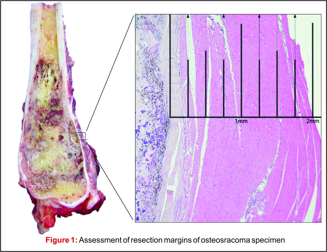
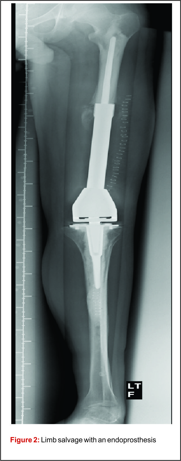
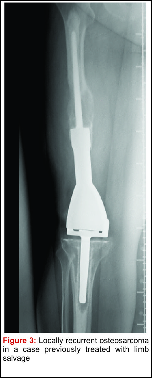
Depending on the extent of the resection, the surgical resections can be categorised as Osteo-articular resections and Intercalary resections. Reconstruction after osteoarticular resections is mainly done by megaprosthesis (non biological). If you want to retain joint mobility the biological options available are limited to osteoarticular allografts. Though these maintain bone stock and provide a better attachment for surrounding soft tissue resulting in increased stability of the construct the long term results with osteoarticular allografts are disappointing .Fracture, arthritis, non unions, infections and repeated surgery are not uncommon. Studies have reported 60-70 % adverse events, overall 5 year survival of 69 % and 79 % for allograft and articulate surface respectively[2,3]. A composite of allograft and prosthesis has been widely used, where allograft helps to maintain the stock and prosthesis provides the articular surface (Fig. 1).   
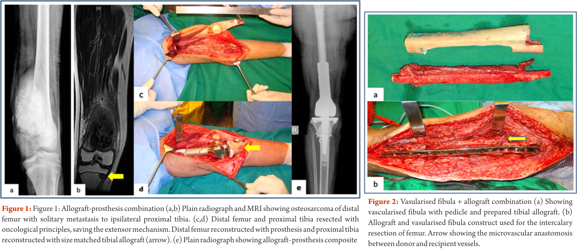
The functional outcomes with composite reconstruction are comparable with prosthetic reconstruction alone but associated with higher complication like nonunion and fracture. This method can have limited use in selected young patients with expected long term survival and require good bone stock for revision surgeries [4,5,6]. Allografts require sophisticated bone banks for procurement and storage and these are not available in most of the developing countries. Bone donations are not as frequent as other organ donations making procuring of size matched allografts even more challenging. Allografts may also be associated with risk of transmission of disease. Though strut allografts alone can be used for the reconstruction of intercalary defects and knee arthrodesis but studies have shown higher rate of complication like fracture, non union and resorption of grafts. Study by Bus MP et al has demonstrated a complication rate of 76 % and 70 % chance for reoperation due to graft failure. Thus strut allografts alone have limited use and are generally preferred for the upper limb or small defects (< 15cms). To overcome the above complication strut allograft may be combined with vascular fibular grafts [7,8]. Fibula is the most widely used autograft for reconstruction. It can be used as a vascularised or non vascularised graft. Proximal fibular head (with articular surface) has been used to reconstruct the articular surface of proximal humerus and distal radius. While isolated vascularized fibula may be adequate for reconstruction of upper limb defects where weight bearing is not an issue, lower limb reconstructions involving femur or the knee generally require a combination of vascularised fibula with strut allografts (Fig 2). Isolated use of fibula autograft or strut allografts have higher failure rates in large lower limb bone defects [9,10,11]. Small osteoarticular defects (up to 5 cm) like after the resection of distal radius lesions can also be reconstructed with iliac crest autograft. Certain anatomical sites have an inherent advantage and ease for reconstruction. Use of the neighbouring bone in forearm and leg provides a vascularised graft after resection of the radius and tibia. This serves as an easy and effective method of reconstruction. Shifting the distal ulna after an osteotomy at an appropriate level into the defect along with its soft tissue attachment and stabilizing it to the radius proximally and carpal bone distally (wrist arthordesis) provides an excellent method of reconstructing the bone defects after resection of distal radius tumors (Fig. 3). This method provides a stable wrist while maintaining forearm rotations (pronation- supination)[12]. Similarly in tibial lesions the fibula is mobilized medially into tibial defect and stabilized. This can be done both, for intercalary resections of the tibia where fibula is shifted after a double osteotomy and distal intrarticular resections where the transposed fibula is stabilized to talus to create an ankle arthrodesis This procedures avoids the requirement of a complex micro vascular procedure, reduces the operative time and also facilitates ease of soft tissue closure as transportation of fellow bone in to the defect will result in volume reduction of the tissues [13]. Reimplanting sterilized tumor host bone is widely used after intercalary resections. Patients own resected bone is sterilized and used to fill the defect. The resected tumor grafts can be sterilized by various methods like radiotherapy (extra-corporeal radiotherapy), pasteurization, autoclaving, liquid nitrogen and microwave. This technique has various advantages over the use of allograft. It does not require a bone bank, provides size matched graft (as it has been taken from the same defect) and has no risk of transmitted disease. After resection of the tumor the tumor bearing bone is taken on a separate table and soft tissues are removed under aseptic precautions. Certain soft tissues like ligaments may be retained on the bone graft in order to facilitate reconstruction. Sterilized bones are implanted back in the defect and stabilized with intramedullary nails or plates (Fig. 4).  Reimplanted bone acts a scaffold for creeping substitution and incorporation. To enhance incorporation and the union at osteotomy sites they can be combined with a vasclarised fibula ( Capanna technique). Puri et al documented a mean union time of 7 months for osteotomy sites and an excellent MSTS score of 29 with extracorporeal radiotherapy [14,15]. To overcome the adverse events like nonunion, fracture and collapse with the use of liquid nitrogen to sterlise tumor bone (fresh frozen autograft), pedical autograft technique was developed. In this technique an osteotomy is done at one end or joint disarticulation done and the whole specimen is treated with liquid nitrogen with other end in continuity with the main bone. It is then stabilized back with internal fixation or athroplasty. As bony continuity is maintained at one end, it is presumed to have early blood flow recovery and faster union and less complication compared with frozen autograft [16,17]. The main drawback of sterilized bones are inadequate mechanical strength resulting in graft fracture and implant failure. To enhance incorporation and to overcome inadequate mechanical strength they can be combined with a vasclarised fibula ( Capanna technique). For surface lesions like periosteal, parosteal and high grade surface osteosarcomas where medullary canal is not involved, bone preserving hemicortical excision may be considered. Meticulous planning with MRI and CT scans are required to obtain adequate margins and preserve good native bone. Computer assisted navigation surgery is advantageous while performing such technically demanding bone preserving surgeries. Various options are available to fill the bone defect after hemicortical excision (sterlised resected bone, strut allografts or small defects can be filled with autografts) [18,19]. Rotationplasty involves converting ankle joint to knee joint by segmental resection and rotating the foot externally to 180 degrees. This is an alternative method for reconstruction especially in children with growth potential where cost constraints may preclude the use of expensive growing prosthesis. This worthies also useful in converting hind quarter or above knee amputations to a functional below knee like amputation in adult patients where conventional resection and reconstructions are not possible due to large or previously inappropriately treated lesions In distal femur and proximal tibia lesions, a segment of involved bone along with knee joint and involved soft tissues is removed only sparing the neurovascular bundle. Here the two segments are connected only with neurovascular bundle. The distal fragment is externally rotated 180 degrees and distal part of femur is stabilized to proximal tibia with appropriate implants, in such a way that the ankle comes to the level of opposite knee joint. In the cases with involvement of whole femur, proximal tibia is articulated with the hip joint with or without use of prosthesis after external 180 dgree rotation. Adequate soft tissue reconstructions and an intense rehabilitation protocol ensures an excellent functional outcome in these cases where the ankle will act like knee joint, dorsiflexion of ankle acts as flexion and plantar flexion acts as extension of knee joint (Fig. 5).  
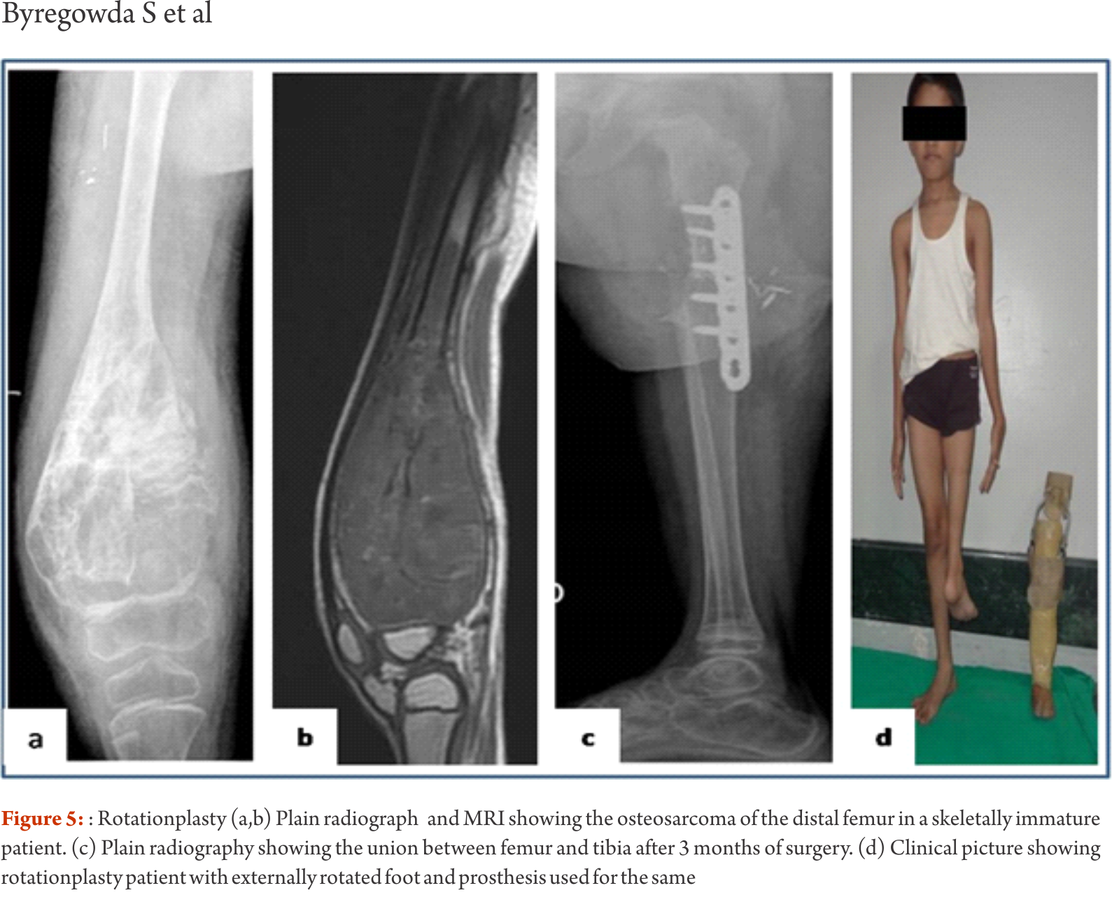
This procedure can also be used as salvage surgery following infected and failed limb salvage reconstruction. Studies have shown excellent oncological and functional outcome with this procedure. Rotationplasty offers a durable reconstruction option. It is not associated with phantom limb pain or sensations which are common following amputations. The main drawback of the procedure is the cosmetic deformity due to posterior rotated foot [20,21]. Distraction osteogensis using ilizarov method has been used for bone defects in tumor resection. It can be combined with live fibula grafts. The disadvantages are prolonged duration of treatment, high incidence of pin tract infections due to immune compromised state of patient receiving chemotherapeutic agents [22,23]. Due to these complications it is not a popular method and is used rarely. Masquelet technique is a two stage procedure for reconstruction of bone defects. In the first stage the defect is filled with bone cement and stabilized. This leads to the formation of a biological membrane over the cement spacer. In second stage procedure the biological membrane is opened, cement spacer removed, filled with cortico-cancellous bone graft and biological membrane sutured to create close content. The procedure was described in children. The ideal time for stage two is between 6 to 8 weeks, though in oncology we wait for completion of adjuvant treatment. Advantage is it makes primary surgery short and rapid uptake of graft due to biological membrane after the second procedure. The disadvantage with procedure is requirement of two surgical interventions [24].


Conclusions

Reconstruction following tumor resection is a challenging task. Different biological and non biological methods are available. Selection of a reconstruction procedure should be tailored to the individual patient based on the bone affected, amount of resection, requirement of patient and expertise and infrastructure available at treating centre. Biological methods are more cost effective and provide durable reconstruction options in properly selected extremity osteosarcoma patients.


References

1. Ghert M, Deheshi B, Holt G, Randall RL, Ferguson P, Wunder J et al.Prophylactic antibiotic regimensin tumour surgery (PARITY): protocol for a multicentre randomised controlledstudy. BMJ Open. 2012 Nov 28;2(6).
2. Muscolo DL, Ayerza MA, Farfalli G, Aponte-Tinao LA. Proximal tibia osteoarticular allografts in tumor limb salvage surgery. Clin OrthopRelat Res. 2010 May;468(5):1396-404.
3. Toy PC, White JR, Scarborough MT, Enneking WF, Gibbs CP. Distal femoral osteoarticular allografts: long-term survival, but frequent complications. Clin OrthopRelat Res. 2010 Nov;468(11):2914-23.
4. Donati D, Colangeli M, Colangeli S, Di Bella C, Mercuri M. Allograft-prosthetic composite in the proximal tibia after bone tumor resection. Clin OrthopRelat Res. 2008;466:459–65.
5. Van de Sande MA, Dijkstra PD, Taminiau AH. Proximal humerus reconstruction after tumour resection:Biological versus endoprosthetic reconstruction. IntOrthop. 2011;35:1375–80.
6. Campanacci L, Manfrini M, Colangeli M, Alí N, Mercuri M. Long-term results in children with massive bone osteoarticular allografts of the knee for high-grade osteosarcoma. J PediatrOrthop. 2010 Dec;30(8):919-27.
7. Bus MP, Dijkstra PD, van de Sande MA, Taminiau AH, Schreuder HW, Jutte PC, van der Geest IC et al. Intercalary allograft reconstructions following resection of primary bone tumors: a nationwide multicenter study. J Bone Joint Surg Am. 2014 Feb 19;96(4):e26.
8. Aponte-Tinao LA, Ayerza MA, Muscolo DL, Farfalli GL. Allograft reconstruction for the treatment of musculoskeletal tumors of the upper extremity. Sarcoma.2013;2013:925413.
9. Scaglioni MF, Chang EI, Gur E, Barnea Y, Meller I, Kollander Y, Bickels J, Dadia S, Zaretski A. The role of the fibula head flap for joint reconstruction after osteoarticular resections. J PlastReconstrAesthet Surg. 2014 May;67(5):617-23.
10. Campanacci DA, Puccini S, Caff G, Beltrami G, Piccioli A, Innocenti M, Capanna R. Vascularised fibular grafts as a salvage procedure in failed intercalary reconstructions after bone tumour resection of the femur. Injury. 2014 Feb;45(2):399-404.
11. Hilven PH, Bayliss L, Cosker T, Dijkstra PD, Jutte PC, Lahoda LU, Schaap GR, Bramer JA, van Drunen GK, Strackee SD, van Vooren J, Gibbons M, Giele H, van de Sande MA. The vascularised fibular graft for limb salvage after bone tumour surgery: a multicentre study. Bone Joint J. 2015 Jun;97-B(6):853-61.
12. Puri A, Gulia A, Agarwal MG, Reddy K. Ulnar translocation after excision of a Campanacci grade-3 giant-cell tumour of the distal radius: an effective method of reconstruction. J Bone Joint Surg Br. 2010 Jun;92(6):875-9.
13. Puri A, Subin BS, Agarwal MG. Fibular centralisation for the reconstruction of defects of the tibial diaphysis and distal metaphysis after excision of bone tumours. J Bone Joint Surg Br. 2009 Feb;91(2):234-9.
14. Mottard S, Grimer RJ, Abudu A, Carter SR, Tillman RM, Jeys L, Spooner D. Biological reconstruction after excision, irradiation and reimplantation of diaphyseal tibial tumours using an ipsilateralvascularised fibular graft. J Bone Joint Surg Br. 2012 Sep;94(9):1282-7.
15. Puri A, Gulia A, Jambhekar N, Laskar S. The outcome of the treatment of diaphyseal primary bone sarcoma by resection, irradiation and re-implantation of the host bone: extracorporeal irradiation as an option for reconstruction in diaphyseal bone sarcomas. J Bone Joint Surg Br. 2012 Jul;94(7):982-8.
16. Igarashi K, Yamamoto N, Shirai T, Hayashi K, Nishida H, Kimura H, Takeuchi A, Tsuchiya H. The long-term outcome following the use of frozen autograft treated with liquid nitrogen in the management of bone and soft-tissue sarcomas. Bone Joint J. 2014 Apr;96-B(4):555-61.
17. Shimozaki S, Yamamoto N, Shirai T, Nishida H, Hayashi K, Tanzawa Y, Kimura H, Takeuchi A, Igarashi K, Inatani H, Kato T, Tsuchiya H. Pedicle versus free frozen autograft for reconstruction in malignant bone and soft tissue tumors of the lower extremities. J Orthop Sci. 2014 Jan;19(1):156-63.
18. Deijkers RL, Bloem RM, Hogendoorn PC, Verlaan JJ, Kroon HM, TaminiauAH.Hemicortical allograft reconstruction after resection of low-grade malignant bone tumours. J Bone Joint Surg Br. 2002 Sep;84(7):1009-14.
19. Agarwal M, Puri A, Anchan C, Shah M, Jambhekar N. Hemicortical excision for low-grade selected surface sarcomas of bone. Clin OrthopRelat Res. 2007 Jun;459:161-6.
20. Agarwal M, Puri A, Anchan C, Shah M, Jambhekar N. Rotationplasty for bone tumors: is there still a role? Clin OrthopRelat Res. 2007 Jun;459:76-81.
21. Gradl G, Postl LK, Lenze U, Stolberg-Stolberg J, Pohlig F, Rechl H, Schmitt-Sody M, von Eisenhart-Rothe R, Kirchhoff C. Long-term functional outcome and quality of life following rotationplasty for treatment of malignant tumors. BMC MusculoskeletDisord. 2015 Sep 24;16:262.
22. Demiralp B, Ege T, Kose O, Yurttas Y, Basbozkurt M. Reconstruction of intercalary bone defects following bone tumor resection with segmental bone transport using an Ilizarov circular external fixator. J Orthop Sci. 2014 Nov;19(6):1004-11.
23. Khira YM, Badawy HA. Pedicled vascularized fibular graft with Ilizarov external fixator for reconstructing a large bone defect of the tibia after tumor resection. J OrthopTraumatol. 2013 Jun;14(2):91-100.
24. Chotel F, Nguiabanda L, Braillon P, Kohler R, Bérard J, Abelin-Genevois K. Induced membrane technique for reconstruction after bone tumor resection in children: a preliminary study. OrthopTraumatolSurg Res. 2012 May;98(3):301-8.


How to Cite this article: Byregowda S, Puri A, Gulia A. Biological Methods of Reconstruction After Excision of Extremity Osteosarco. Journal of Bone and Soft Tissue Tumors May- Aug 2016;2(2):5-9 .


                                                     (Abstract    Full Text HTML)      (Download PDF)


Evaluation of Osteogenic Sarcoma

Volume 2 | Issue 1 | Jan-Apr 2016 | Page 8-12 | Mandip Shah, Chetan Anchan.


Authors: Mandip Shah[1], Chetan Anchan[2]

[1] SPARSH orthopedic Oncology Clinic, 9th floor, Medicare building, Ellisbridge, Ahmedabad. India
[2]Bombay Hospital & Medical Research Centre, Mumbai, India

Address of Correspondence
Dr. Mandip C Shah (M.S Ortho)
SPARSH orthopedic Oncology Clinic
9th floor, Medicare building, Ellisbridge, Ahmedabad, 380006
Email: drmandip@gmail.com


Abstract

Primary malignant bone tumors are very rare diseases and the initial symptoms and signs can be vague and nonspecific resulting in such patients receiving, at best, some symptomatic care, with the expectation that the problem would resolve naturally. Often the patient may wait out a period of weeks or months hoping that the problem would settle on its own with some home remedy. The foundation of optimal outcome in the treatment of any malignant disease is early detection and correct diagnosis. Osteosarcoma (also called as osteogenic sarcoma) is a high grade malignant disease which is fatal unless treated in time. Early detection and correct diagnosis can make a big difference in the outcome of treatment of these diseases. Awareness of these conditions and the knowledge of vulnerable age groups is the perfect start for achieving this goal. A detailed history of the presenting complaints and a thorough clinical evaluation of the patient will provide vital clues that should alert the clinician to a possible bone tumor. Radiographs and MRI form the mainstay of radiological investigation of bone tumors. Besides aiding in detection of the bone tumor, radiographs are of vital diagnostic value; whereas MRI provides very detailed anatomical information of the extent of the disease. No diagnosis of a malignant bone tumor is complete without histological confirmation of the disease and therefore biopsy is the final step in the diagnostic evaluation of a suspected malignant bone tumor. As for all malignant tumors, staging investigations must be done before starting treatment for osteosarcoma.
Keywords: Osteogenic Sarcoma, diagnosis, evaluation.


Introduction

Osteosarcoma is a malignant tumor of mesodermal origin where the tumor cells produce bone or osteoid [1]. It is the most common primary malignant bone tumor, excluding hematopoietic bone tumors [1, 2]. Despite the simple and clear definition of this disease, the term osteosarcoma represents a family of tumors with significant diversity in its histological features, grade and clinical behavior [1]. However, it is a very rare disease and represents less than 1% of all cancers diagnosed in the United States [4]. It is seen most frequently in children and adolescents peaking in the second decade, which coincides with the growth spurt [3]. In these young patients, it chiefly affects the metaphysis of long bones. The most commonly involved region is the knee with the distal femur being the most affected, followed by the proximal tibia [3]. Besides the appendicular skeleton, osteosarcoma can affect other bones too; including the skull, axial, and very rarely, the acral bones. Although the majority of osteosarcomas occur in children and adolescents, there is a second spike in its incidence which is seen in the elderly – above the age of 60 years [5]. Unlike in the younger patients where most of the osteosarcomas arise de novo, a large number of osteosarcomas in the elderly arise in preexisting bone pathologies like Paget’s disease, fibrous dysplasia and in areas previously treated with radiation for some other cause [6, 7]. Males are more frequently affected than females. The overall world male to female ratio of osteosarcoma, in the age group of 0-24 years is 1.43:1 [8]. This difference steadily decreases with increasing age [8]. Osteosarcoma is a high grade malignant tumor which is fatal unless detected and diagnosed in time, and treated appropriately. Due to the rarity of this disease and lack of very obvious early clinical diagnostic features, there is often a delay in its detection and diagnosis; adversely affecting the outcome of treatment. Early detection and correct diagnosis gives the patient the best start to a long and difficult fight. In this article we describe a simple, logical and practical approach to evaluating a patient for a suspected bone tumor.

Evaluation of Osteosarcoma
A systematic approach is involved in the evaluation of any suspected bone neoplasm so as to reach a correct diagnosis, following which optimal treatment can be planned. As for most bone tumors, in cases of suspected osteosarcoma, this involves detailed clinical, radiological and histological evaluation.

Clinical evaluation
The three chief presenting symptoms of any bone tumor are pain, swelling and disability (Fig 1). Of these, pain is the most common presenting complaint in osteosarcoma, which, to begin with, may be experienced during activity that loads the affected bone. The pain may be in the form of a dull ache or such non-specific nature which could be attributed to more common causes like bone/muscle/ligament injury, articular pathologies etc. The duration of this pain may range from days to months. Special attention must be paid to patients in the vulnerable age group, especially when the complaint is unilateral, localized, persistent or progressive. Some individuals may associate the onset of the disease with some past injury. However, there is no evidence to substantiate that injury can lead to genesis of osteosarcoma.
Unexplained musculoskeletal pain should be taken very seriously, especially in children and adolescents, and should not be dismissed without proper investigation. In general, one must rule out a neoplastic cause for the musculoskeletal pain if one or more of the points mentioned below are noted.
1) Unilateral and localized extremity pain without a known cause
2) Pain intensity/duration/evolution in conflict with assumed routine cause
3) Pain with swelling
4) Pain since weeks/months
5) Persistent or progressively increasing pain
6) Pain, only temporarily / not relieved – with conservative care (rest and analgesics)
7) Pain causing disability, or affecting activity which is considered normal for the patient
8) Pain aggravated/triggered with activity
9) Rest/night pain

Figure 1

The next common presenting complaint is swelling in the affected region. This swelling may be visible or/and palpable – depending on the size and location of the tumor. It is unusual for a patient of osteosarcoma to present with a painless swelling, with the possible exception of parosteal osteosarcoma. Unlike pain, which is far more likely to be due to some injury or many such routine causes, a swelling is clearly an indication of a pathology, the significance of which should be investigated without further delay. Again, one must be aware that there are many causes of bony swelling ranging from infection to various types of benign and malignant tumors, and tumor like conditions. It is useful to get answers to the following questions when a patient presents with a bony swelling.
1) Location and size of swelling?
2) Is the swelling painful or painless?
3) Did the pain lead to discovery of the swelling or an existing swelling became painful?
4) Duration – Days/weeks/months/years?
5) Rate of growth?
6) Solitary or multiple?
Pain or/and swelling may result in some form of disability. Pain in the lower limb may affect ambulation or cause limitation of range or function across the adjacent joint. Rarely, patients with osteosarcoma may present with a pathological fracture. Pathological fracture is uncommon in osteosarcoma as majority of these patients would have sought medical attention before such an event occurred [9]. The risk of pathological fracture is higher in telangiectatic variant of osteosarcoma as it is a lytic expansile disease. Pathological fracture in children and adolescents is far more likely to be due to benign conditions like simple bone cyst, fibrous dysplasia, aneurysmal bone cyst, etc. Nevertheless, an occasional telangiectatic osteosarcoma can present in a similar way. Therefore, it becomes essential that a clear diagnosis of the cause of the fracture is established before deciding on the treatment. To identify a pathological fracture, one must rely a lot on the circumstances of the fracture rather than the X-ray alone. One must seek answers to the following questions:
1) How did the fracture occur? Was the cause significant or trivial?
2) Did the patient have complaints of pain/swelling/disability in the affected region prior to the fracture?
3) Has the patient suffered similar fractures in the past in the same location or in other bones?

There are generally no systemic or constitutional symptoms due to osteosarcoma, unless the disease is very advanced with extensive metastases. Lungs are the most common site for metastasis and these patients mainly present with breathlessness [10]. Some patients may present with bone metastases, which is the most common site for extra-pulmonary metastasis [10]. Regional nodal metastases and systemic metastasis to other organs/tissue is rare [10].

Clinical Evaluation
A detailed clinical examination is the next step in the evaluation of a patient with suspected bone tumor. A detailed local examination assessing the exact location, size and extent of the lesion should be done. The findings could range from subtle signs like raised local temperature/deep tenderness/vague swelling, to a very obvious painful, tender and large bony swelling with stretched hypervascular overlying skin and restriction of associated joint function. One must also make a note of the function of the adjacent joint and any distal neuro-vascular deficit. Although nodal metastasis is very rare in osteosarcoma, as a routine practice, regional draining nodes should be examined.

Blood investigations
There are no specific serum markers for osteosarcoma. Patients with high pre-treatment Lactate Dehydrogenase (LDH) levels have been reported to have 20% lower disease free survival as compared to those with normal LDH levels [12]. Similarly, a high pretreatment level of serum Alkaline Phosphatase has been reported to be an independent adverse prognostic marker in the outcome of treatment of non-metastatic osteosarcoma of extremities [13].

Radiological Evaluation
The next logical step in the work-up of a suspected bone tumor is imaging. MRI and CT scan have revolutionized medical imaging of human body and have contributed hugely to the success in the treatment of musculoskeletal tumors. However, when it comes to diagnosing bone tumors, the imaging modality that matters the most is the plain radiograph. With few exceptions, all other imaging modalities help mainly in understanding the anatomical extent of the disease and are of limited/selective diagnostic value.

Radiograph
A good quality plain radiograph in two perpendicular planes screening the entire bone should be taken. Conventional osteosarcoma can have varying appearance on the plain X-ray. It appears like an ill-defined cloudy/fluffy radiodensity in the bone which may show a mixture of lytic and sclerotic areas. The borders of this lesion are ill-defined and it appears to permeate through the normal bone around. It does not have a precisely identifiable border on the X-ray and there is a wide zone where the disease merges with the normal bone. This is described as a “wide zone of transition” and is a sign of an aggressive disease. Once the disease breaches the cortex, it lifts up the periosteum which elicits a periosteal reaction which may have varying appearances described as a sunburst /spiculated/lamellated reaction or as a Codman triangle. All such patterns of periosteal reaction, which is described as an interrupted periosteal reaction, are a very important sign of a potentially malignant disease. Large osteosarcomas can have soft tissue extension of the disease which appears as a soft tissue shadow on the X-ray and which may show cloudy/fluffy radiodensities within it. Besides these classic X-ray findings of a conventional osteosarcoma, many of the rare variants of osteosarcoma have X-ray characteristics which are unique to that particular sub-type and could help in suspecting/identifying them [11] (Fig 2).

Figure 2

MRI
MRI is the investigation of choice in suspected case of osteosarcoma for local staging [14, 15]. One must insist on a contrast study screening of whole involved bone to rule out any skip lesion [16]. MRI must ideally be done before the biopsy as it helps in planning the biopsy approach and also in targeting representative areas within the lesion, avoiding areas of tumor necrosis. Also, doing an invasive procedure before the MRI may alter the MRI findings by causing procedure related artifacts and edema. MRI gives useful information on intra medullary and extramedullary extent of disease, presence of any skip lesion, proximity of the tumor to the neurovascular structures and involvement of joint / physeal plate etc (Fig. 3,4,5). An additional MRI study is usually advised after the completion of neoadjuvant chemotherapy, just prior to the surgery for local management of the osteosarcoma, Post chemotherapy response prediction can be assisted with MRI as well. Reduction in the size of the soft tissue mass/vascularity/reactive zone and intramedullary edema, thickening of the peritumoral capsule and presence of necrosis are some of the signs of good response to chemotherapy. Assessment of chemotherapy response is best done by contrast enhanced, diffusion weighted study [17,18].

Figure 3, 4, 5, 6

Histopathological Evaluation
Although, the diagnosis of osteosarcoma can be assumed with a fair degree of certainty based on the clinical and radiological findings, under no circumstances the treatment can be started without histological confirmation. Osteomyelitis, osteoblastoma, bone metastasis, lymphoma, GCT, ABC, are the radiological differentials to osteosarcoma. On the other hand, one cannot rely only on biopsy alone for diagnosis of osteosarcoma – the classic example is of callus which can be indistinguishable from osteosarcoma on histology. Hence it is very important to correlate clinical, radiological and histological information to reach a diagnosis of any bone tumor. Biopsy is a procedure where a representative sample of the disease tissue is procured for histological studies. There are many ways this sample can be obtained. The routine procedures are open biopsy, needle biopsy and fine needle aspiration cytology (FNAC). Before doing a biopsy, it is advisable to complete all the radiological imaging studies. The most important step in planning a biopsy of any bone tumor is to decide on the approach. This is very important because, during the definitive surgery of a malignant bone tumor, the entire biopsy tract including the skin scar is excised en masse with the tumor. Therefore, it is very essential that the biopsy incision is placed in the line of the incision of the future surgery [19]. Open biopsy is a surgical procedure where tissue samples are obtained through a minor surgical procedure. The incision should be just adequate to obtain the deeper tissue and should be parallel to the long axis of the limb, in a location that would allow its easy excision along with the tumor at the time of definitive surgery. Needle biopsy is a procedure where tissue samples are obtained using a bone biopsy needle through a small stab incision. There are several advantages of needle biopsy over open biopsy. It causes limited contamination of the biopsy tract as it has a small footprint, which makes excision of the biopsy tract much easier during definitive surgery and also results in much less loss of skin as a result of the same. Besides this, it has several advantages like faster recovery, less hospital stay, lower cost etc. Also, the longer reach of the needle makes it easier to sample different regions of the tumor. As with open biopsy, the placement of the biopsy incision is important. Also, sampling of different regions of the lesion should be done through the same incision by just changing the angle of the needle and not through another skin incision. The only relative disadvantage of this procedure as compared to open biopsy is perhaps the smaller quantity of tissue sample that may be obtained, which could prove challenging to the pathologist to work on. However, in experienced hands this is generally not a problem. Frozen section may be used to confirm that the tissue sample obtained is representative. However, it should not be relied on to make a definitive diagnosis of bone tumors. FNAC as a procedure has many advantages, being minimally invasive and practically without morbidity, and with the least risk of tumor seeding along the biopsy tract. There are many reports of bone tumor diagnosis using FNAC. However, it has some limitations especially related to adequate representative tissue sampling and hence is not ideal for a definitive diagnosis of bone tumors like osteosarcoma [20].

Staging in Osteosarcoma
Cancer staging is a process to know the magnitude of the primary tumor and possible spread of the disease in a particular patient. It helps to understand the severity of the disease and hence the prognosis and thus aids in optimal treatment planning. Staging any cancer is therefore mandatory before starting its treatment. The most common site for metastasis in osteosarcoma is lung, followed by the skeletal system. At presentation, the reported incidence of lung metastasis is 15-20% whereas for skeletal metastasis it is 4%. Staging investigations includes High Resolution CT scan of thorax (plain) + Tc-99m methylene diphosphonate (Tc-99m MDP) Bone scan. Nowadays, 18 Fluoro Deoxy Glucose PET-CT scan is showing great promise as an alternative staging investigation. Plain chest radiograph can only detect large lung metastasis. For detection of early smaller lung lesions, a high resolution CT scan of thorax without contrast is recommended [21]. Typically metastases appear of soft tissue attenuation, dull, well circumscribed rounded lesions, more often in the periphery of the lung. Patients who present with metastatic pulmonary disease have a poorer prognosis. However, cure can be achieved in a small number of patients who respond well to chemotherapy and undergo pulmonary metastatectomy [22, 23]. (Tc-99m MDP) Triple-phase, whole-body bone scintigraphy still remains standard of care for determining the sites of metastatic disease in the skeletal system [24]. It may also detect skip lesions, although MRI is more accurate for this purpose. Whole-body turbo STIR MRI is also a reliable method for screening patients with suspected skeletal metastases. It is more specific than bone scan. This technique is also advantageous in that it reveals extraskeletal organ and soft tissue metastases [25]. Longer study time and cost are the limiting factors. Functional or metabolic imaging in form of 18 Fluoro Deoxy Glucose PET-CT scan is much more sensitive and specific than Tc-99m MDP bone scan in picking up the skeletal metastasis in osteosarcoma [26]. Moreover it gives valuable information on viable disease representation in proposed site for biopsy and some idea of the grade of the sarcoma. As it remains unaffected by presence of metallic prosthesis and radiation beam hardening artifacts, it is extremely valuable in detecting and defining a suspected recurrence [27]. However its scarce availability and prohibitive cost at present, makes it a difficult investigation to recommend in every case. Most popular staging system for bone and soft tissue sarcomas has been the Enneking’s staging system (Table 1). It is based on histological grade of sarcoma, local extent of disease i.e. intra or extra- compartmental involvement and presence or absence of metastasis [28]. American Joint committee on Cancer (AJCC) has also developed a staging system for sarcomas. (Table 2) It takes into the consideration the size of sarcoma, tumor grade, presence, and location of metastases [29].

Table 1


Conclusion

Osteosarcoma is a high grade malignant disease which is fatal unless treated appropriately, in time. Effective treatment is available for this disease with a high cure rate. However, despite the availability of such treatment in developing countries, the cure rates for osteosarcoma are much lower as compared to the western population. One of the most significant points of failure is timely detection and diagnosis of this condition. Awareness of this disease and the knowledge of the vulnerable age group can go a long way in improving the prospects for osteosarcoma patients in developing countries. Time tested clinical skills along with readily available radiological imaging modalities and histopathology will help us reach accurate diagnosis and staging in most cases of osteosarcoma.


References

1. Klein MJ, Siegal GP. Osteosarcoma: anatomic and histologic variants. Am J Clin Pathol. 2006 Apr; 125(4):555-81.
2. Dorfman HA, Czerniak B. Bone Cancers. Cancer supplement. 1995;75(1):203–10.
3. Ottaviani G, Jaffe N. The epidemiology of osteosarcoma. Cancer Treat Res. 2009;152:3-13.
4. Lisa Mirabello, Rebecca J. Troisi, and Sharon A. Savage. Osteosarcoma incidence and survival rates from 1973 to 2004: Data from the Survei llance, Epidemiology, and En d R esults Program. Cancer. 2009 Apr 1; 115(7): 1531–1543.
5. Unni KK. Dahlin’s bone tumors: general aspects and data on 11,087 cases. 5. Philadelphia: Lippincott-Raven; 1996. pp. 143–83.
6. Huvos AG. Osteogenic sarcoma of bones and soft tissues in older persons. A clinicopathologic analysis of 117 patients older tha n 60 years. Cancer . 1986 Apr 1;57(7):14 -42 9
7. Jhala DN, Eltoum I, Carroll AJ, et al. Osteosarcoma in a patient with McCune-Albright syndrome and Mazabraud’s syndro me: a case rep ort emp hasizin g the cytological and cy togenetic findings. Hum Pathol. 2003;34:1354-1357
8. Lisa Mirabello, Rebecca J. Troisi, and Sharon A. Savage. International osteosarcoma incidence patterns in children an d adolesce nts, middle ages, and elderly persons. Int J Cancer. 2009 Jul 1; 125(1): 229–234.
9. Lee RK1, Chu WC, Leung JH, Cheng FW, Li CK. Pathological fracture as the presenting feature in pediatric osteosarcoma . Pediatr Bloo d Cance r. 2013 Jul;60(7):1118-21.
10. Jeffree GM, Price CH, Sissons HA. The metastatic patterns of osteosarcoma. Br J Cancer. 1975 Jul; 32(1): 87–107
11. Yarmish G1, Klein MJ, Landa J, Lefkowitz RA, Hwang S. Imaging characteristics of primary osteosarcoma: nonconventional subtypes. Radiographics. 2010 Oct;30(6):1653-72.
12. Bacci G, Longhi A, Ferrari S, Briccoli A, Donati D, De Paolis M, Versari M. Prognostic significance of serum lactate dehydrogenase in osteosarcoma of the extremity: experience at Rizzoli on 1421 patients treated over the last 30 years. Tumori. 2004 Sep-Oct;90(5):4 78-8 4
13. Bacci G, Longhi A, Versari M, Mercuri M, Briccoli A, Picci P. Prognostic factors for osteosarcoma of the extremity treated with neoadjuvant chemotherapy: 15-year experience in 789 patients treated at a single institution. Cancer. 2006 Mar 1;106(5): 1154-61.
14. Rubin DA. Magnetic resonance imaging: Practical considerations. In: Resnick D, Kransdorf MJ, editors. Bone and joint imaging. 3rd ed. Philadelphia Pennsylvania: Elsevier Saunders; 2005. pp. 118–32.
15. Bohndorf K, Reiser M, Lochner B, Feaux DL, Steinbrich W. Magnetic resonance imaging of primary tumors and tumor-like lesions of bone. Skeletal Radiol. 1986;15:511–7.
16. Skip Metastases in Osteosarcoma: Experience of the Cooperative Osteosarcoma Study Group JCO April 1, 2006 vol. 24 no. 10 1535-1541 Leo Kager, Andreas Zoubek, Ulrike Kastner et al.
17. Holscher HC, Bloem JL, Vanel D, Hermans J, Nooy MA, Taminiau AH, et al. Osteosarcoma: Chemotherapy induced changes at MR imaging. Radiology. 1992;182:839–44.
18. Uhl M, Saueressig U, van Buiren M, Kontny U, Niemeyer C, Köhler G, et al. Osteosarcoma: Preliminary results of in vivo assessment of tumor necrosis after chemotherapy with diffusion- and perfusion-weighted magnetic resonance imaging. Invest Radiol. 2006;41:618–23.
19. Liu PT, Valadez SD, Chivers FS, Roberts CC, Beauchamp CP. Anatomically Based Guidelines for Core Needle Biopsy of Bone Tumors: Implications for Limb-sparing Surgery Radiographics. 2007 Jan-Feb;27(1):189-205; discussion 206.
20. Jorda M1, Rey L, Hanly A, Ganjei-Azar P. Fine-needle aspiration cytology of bone: accuracy and pitfalls of cytodiagnosis. Cancer. 2000 Feb 25;90(1):47-54.
21. Picci P, Vanel D, Briccoli A et al. Computed tomography of pulmonary metastases from osteosarcoma: the less poor technique. A study of 51 patients with histological correlation. Ann Oncol 2001; 12: 1601–1604.
22. Rasalkar DD1, Chu WC, Lee V, Paunipagar BK, Cheng FW, Li CK. Pulmonary metastases in children with osteosarcoma: characteristics and impact on patient survival. Pediatr Radiol. 2011 Feb;41(2):227-36.
23. Bacci G, Picci P, Briccoli A, Avella M, Ferrari S, Femino FP, et al. Osteosarcoma of the extremity metastatic at presentation: results achieved in 26 patients treated with combined therapy (primary chemotherapy followed by simultaneous resection of the primary and met astatic lesions). Tumori. 1992;78:200–6.
24. Schneider R. Radionuclide technique. In: Resnick D, Kransdorf MJ, editors. Bone and joint imaging. 3rd ed. Philadelphia Pennsylvania: Elsevier Saunders; 2005. pp. 86–117.
25. Frat, Ali, Ağldere, Muhtesem , Gençoğlu, Arzu et al. Value of Whole-Body Turbo Short Tau Inversion Recovery Magnetic Resonance Imaging With Panoramic Table For Detecting Bone Metastases: Comparison With 99MTc-Methylene Diphosphonate Scintigraphy; Journal of Comp uter Assisted Tomography: January/February 2006 – Volume 30 – Issue 1 – pp 151-156
26. Byun BH, Kong CB, Lim SM et al. Comparison of (18) F-FDG PET/CT and (99 m)Tc-MDP bone scintigraphy for detection of bone metastasis in osteosarcoma. Skeletal Radiol. 2013 Dec; 42(12):1673-81.
27. Benz MR, Tchekmedyian N, Eilber FC et al. Utilization of positron emission tomography in the management of patients with sarcoma. Curr Opin Oncol 2009; 21: 345 –351.
28. Enneking WF, Spanier SS, Goodman MA. A system for the surgical staging of musculoskeletal sarcoma. Clin Orthop. 1980;153:106–20.
29. Edge SB, Byrd DR, Compton CC, Fritz AG, Greene FL, Trotti A, editors. AJCC cancer staging manual (7th ed). New York, NY: Springer; 2010.


How to Cite this article: Shah M, Anchan C. Evaluation of Osteogenic Sarcoma. Journal of  Bone and Soft Tissue Tumors Jan-Apr 2016;2(1):8-12.

Photo 1


(Abstract    Full Text HTML)      (Download PDF)


How important are surgical margins in Osteosarcoma?

 Volume 2 | Issue 1 | Jan-Apr 2016 | Page 22-26 |Thomas P Cloake, Lee M Jeys.


Authors: Thomas P Cloake[1], Lee M Jeys[2].

[1]The Royal Orthopaedic Hospital, Bristol Road South, Birmingham, B31 2AP, UK.
[2]School of Health and Life Sciences, Aston University, Aston Triangle, Birmingham, B4 7ET, UK.

Address of Correspondence
Professor Lee M. Jeys
Professor of Health and Life Sciences
Aston University, Aston Triangle, Birmingham, B4 7ET, UK.
E-mail: lee.jeys@nhs.net


Abstract

Surgical resection combined with chemotherapy is the mainstay of treatment of osteosarcoma. Traditionally, surgical margins were based upon tumour grade and classified into marginal, wide or radical resection. The definition of these margins, however, remains subjective and recent research has questioned the need for wide or radical margins. Advances in surgical technique and the use of neo-adjuvant chemotherapy have led to an improvement in outcome. By reducing tumour burden, chemotherapy has provided surgeons with the option of limb salvage surgery rather than radical resection. Surgical margins and response to chemotherapy are now considered the two most important predictors of outcome in osteosarcoma. This review focuses on surgical margins with respect to limb salvage surgery and discusses the importance of response to chemotherapy.
Keywords: osteogenic sarcoma, osteosarcoma, surgical margins, chemotherapy, limb salvage surgery.


Introduction
Osteosarcoma is a high grade, primary tumour of bone in which the tumour cells produce osteoid [1]. It is the most common primary bone tumour, with an annual incidence rate of 5.0 per million [2]. Osteosarcoma is predominantly a disease of the young with a peak incidence in the second decade and displays a male predominance which is most pronounced at a younger age [3]. The treatment of osteosarcoma is challenging. The use of neo-adjuvant chemotherapy regimes combined with surgical resection has led to an improvement in outcome. Nevertheless, despite recent advances in surgical technique and chemotherapy agents, the survival rate has plateaued over the last 30 years [4]. There has been much research into prognostic factors that may help predict outcome in osteosarcoma, a number of these have been identified (see Table 1). Authors have suggested the most important, independent risk factors are the response to adjuvant chemotherapy and resection margins [5-7]. This review considers the impact of resection margins with a focus on limb salvage surgery and discusses the significance of response to chemotherapy.

Table 1

Resection margins
There has been much debate around the margin of clearance required for surgical treatment of osteosarcoma.  Enneking et al. were the first group to formally stage osteosarcoma into three distinct grades according to biologic aggressiveness, tumour site and distant metastases [8].  The authors suggested this system be used in surgical planning and inform the use of marginal, wide or radical resection margins. Nonetheless the definition of marginal or wide resection remains subjective and may vary between surgeons or units and has never been objectively defined (Fig 1).  Kawaguchi et al. developed this concept by giving distinct numerical values for desired resection margin according to the grade to tumour suggesting a 2cm margin was required for low-grade tumours and a 3cm margin was needed for high-grade neoplasms such as osteosarcoma [9].More contemporary studies have failed to reach a consensus on a numerical value for an adequate resection margin. Li et al. reported there was no difference in local recurrence when wide (>5mm) margins and close (<5mm) margins were used [10]. Bispo et al. failed to detect a difference in local recurrence using a margin of 2mm [11]. Betrand et al. found surgical margin to be the only independent risk factor for local recurrence and suggested a margin of 1mm may be adequate [12].  These papers suggest resection does not require a strict numerical margin, however efforts should be made to ensure no margins are intralesional. However, international consensus is in equipoise regarding margins, and this has made interpreting research articles very difficult. Even within units, tumour clear margins and ‘wide’ margins have become interchangeable when in reality they may be completely different and may lead to inappropriate treatment for patients. In the oncological world, the concept of patient specific treatment or ‘personalised medicine’ is gaining popularity and what is correct for one patient, may not be suitable for another patient, even with the same tumour type.

Figure 1

Limb salvage surgery

Prior to the advent of effective chemotherapy, the surgical treatment for osteosarcoma involved early radical amputation or disarticulation of the affected limb. Whilst ensuring complete removal of the tumour, performing this radical surgery on young patients caused loss of function and permanent disability, without improving patient survival. Limb salvage surgery (LSS) aims to resect the tumour, whilst maintaining function of the preserved limb, all with minimal risk to the patient (Fig 2).


Figure 2 Figure 3


The emergence of efficacious chemotherapy regimes, which acted to reduce tumour burden and reduce metastatic spread, and enhanced imaging techniques such as CT and MRI have led to the increased use of LSS [13-15]. By definition, the use of LSS requires preservation of limb neurovascular structures and narrower surgical margins when compared to amputation. Preservation of tissue during tumour resection has led to the inevitable decrease in resection margins, which potentially risks causing an increase in local recurrence (Fig 3). There are conflicting reports on the rate of local recurrence in LSS with some studies reporting an increase [15-17] and others a decrease [18], when compared to amputation. Considering local recurrence is associated with poor outcome, much work has been done to examine the impact of LSS on survival. Simon et al. were one of the first groups to investigate outcomes following LSS in a multi-centre retrospective review of 227 patients. They reported LSS had a comparable survival rate with amputation at 5 years follow up [19] and provided the impetus for increased uptake of LSS amongst surgeons. A large study by Bacci et al. retrospectively compared the outcome in patients who underwent LSS to amputation. The authors report that whilst LSS was associated with reduced resection margins, local recurrence and 5-year disease free survival were comparable to amputation [20]. These results are confirmed by a number of other groups, with each describing a survival rate equal to or better than that of amputation [15,17,18,21-27].It is important to consider, however, these studies are limited by their retrospective nature. Without robust methods of randomisation, treatment decisions have been based on individual patient and tumour characteristics, local practice and patient choice, leaving them open to the influence of selection bias. Postoperative quality of life is an important outcome measure in osteosarcoma. As patients with osteosarcoma are young and can expect a prolonged period of survival following treatment, the demands put upon a salvaged limb or prosthesis can be great. It is essential, therefore to ensure there is minimal risk of technical failure, the limb provides adequate function for the individual patient and has an acceptable cosmesis for both the patient and their care givers. Measurement of quality of life in children is difficult and there have been relatively few studies assessing this outcome measure. Using objective quality of life scores, LSS and amputation groups report reduced quality of life compared to population norms [29,30]. A meta-analysis comparing quality of life in patients who underwent LSS and amputation found there was no significant difference between the 2 groups. Taking into consideration all the above evidence LSS remains a safe and effective management option and when used in combination with adjuvant chemotherapy offers a good survival outcome.

Chemotherapy/chemonecrosis

The introduction of chemotherapy regimes alongside surgical resection has led to a dramatic improvement in survival. The use of chemotherapy in the treatment of osteosarcoma began in the 1970s with the use of doxorubicin and high dose methotrexate regimens [31]. Administration of chemotherapy agents before surgical resection as neo-adjuvant therapy enhanced survival from10 – 20% to 70% [32].Current modern chemotherapy regimes are based on combination therapy using methotrexate, adriamycin/doxirubicin and cisplatin. Poor response to chemotherapy has been identified as an important independent risk factor for poor prognosis. Histological evaluation of surgical resection specimens permits the classification of response to chemotherapy as good (>90% tumour necrosis) and poor (<90% tumour necrosis). Patients who display poor response are consistently reported to have worse outcome [33,34]. A number of strategies have been employed to improve results in poor responders. Evidence suggests modification of chemotherapy regime may improve results. Several groups have showed intensification of pre-operative chemotherapy enhances tumour response [35-37] and may improve survival [38-40]. This benefit however, is limited and intensification of chemotherapy beyond a certain level does not improve outcome [36,41-43]. The use of high dose, intensive treatment to induce a good response early in the disease process has also been shown not to convey overall survival benefit [38,42-44]. Further work is therefore required to optimize tumour response and improve outcome in patients with poor chemotherapy response. A recent, large, multi-national study EURAMOS-1 investigated the effect of adding the additional agents, ifosfamide and etoposide, to salvage poor response to chemotherapy, as well as evaluating the addition of pegylated interferon for good responding tumours [45]. The published initial results suggest that the addition of interferon for good responding tumours appears beneficial, however, it was poorly tolerated and frequently refused by patients. Current practice involves assessing tumour response using resection specimens following surgery, after the completion of neo-adjuvant chemotherapy, to advise further treatment[45]. Considering tumour response to chemotherapy is such a significant prognostic factor, measuring response early in the disease process may inform further management choices. Non-invasive imaging techniques such as CT [46], MRI [47-49] and F-FDG PET [50,51] have all be used to investigate response to neo-adjuvant chemotherapy. A combination of F-FDG PET and CT (F-FDG PET-CT) scanning is widely used for the detection of many cancers. Meta-analysis of the current evidence for its use in osteosarcoma has shown F-FDG PET-CT to be a valuable modality to assess chemotherapy-induced necrosis [52]. Newer techniques for evaluating response to chemotherapy prior to surgery, such as functional MRI (fMRI) are also promising and may inform surgeon’s decisions in planning surgical margins.
Patients with poor response to chemotherapy present a complex management challenge. There have been few studies presenting evidence to guide the surgical management of these patients. Bacci et al. suggested that amputation should be considered in the setting of poor response to chemotherapy due to its significant correlation with local recurrence rates [20]. Recent work in Birmingham investigated the influence of resection margins on survival in patients with poor response to chemotherapy [28]. The authors showed there was no survival benefit gained from amputation when compared to LSS with close margins, irrespective of the risk of developing local recurrence [28]. These data demonstrate resection with preservation of the limb to be a safe surgical option even in patients with poor chemonecrosis.

Predicting outcome
The current classification systems used to grade osteosarcoma, pioneered by Enneking, incorporate tumour characteristics including the presence of metastases to guide surgical management and predict prognosis [8]. However, despite the widely accepted importance of response to chemotherapy in prognosis, the current classification fails to reflect this.  In a recent presentation at International Society of Limb Salvage (ISOLS 2015), Jeys et al introduced The Birmingham Classification, which uses numerically defined tumour margins and response to chemotherapy to predict both local recurrence and survival. In this series, chemotherapy response was reported to show a significant effect on the rate of local recurrence and overall survival. It was also reported that a margin of 2mm was a statistically significant cut off value for predicting local recurrence.  Furthermore, combining resection margins (greater or lesser then 2mm) with response to chemotherapy (good, >90% or poor, <90%) was more effective in predicting local recurrence and survival than other staging systems.  This classification, however, requires further validation on a multi-centre basis.


Conclusion

Osteosarcoma continues to present a number to treatment challenges. Although surgical resection margins are an important predictor of outcome, limb salvage surgery with close margins has been shown to be a safe and effective surgical option.  Response to chemotherapy is an important independent predictor of survival.  A distinct group of poor responders exist, who despite modification to chemotherapy regimes and complete surgical excision of the tumour continue to have a poor outcome.  Current classification systems have so far failed to reflect important prognostic indicators, the Birmingham Classification represents a new, robust system for classifying osteosarcoma and predicting outcome.


References

1. Fletcher C, Bridge J, Hogendoorn P, Mertens F. WHO Classification of Tumours of Soft Tissue and Bone. Fourth Edition ed.; 2013. p. 247.
2. Ottaviani G, Jaffe N. The epidemiology of osteosarcoma. Cancer Treat Res 2009;152:3-13.
3. Dorfman HD, Czerniak B. Bone cancers. Cancer 1995 Jan 1;75(1 Suppl):203-210.
4. Allison DC, Carney SC, Ahlmann ER, Hendifar A, Chawla S, Fedenko A, et al. A meta-analysis of osteosarcoma outcomes in the modern medical era. Sarcoma 2012;2012:704872.
5. Bacci G, Longhi A, Versari M, Mercuri M, Briccoli A, Picci P. Prognostic factors for osteosarcoma of the extremity treated with neoadjuvant chemotherapy: 15-year experience in 789 patients treated at a single institution. Cancer 2006 Mar 1;106(5):1154-1161.
6. Bielack SS, Kempf-Bielack B, Delling G, Exner GU, Flege S, Helmke K, et al. Prognostic factors in high-grade osteosarcoma of the extremities or trunk: an analysis of 1,702 patients treated on neoadjuvant cooperative osteosarcoma study group protocols. J Clin Oncol 2002 Feb 1;20(3):776-790.
7. Picci P, Sangiorgi L, Rougraff BT, Neff JR, Casadei R, Campanacci M. Relationship of chemotherapy-induced necrosis and surgical margins to local recurrence in osteosarcoma. J Clin Oncol 1994 Dec;12(12):2699-2705.
8. Enneking WF, Spanier SS, Goodman MA. A system for the surgical staging of musculoskeletal sarcoma. Clin Orthop Relat Res 1980 Nov-Dec;(153)(153):106-120.
9. Kawaguchi N, Ahmed AR, Matsumoto S, Manabe J, Matsushita Y. The concept of curative margin in surgery for bone and soft tissue sarcoma. Clin Orthop Relat Res 2004 Feb;(419)(419):165-172.
10. Li X, Moretti VM, Ashana AO, Lackman RD. Impact of close surgical margin on local recurrence and survival in osteosarcoma. Int Orthop 2012 Jan;36(1):131-137.
11. Bispo Junior RZ, Camargo OP. Prognostic factors in the survival of patients diagnosed with primary non-metastatic osteosarcoma with a poor response to neoadjuvant chemotherapy. Clinics (Sao Paulo) 2009;64(12):1177-1186.
12. Bertrand TE, Cruz A, Binitie O, Cheong D, Letson GD. Do Surgical Margins Affect Local Recurrence and Survival in Extremity, Nonmetastatic, High-grade Osteosarcoma? Clin Orthop Relat Res 2015 May 27.
13. Ayerza MA, Farfalli GL, Aponte-Tinao L, Muscolo DL. Does increased rate of limb-sparing surgery affect survival in osteosarcoma? Clin Orthop Relat Res 2010 Nov;468(11):2854-2859.
14. Kotz R, Dominkus M, Zettl T, Ritschl P, Windhager R, Gadner H, et al. Advances in bone tumour treatment in 30 years with respect to survival and limb salvage. A single institution experience. Int Orthop 2002;26(4):197-202.
15. Tsuchiya H, Tomita K. Prognosis of osteosarcoma treated by limb-salvage surgery: the ten-year intergroup study in Japan. Jpn J Clin Oncol 1992 Oct;22(5):347-353.
16. Brosjo O. Surgical procedure and local recurrence in 223 patients treated 1982-1997 according to two osteosarcoma chemotherapy protocols. The Scandinavian Sarcoma Group experience. Acta Orthop Scand Suppl 1999 Jun;285:58-61.
17. Rougraff BT, Simon MA, Kneisl JS, Greenberg DB, Mankin HJ. Limb salvage compared with amputation for osteosarcoma of the distal end of the femur. A long-term oncological, functional, and quality-of-life study. J Bone Joint Surg Am 1994 May;76(5):649-656.
18. Sluga M, Windhager R, Lang S, Heinzl H, Bielack S, Kotz R. Local and systemic control after ablative and limb sparing surgery in patients with osteosarcoma. Clin Orthop Relat Res 1999 Jan;(358)(358):120-127.
19. Simon MA, Aschliman MA, Thomas N, Mankin HJ. Limb-salvage treatment versus amputation for osteosarcoma of the distal end of the femur. 1986. J Bone Joint Surg Am 2005 Dec;87(12):2822.
20. Bacci G, Ferrari S, Lari S, Mercuri M, Donati D, Longhi A, et al. Osteosarcoma of the limb. Amputation or limb salvage in patients treated by neoadjuvant chemotherapy. J Bone Joint Surg Br 2002 Jan;84(1):88-92.
21. Mavrogenis AF, Abati CN, Romagnoli C, Ruggieri P. Similar survival but better function for patients after limb salvage versus amputation for distal tibia osteosarcoma. Clin Orthop Relat Res 2012 Jun;470(6):1735-1748.
22. Lindner NJ, Ramm O, Hillmann A, Roedl R, Gosheger G, Brinkschmidt C, et al. Limb salvage and outcome of osteosarcoma. The University of Muenster experience. Clin Orthop Relat Res 1999 Jan;(358)(358):83-89.
23. Bacci G, Ruggieri P, Bertoni F, Ferrari S, Longhi A, Biagini R, et al. Local and systemic control for osteosarcoma of the extremity treated with neoadjuvant chemotherapy and limb salvage surgery: the Rizzoli experience. Oncol Rep 2000 Sep-Oct;7(5):1129-1133.
24. Eilber FR, Mirra JJ, Grant TT, Weisenburger T, Morton DL. Is amputation necessary for sarcomas? A seven-year experience with limb salvage. Ann Surg 1980;192(4):431-438.
25. Jiang F, Shi Y, Li GJ, Zhou F. A meta-analysis of limb-salvage versus amputation in the treatment of patients with Ennekingdouble daggerU pathologic fracture osteosarcoma. Indian J Cancer 2015 Feb;51 Suppl 2:e21-4.
26. Yin K, Liao Q, Zhong D, Ding J, Niu B, Long Q, et al. Meta-analysis of limb salvage versus amputation for treating high-grade and localized osteosarcoma in patients with pathological fracture. Exp Ther Med 2012 Nov;4(5):889-894.
27. Schrager J, Patzer RE, Mink PJ, Ward KC, Goodman M. Survival outcomes of pediatric osteosarcoma and Ewing’s sarcoma: a comparison of surgery type within the SEER database, 1988-2007. J Registry Manag 2011 Autumn;38(3):153-161.
28. Reddy KI, Wafa H, Gaston CL, Grimer RJ, Abudu AT, Jeys LM, et al. Does amputation offer any survival benefit over limb salvage in osteosarcoma patients with poor chemonecrosis and close margins? Bone Joint J 2015 Jan;97-B(1):115-120.
29. Bekkering WP, Vliet Vlieland TP, Koopman HM, Schaap GR, Schreuder HW, Beishuizen A, et al. Quality of life in young patients after bone tumor surgery around the knee joint and comparison with healthy controls. Pediatr Blood Cancer 2010 May;54(5):738-745.
30. Eiser C, Darlington AS, Stride CB, Grimer R. Quality of life implications as a consequence of surgery: limb salvage, primary and secondary amputation. Sarcoma 2001;5(4):189-195.
31. Rosen G, Murphy ML, Huvos AG, Gutierrez M, Marcove RC. Chemotherapy, en bloc resection, and prosthetic bone replacement in the treatment of osteogenic sarcoma. Cancer 1976 Jan;37(1):1-11.
32. Anninga JK, Gelderblom H, Fiocco M, Kroep JR, Taminiau AH, Hogendoorn PC, et al. Chemotherapeutic adjuvant treatment for osteosarcoma: where do we stand? Eur J Cancer 2011 Nov;47(16):2431-2445.
33. Winkler K, Beron G, Delling G, Heise U, Kabisch H, Purfurst C, et al. Neoadjuvant chemotherapy of osteosarcoma: results of a randomized cooperative trial (COSS-82) with salvage chemotherapy based on histological tumor response. J Clin Oncol 1988 Feb;6(2):329-337.
34. Goorin AM, Schwartzentruber DJ, Devidas M, Gebhardt MC, Ayala AG, Harris MB, et al. Presurgical chemotherapy compared with immediate surgery and adjuvant chemotherapy for nonmetastatic osteosarcoma: Pediatric Oncology Group Study POG-8651. J Clin Oncol 2003 Apr 15;21(8):1574-1580.
35. Fuchs N, Bielack SS, Epler D, Bieling P, Delling G, Korholz D, et al. Long-term results of the co-operative German-Austrian-Swiss osteosarcoma study group’s protocol COSS-86 of intensive multidrug chemotherapy and surgery for osteosarcoma of the limbs. Ann Oncol 1998 Aug;9(8):893-899
36. Smeland S, Muller C, Alvegard TA, Wiklund T, Wiebe T, Bjork O, et al. Scandinavian Sarcoma Group Osteosarcoma Study SSG VIII: prognostic factors for outcome and the role of replacement salvage chemotherapy for poor histological responders. Eur J Cancer 2003 Mar;39(4):488-494.
37. Winkler K, Bielack SS, Delling G, Jurgens H, Kotz R, Salzer-Kuntschik M. Treatment of osteosarcoma: experience of the Cooperative Osteosarcoma Study Group (COSS). Cancer Treat Res 1993;62:269-277.
38. Fuchs N, Bielack SS, Epler D, Bieling P, Delling G, Korholz D, et al. Long-term results of the co-operative German-Austrian-Swiss osteosarcoma study group’s protocol COSS-86 of intensive multidrug chemotherapy and surgery for osteosarcoma of the limbs. Ann Oncol 1998 Aug;9(8):893-899
39. Ferrari S, Mercuri M, Picci P, Bertoni F, Brach del Prever A, Tienghi A, et al. Nonmetastatic osteosarcoma of the extremity: results of a neoadjuvant chemotherapy protocol (IOR/OS-3) with high-dose methotrexate, intraarterial or intravenous cisplatin, doxorubicin, and salvage chemotherapy based on histologic tumor response. Tumori 1999 Nov-Dec;85(6):458-464.
40. Bacci G, Ferrari S, Bertoni F, Ruggieri P, Picci P, Longhi A, et al. Long-term outcome for patients with nonmetastatic osteosarcoma of the extremity treated at the istituto ortopedico rizzoli according to the istituto ortopedico rizzoli/osteosarcoma-2 protocol: an updated report.J Clin Oncol 2000 Dec 15;18(24):4016-4027.
41. Ferrari S, Smeland S, Mercuri M, Bertoni F, Longhi A, Ruggieri P, et al. Neoadjuvant chemotherapy with high-dose Ifosfamide, high-dose methotrexate, cisplatin, and doxorubicin for patients with localized osteosarcoma of the extremity: a joint study by the Italian and ScandinavianSarcoma Groups. J Clin Oncol 2005 Dec 1;23(34):8845-8852.
42. Lewis IJ, Nooij MA, Whelan J, Sydes MR, Grimer R, Hogendoorn PC, et al. Improvement in histologic response but not survival in osteosarcoma patients treated with intensified chemotherapy: a randomized phase III trial of the European Osteosarcoma Intergroup. J Natl Cancer Inst 2007Jan 17;99(2):112-128.
43. Meyers PA, Gorlick R, Heller G, Casper E, Lane J, Huvos AG, et al. Intensification of preoperative chemotherapy for osteogenic sarcoma: results of the Memorial Sloan-Kettering (T12) protocol. J Clin Oncol 1998 Jul;16(7):2452-2458.
44. Eselgrim M, Grunert H, Kuhne T, Zoubek A, Kevric M, Burger H, et al. Dose intensity of chemotherapy for osteosarcoma and outcome in the Cooperative Osteosarcoma Study Group (COSS) trials. Pediatr Blood Cancer 2006 Jul;47(1):42-50.
45. Whelan JS, Bielack SS, Marina N, Smeland S, Jovic G, Hook JM, et al. EURAMOS-1, an international randomised study for osteosarcoma: results from pre-randomisation treatment. Ann Oncol 2015 Feb;26(2):407-414.
46. Wellings RM, Davies AM, Pynsent PB, Carter SR, Grimer RJ. The value of computed tomographic measurements in osteosarcoma as a predictor of response to adjuvant chemotherapy. Clin Radiol 1994 Jan;49(1):19-23.
47. Bajpai J, Gamnagatti S, Kumar R, Sreenivas V, Sharma MC, Khan SA, et al. Role of MRI in osteosarcoma for evaluation and prediction of chemotherapy response: correlation with histological necrosis. Pediatr Radiol 2011 Apr;41(4):441-450.
48. Uhl M, Saueressig U, Koehler G, Kontny U, Niemeyer C, Reichardt W, et al. Evaluation of tumour necrosis during chemotherapy with diffusion-weighted MR imaging: preliminary results in osteosarcomas. Pediatr Radiol 2006 Dec;36(12):1306-1311.
49. Hayashida Y, Yakushiji T, Awai K, Katahira K, Nakayama Y, Shimomura O, et al. Monitoring therapeutic responses of primary bone tumors by diffusion-weighted image: Initial results. Eur Radiol 2006 Dec;16(12):2637-2643.
50. Im HJ, Kim TS, Park SY, Min HS, Kim JH, Kang HG, et al. Prediction of tumour necrosis fractions using metabolic and volumetric 18F-FDG PET/CT indices, after one course and at the completion of neoadjuvant chemotherapy, in children and young adults with osteosarcoma. Eur J Nucl MedMol Imaging 2012 Jan;39(1):39-49.
51. Cheon GJ, Kim MS, Lee JA, Lee SY, Cho WH, Song WS, et al. Prediction model of chemotherapy response in osteosarcoma by 18F-FDG PET and MRI. J Nucl Med 2009 Sep;50(9):1435-1440.
52. Hongtao L, Hui Z, Bingshun W, Xiaojin W, Zhiyu W, Shuier Z, et al. 18F-FDG positron emission tomography for the assessment of histological response to neoadjuvant chemotherapy in osteosarcomas: a meta-analysis. Surg Oncol 2012 Dec;21(4):e165-70.
53. Duchman KR, Gao Y, Miller BJ. Prognostic factors for survival in patients with high-grade osteosarcoma using the Surveillance, Epidemiology, and End Results (SEER) Program database. Cancer Epidemiol 2015 Aug;39(4):593-599.
54. Ferrari S, Bertoni F, Mercuri M, Picci P, Giacomini S, Longhi A, et al. Predictive factors of disease-free survival for non-metastatic osteosarcoma of the extremity: an analysis of 300 patients treated at the Rizzoli Institute. Ann Oncol 2001 Aug;12(8):1145-1150.


How to Cite this article: Cloake T, Jeys L.How important are surgical margins in Osteosarcoma? . Journal of  Bone and Soft Tissue Tumors Jan-Apr 2016;2(1):22-26.

Dr. Thomas P Cloake

Dr. Thomas P Cloake

Prof Lee M Jeys

Prof Lee M Jeys


 (Abstract    Full Text HTML)      (Download PDF)


The Current Role of Radiation Therapy for Osteogenic Sarcoma

Volume 2 | Issue 1 | Jan-Apr 2016 | Page 33-35  Sangeeta Kakoti, Nehal Khanna, Siddhartha Laskar.


Authers: Sangeeta Kakoti[1] Nehal Khanna[1], Siddhartha Laskar[1]

[1]Department of Radiation Oncology, Tata Memorial Hospital, Mumbai. India

Address of Correspondence
Dr.Siddhartha Laskar
Professor, Department of Radiation Oncology, Tata Memorial Hospital, Dr Ernest Borges Marg, Parel, Mumbai – 400012, India.
Email: laskars2000@yahoo.com


Abstract

Osteosarcomas are known to be relatively radio-resistant, definitive radiotherapy has a role in cases that are unresectable or have poor prognostic factors. Neo-adjuvant Chemotherapy followed by local therapy (surgery alone and/or radiotherapy) and maintenance chemotherapy remain the current standard of care for treatment of non-metastatic high grade osteosarcoma. New technologies like particle beam therapy using proton and carbon ions and use of high precision radiation therapy techniques have further improved the results of definitive radiation therapy. Current review traces the advent of radiotherapy, its current role in management of osteosarcoma and future trends in the field.
Keywords: Osteogenic Sarcoma, Radiotherapy, Management.


Introduction

Osteosarcoma (OGS), an osteoid-producing malignant mesenchymal tumour, accounts for 20-45% of all skeletal malignancies. It has a bimodal age distribution with peak incidence at 10-19 years and over 60 years (secondary to prior radiotherapy exposure, Paget’s disease etc). Male to female ratio is approximately 1.6:1. The most common sites of involvement are femur (50%), followed by tibia, humerus, pelvis, jaw, fibula and ribs. The major histological variants are conventional osteosarcoma (osteoblastic, fibroblastic or chondroblastic, according to the predominant type of matrix produced), teleangiectatic and small cell osteosarcoma. Patients commonly present with bony pain and local swelling. Patients may also present with symptoms of metastatic disease like dyspnoea, hemoptysis, or bone pain. Diagnostic investigations include Plain radiograph (characteristic ‘Sun burst’ appearance and ‘Çodman’s triangle’), MRI of local part, CT scan of chest, Bone scan (to look for skip lesions) and a histopathological examination. Tumors are staged according to either the AJCC or Enneking (MSTS) systems. Prognostic factors impacting survival [1] include presence of metastasis, response to Neoadjuvant chemotherapy (NACT), histologic type, age (each decade increases mortality rate by 7 fold), tumour location (tumours of tibia fare better than those of femur) and choice of therapy (post operative radiotherapy and amputation was associated with 92% and 76% increased relative risk of death respectively, may be confounded by advanced disease status). Prior to the extensive use of chemotherapy for treating patients with osteosarcoma, aggressive surgery was considered the treatment of choice, resulting in five year overall survival rate of 10-20% [2]. A meta-analysis by Kassir et al [3] on head and neck Osteosarcoma showed that surgical cut margin status was the sole prognostic factor and there was no survival benefit by adding radiation therapy and/or chemotherapy. But subsequently there have been great leaps in the success of osteosarcoma management. Incorporation of highly active chemotherapeutic agents resulted in significant improvement in outcomes to the tune of 60-75% [4]. The MIOS trial [5] reporting 5% versus 65% overall survival rates in patients randomised to surgery versus surgery and chemotherapy respectively, formed the basis for multimodality therapy in these tumours.

Role of Radiotherapy in management of Osteosarcoma
Osteosarcoma was always thought to be a radio-resistant tumour and hence radiotherapy was initially not included in the standard management regimens. Sir Stanford Cade a British surgeon radiotherapist in 1931 treated 133 patients with radiation therapy with an intention to avoid futile amputation in patients developing lung metastases in subsequent 6-9 months [6]. Following completion of therapy (60 Gy over six weeks) the resected specimen revealed 100% tumour necrosis in all patients.

1) Radiotherapy in definitive setting
There are no randomized trials comparing surgery versus radiotherapy (RT) as primary local therapy for osteosarcoma and is unlikely to be one in future due to ethical issues. However there are a few single arm series showing encouraging results. Machak et al [7] treated 31 patients with extremity osteosarcomas with definitive radiotherapy to a median dose of 60 Gy (range, 40–68 Gy). The 5-year local control (LC), metastasis-free and overall survival (OS) rates were 56%, 62%, and 61%, respectively. Similarly, Caceres et al [8] also noted a complete pathological response in 80% patients with limb OGS treated by chemotherapy and 60 Gy of RT. Excellent functional outcomes was noted in 86% of the patients. In 13 patients treated with definitive RT to median dose of 60 Gy, at a median follow up of 161 months, 3 year LC and OS was 70% and 75% respectively [9]. Subsequently, in the COSS registry of 175 patients [10] treated from 1980 to 2007, at a median follow up of 1.5 years (0.2-23 years), the overall survival rates after RT for treatment of primary tumors, local recurrence, and metastases were 55%, 15%, and 0% respectively. Local control rates for combined surgery and RT were significantly better than those for RT alone (48% vs. 22%). Feasibility of Stereotactic body radiotherapy (SBRT) for recurrent OGS lesions was evaluated by Brown et al [11]. Median dose delivered was 40 Gy in 5 fractions (range, 30-60 Gy in 3-10 fractions; total of 14 patients). Two grade 2 and 1 grade 3 late toxicities occurred (in the setting of concurrent chemotherapy and reirradiation); consisting of myonecrosis, avascular necrosis with pathologic fracture, and sacral plexopathy [11]. Efficacy and long term toxicity are yet to be determined. Gaitan-Yanguas showed a dose-response relationship with no lesion controlled at doses of 30 Gy, and all lesions controlled with doses of >90 Gy [12].
Approximately 25% of pelvic and 10% of head and neck osteosarcomas are not resectable and hence are candidates for definitive radiotherapy. In our institute, we prescribe 70.2 Gy in 39 fractions over 8 weeks.

2) Radiotherapy in preoperative setting
Preoperative radiotherapy is gradually evolving to facilitate function preserving less mutilating surgeries. Dincsbas et al [13] treated 44 patients with preoperative RT to a dose of 35 Gy in 10 fractions followed by limb sparing surgery. The tumor necrosis rate was 90% in 87% of the patients. At a median follow-up of 44 months, the 5-year LC and OS were 97.5% and 48.4% respectively. They documented subcutaneous fibrosis in 16%, joint movement restriction in 20%, and osteo-radionecrosis and pathologic fracture in 4% patients. Chambers et al [14] reported an OS of 73% at 5 years of 33 patients treated with preoperative RT and resection for craniofacial OGS.

3) Radiotherapy in adjuvant setting
Delaney et al [15] reported 41 patients with osteosarcoma involving various sites (primary, recurrent as well as metastatic) in different settings to a dose of 10 to 80 Gy (median 66 Gy) preceded by gross total tumor resection in 65.8%, subtotal resection in 21.9% and biopsy only in 12.2%. The local control rates according to the extent of resection were 78.4%, 77.8% and 40% respectively. The overall survival rates in corresponding groups were 74.45%, 74.1% and 25% respectively. The authors concluded that adjuvant RT can help provide local control of osteosarcoma for patients in whom surgical resection with widely negative margins is not possible. Dose response relationship was not found to be significant. Caveat of the study was that the patient population as well as the treatment parameters including dose and timing of radiation (some received preoperative followed by postoperative RT) was very heterogeneous.
Guadagnolo et al [16] reported that the addition of adjuvant RT in head and neck osteosarcoma definitely improves local control for those with positive or uncertain margins. Laskar et al reported the outcomes of patients with head and neck osteosarcomas treated at the Tata Memorial Hospital, Mumbai [17]. The authors highlighted the impact of post-operative adjuvant radiotherapy, even after R0 resection or in patients with adverse prognostic factors (large tumour size, lymphovascular invasion, soft tissue infiltration etc). The patients receiving adjuvant RT at TMH were prescribed a dose of 64.8 Gy in 36 fractions over 7 weeks. The authors reported local control rate of 36%. High dose intra-operative EBRT with kV X rays or electrons is emerging as yet another experimental option. Hong et al reported outcome of extracorporeal irradiation (ECI) in the management of 16 pts with a variety of tumours (OGS being in 4 of them) to a dose of 50 Gy in single fraction. At a median follow-up of 19.5 months, there were no cases of local recurrence or graft failure. One patient required amputation due to chronic osteomyelitis [18]. Puri et al reported the outcomes of patients treated at the Tata Memorial Hospital, Mumbai, using extracorporeal irradiation [19]. Thirty-two patients (16 Ewing’s sarcoma and 16 OGS) with a mean age of 15 years (2 to 35 years) underwent this procedure. There were three local recurrences. All were associated with disseminated disease and the recurrences were in soft- tissue remote from the irradiated graft. There were no local recurrences involving the irradiated bone. The OS for patients with osteosarcoma was 65% with excellent functional outcome.

4) Radiotherapy in palliative setting

There is little data regarding dose fractionation and efficacy of radiotherapy for palliation of advanced osteosarcoma. Considering the similar mechanisms of pain and inflammation like bony metastases, data from the later are often extrapolated [20] and single fraction or protracted fractionation have both been equally used. Oligo-metastatic OGS is treated with curative intent. Metastatectomy is the gold standard as a component of the curative regimen with a documented 5 year OS of approx 22%. Stereotactic body radiotherapy (SBRT) to limited lung metastases is an equally efficacious emerging non invasive option. In a series of 46 patients with oligometastatic disease to lungs from sarcomas, at a median follow up of 22 months after median dose of 10-48 Gy in 1-5 fractions, 31% of patients survived for more than 3 years [21]. In a multicentric phase I/II trial treating 38 patients with oligometastases to a median dose of 38-60 Gy in 3 fractions, LC at two years was 96% and median survival was 19 months. Incidence of grade III-IV toxicity was 8% [22].

5) Particle therapy for osteosarcoma
Ciernik et al, treated 55 patients (42% received definitive RT) with osteosarcoma of all sites [23] using combination of photons and protons to a mean dose of 68.4 Gy. With a median follow-up of 27 months, LC at 3 and 5 years were 82% and 72% respectively. The 5-year DFS and OS was 65% and 67% respectively. Prognostic factors found to have a significant impact on disease control were grade and bulk of the tumour. The extent of surgical resection did not correlate with outcome. Grade 3 to 4 late toxicity was seen in 30.1 % of patients. In another series of 30 patients with unresectable OGS of the trunk treated with definitive Carbon ion therapy to a dose of 52.8–73.6 Gy, the 3 and 5 year LC at a median follow up of 33 months was 62% and 49% respectively. The corresponding OS was 53% and 29%. Severe skin/soft tissue reaction was reported in 5 patients [24]. With neutrons, local control rates of 55% were documented in patients with unresectable OGS of different sites [25]. A median prescribed dose of 66 Gy has been tried in a series to patients with paraspinal osteosarcomas with a resultant LC of 74%. There were no reported late toxicities [26].

6) Role of brachytherapy
There is very limited role of brachytherapy in osteosarcomas. A new treatment strategy based on direct injections of 90Y-hydroxide into the tumor bed is under preclinical trial   [27].


 Conclusion

Neo-adjuvant Chemotherapy followed by local therapy (surgery alone and/or radiotherapy) and maintenance chemotherapy remain the current standard of care for treatment of non-metastatic high grade osteosarcoma. Although osteosarcomas are considered to be relatively radio-resistant, definitive radiation therapy results in significant long term disease control in patients with inoperable disease and postoperatively in patients with poor prognostic factors. The outcomes of definitive treatment using radiation therapy has further been improved by the use of particle beam therapy like protons & carbon ions & escalated doses of photon therapy using modern high precision radiation therapy techniques. Hence, Radiotherapy remains an important option for local treatment of unresectable tumors, following incomplete resection, or as an effective tool for palliation of symptomatic metastases


References

1. Prognostic factors and outcomes for osteosarcoma: An international collaboration. Emil-ios E. Pakos, Andreas D. Nearchou, Robert J. Grimer et al. European Journal of Cancer. 2009; 4 5: 2 3 6 7 –2 3 7 5
2. Osteogenic sarcoma: a study of 600 cases. Dahlin DC, Coventry MBBoneJoint Surg 1967; 49: 101-l 10.
3. Osteosarcoma of the Head and Neck: Meta-analysis of Nonrandomized Studies. Laryn-goscope, 1997; 107: 56-61.
4. Dana-Farber Cancer Institute/The Children’s Hospital adjuvant chemotherapy trials for osteosarcoma: three sequential studies, A. M. Goorin, M. Delorey, and R. D. Gelber, Cancer Treatment Symposia. 1985; 3: 155–159.
5. The effect of adjuvant chemotherapy on relapse-free survival in patients with osteosar-coma of the extremity. M. P. Link, A. M. Goorin, and A. W. Miser. The New England Journal of Medicine. 1986; 314: 1513
6. Osteogenic sarcoma. A study based on 133 patients. Cade S. J R Coll Surg Edinb. 1955; 1: 79-111.
7. Neoadjuvant chemotherapy and local radiotherapy for high-grade osteosarcoma of the ex-tremities. Machak GN, Tkachev SI, Solovyev YN et al Mayo Clin Proc. 2003;78:147-155
8. Local control of osteogenic sarcoma by radiation and chemotherapy. Caceres E, Zaharia M, Valdivia S, et al. Int J Radiat Oncol Biol Phys . 1984;10:35-39
9. Patrick Hundsdoerfer et al, European Journal of Cancer 4 5 (2009) 24 47 –2451
10. Craniofacial osteosarcoma Experience of the cooperative German–Austrian– Swiss osteosarcoma study group. Sven Jasnau, Ulrich Meyer, Jenny Potratz et al. Oral Oncology (2008) 44, 286– 294
11. Stereotactic body radiotherapy for metastatic and recurrent ewing sarcoma and osteosar-coma. Brown LC, Lester RA, Grams MP et al, Sarcoma. 2014:418270
12. A study of the response of osteogenic sarcoma and adjacent normal tissues to radiation. Gaitan-Yanguas M. IJROBP. 1981; 7: 593-595
13. The role of preoperative radiotherapy in non metastatic high-grade osteosarcoma of the extremities for limb-sparing surgery. Dincbas FO, Koca S, Mandel NM et al. Int J Radiat Oncol Biol Phys 2005;62:820-828
14. Osteogenic sarcoma of the mandible, current management. Chambers RG, Mahoney WD. Am Surg . 1970;36:463-471
15. Radiotherapy for local control of osteosarcoma. Thomas F. Delaney, Lily Park, Savelli I Goldberg et al, IJROBP, 2005; 61(2): 492-498.
16. Osteosarcoma of the jaw/craniofacial region: outcomes after multimodality treatment. Guadagnolo BA, Zagars GK, Raymond AK, et al, Cancer 2009;115: 3262-70.
17. Osteosarcoma of the head and neck region: lessons learnt from a single institutional expe-rience of 50 patients, Siddhartha Laskar, Ayan Basu, Mary Ann Muckaden et al, Head & Neck, 2008: 1020-1026.
18. Extracorporeal irradiation for malignant bone tumors. Hong A, Stevens G, Stalley P, et al. Int J Radiat Oncol Biol Phys . 2001;50:441-447
19. The outcome of the treatment of diaphyseal primary bone sarcoma by resection, irradia-tion and re-implantation of the host bone. A Puri, A Gulia, N Jambhekar, S Laskar. J Bone Joint Surg Br 2012;94-B:982–8.
20. Randomized trial of short versus long-course radiotherapy for palliation of painful bone metastases. Hartsell WF, Scott CB, Bruner DW, et al. J Natl Cancer Inst 2005;97:798–804
21. A retrospective study of SBRT of metastases in patients with primary sarcoma. Christina Linder Stragliotto, Kristin Karlsson, Ingmar Lax et al. Med Oncol. 2012; 29:3431–3439
22. Multi-Institutional Phase I/II Trial of Stereotactic Body Radiation Therapy for Lung Me-tastases, Kyle E. Rusthoven, Brian D. Kavanagh, Stuart H. Burri. J Clin Oncol 2009; 27:1579-1584.
23. Proton-Based Radiotherapy for Unresectable or Incompletely Resected Osteosarcoma: I. Frank Ciernik, MD1,2; Andrzej Niemierko, PhD1,3,4; David C. Harmon et al, cancer 2011; 117: 4522–30.
24. Impact of Carbon Ion Radiotherapy on Outcome in Unresectable High-grade Osteosar-coma of the Trunk, T. Kamada, R. Imai, S. Sugawara et al, I. J. Radiation Oncology d Biology d Physics Volume 75, Number 3, Supplement, 2009
25. Fast neutron radiotherapy for sarcomas of soft tissue, bone, and cartilage: Laramore GE, Griffith JT, Boespflug M et al, Am J Clin Oncol, 1989, vol 12, pp 320-326
26. Image-guided intensity-modulated photon radiotherapy using multifractionated regimen to paraspinalchordomas and rare sarcomas. Terezakis SA, Lovelock DM, Bilsky MH et al. Int J RadiatOncolBiol Phys. 2007;69(5):1502-8
27. Dosimetry of a 90Y-hydroxide liquid brachytherapy treatment approach to ca-nine osteosarcoma using PET/CT. Jien Jie Zhou, Arnulfo Gonzalez, Mark W. Lenox. Ap-plied Radiation and Isotopes. 2015; 95: 193-200.


How to Cite this article:Kakoti S, Khanna N, Laskar S. The Current Role of Radiation Therapy for Osteogenic Sarcoma. Journal of  Bone and Soft Tissue Tumors Jan-Apr 2016;2(1): 33-35.

Photo


 (Abstract    Full Text HTML)      (Download PDF)