Tag Archive for: Limb Salvage

Effect of Combination Chemotherapy and Radiotherapy in the Management of a Pathological Fracture in High-grade Osteosarcoma with Limb Salvage Procedure – A Case Report

Original Article | Volume 6 | Issue 3 | JBST September – December 2020 | Page 9-12 | V.R.Ganesan, T.C.Prem Kumar, Sanjay.C, Roopesh Kumar.S DOI: 10.13107/jbst.2020.v06i03.32

Author: V.R.Ganesan[1], T.C.Prem Kumar[1], Sanjay.C[1], Roopesh Kumar.S[1]

[1]Department of Orthopaedic Surgery & Traumatology,
Madurai Medical College & GRH, Madurai, Tamilnadu. India.

Address of Correspondence
Dr. V R Ganesan,
Department of Orthopaedic Surgery & Traumatology,
Madurai Medical College & GRH , Madurai, Tamilnadu. India.
E-mail: sphospital@hotmail.com


Introduction: Osteosarcoma (OS), the most common primary bone tumor, is known to be relatively a radioresistant tumor. Pathological fracture in OS denotes its aggressive biological response and so it was considered a contraindication to limb salvage in earlier days. Radiotherapy has its role only in cases that are inoperable or have poor prognostic factors. In recent years, there have been major advances in the management of pathological fractures in high-grade OS (HGOS). This case report is about the effect of combination chemotherapy and radiotherapy in the management of pathological fracture in HGOS with limb salvage procedure.
Case Report: A 19-year-old male with pain and swelling in his right lower third of thigh and inability to walk for 3 months was diagnosed as a case of OS right distal femur with a pathological fracture. Open biopsy was done which confirmed the diagnosis as HGOS, staged Enneking IIb. He was treated with a combination of chemotherapy, external beam radiotherapy, tumor resection, and modular resection prosthesis.
Results: There were no immediate, early, and late complications. At the end of 1½ years, his functional recovery is good and he has reached more than 70% of the right knee functions. He has no signs of recurrence at present. He has got a better quality of life and functional activity with the prosthesis compared to what an amputated limb can produce.
Conclusion: Pathological fracture in OS is not a contraindication to limb salvage. Radiotherapy can be used in combination with chemotherapy and limb salvage surgery in HGOS with a pathological fracture. This combination treatment helps in increasing the chances of limb sparing surgery with good local control and tumor necrosis rate. The new knowledge that radiotherapy can be effective when used with chemotherapy has shown good result in our case.
Keywords: Osteosarcoma, pathological fracture, external beam radiotherapy, limb salvage, custom modular prosthesis.


Reference:
1. Abudu A, Sferopoulos NK, Tillman RM, Carter SR, Grimer RJ. The surgical treatment and outcome of pathological fractures in localized osteosarcoma. J Bone Joint Surg Br 1996;78:694-8.
2. Ferguson PC, McLaughlin CE, Griffin AM, Bell RS, Deheshi BM, Wunder JS. Clinical and functional outcomes of patients with a pathologic fracture in high-grade osteosarcoma. J Surg Oncol 2010;102:120-4.
3. Scully SP, Ghert MA, Zurakowski D, Thompson RC, Gebhardt MC. Pathologic fracture in osteosarcoma: Prognostic importance and treatment implications. J Bone Joint Surg Am 2002;84:49-57.
4. Glasser DB, Lane JM, Huvos AG, Marcove RC, Rosen G. Survival, prognosis and therapeutic response in osteogenic sarcoma: The Memorial Hospital experience. Cancer 1992;69:698-708.
5. Finn HA, Simon MA. Limb-salvage surgery in the treatment of osteosarcoma in skeletally immature individuals. Clin Orthop Relat Res 1991;262:108-18.
6. Mankin HJ, Mankin CJ, Simon MA. The hazards of the biopsy, revisited: Members of the Musculoskeletal Tumor Society. J Bone Joint Surg 1996;78:656-63.
7. Bacci G, Ferrari S, Longhi A, Donati D, Manfrini M, Giacomini S, et al. Nonmetastatic osteosarcoma of the extremity with pathologic fracture at presentation: Local and systemic control by amputation or limb salvage after preoperative chemotherapy. Acta Orthop Scand 2003;74:449 54.
8. Kim MS, Lee SY, Lee TR, Cho WH, Song WS, Cho SH, et al. Prognostic effect of pathologic fracture in localized osteosarcoma: A cohort/case controlled study at a single institute. J Surg Oncol 2009;100:233 9.
9. Jaffe N, Spears R, Eftekhari F, Robertson R, Cangir A, Takaue Y, et al. Pathologic fracture in osteosarcoma. Impact of chemotherapy on primary tumor and survival. Cancer 1987;59:701-9.
10. Longhi A, Errani C, De Paolis M, Mercuri M, Bacci G. Primary bone osteosarcoma in the pediatric age: State of the art. Cancer Treat Rev 2006;32:423-36.
11. Jeon DG, Lee SY, Kim JW. Bone primary sarcomas undergone unplanned intralesional procedures the possibility of limb salvage and their oncologic results. J Surg Oncol 2006;94:592-8.
12. Xie J, Diener M, Sorg R, Wu EQ, Namjoshi M. Cost-effectiveness of denosumab compared with zoledronic acid in patients with breast cancer and bone metastases. Clin Breast Cancer 2012;12:247-58.
13. Ebeid W, Amin S, Abdelmegid A. Limb salvage management of pathologic fractures of primary malignant bone tumors. Cancer Control 2005;12:57-61.
14. Natarajan MV, Govardhan RH, Williams S, Raja GT. Limb salvage surgery for pathological fractures in osteosarcoma. Int Orthop 2000;24:170-2.
15. Cui Q, Li DF, Liu C, Guo J, Liu SB, Liu YS, et al. Two case-reports of the limb salvage treatment of osteosarcoma consolidated with obvious pathological fractures. Pathol Oncol Res 2011;17:973-9.
16. Godley K, Watts AC, Robb JE. Pathological femoral fracture caused by primary bone tumour: A population-based study. Scott Med J 2011;56:5-9.
17. Chandrasekar CR, Grimer RJ, Carter SR, Tillman RM, Abudu AT, Jeys LM. Outcome of pathologic fractures of the proximal femur in nonosteogenic primary bone sarcoma. Eur J Surg Oncol 2011;37:532-6.
18. Yin K, Liao Q, Zhong D, Ding J, Niu B, Long Q, et al. Metaanalysis of limb salvage versusamputation for treating high grade and localized osteosarcoma in patients with pathological fracture. Exp Ther Med 2012;4:889-94.
19. Scully SP, Temple HT, O’Keefe RJ, Mankin HJ, Gebhardt M. The surgical treatment of patients with osteosarcoma who sustain a pathological fracture. Clin Orthop 1996;324:227-32.
20. Machak GN, Tkachev SI, Solovyev YN, Sinyukov PA, Ivanov SM, Kochergina NV, et al. Neoadjuvant chemotherapy and local radiotherapy for high-grade osteosarcoma of the extremities. Mayo Clin Proc 2003;78:147-55.
21. Caceres E, Zaharia M, Valdivia S, Hilsenbeck S. Tejada F. Local control of osteogenic sarcoma by radiation and chemotherapy. Int J Radiat Oncol Biol Phys 1984;10:35-9.
22. Dincbas FO, Koca S, Mandel NM, Hiz M, Dervişoğlu S, Seçmezacar H, et al. The role of preoperative radiotherapy in non-metastatic high-grade osteosarcoma of the extremities for limb-sparing surgery. Int J Radiat Oncol Biol Phys 2005;62:820-8.


How to Cite this article: Ganesan VR, Kumar TCP, Sanjay C, Kumar SR| Effect of Combination Chemotherapy and Radiotherapy in the Management of a Pathological Fracture in High-grade Osteosarcoma with Limb Salvage Procedure – A Case Report | Journal of Bone and Soft Tissue Tumors | September-December 2020; 6(3): 9-12.

[Full Text HTML] [Full Text PDF] [XML]


An Interesting Reconstruction Option after Resection for a Distal Fibular Osteosarcoma: A Case Report

Case Report | Volume 6 | Issue 1 | JBST  Jan-April 2020 | Page 13-16 | DOI: 10.13107/jbst.2020.v06i01.008

Author:  Manish Pruthi [1], Jagandeep Singh Virk [2], Lokesh Garg [1], Sumit Goyal [4], Anila Sharma [3]


1. Department of Orthopaedic Oncology, Rajiv Gandhi Cancer Institute and Research Centre, New Delhi,Delhi, India,
2. Department of Orthopaedic Oncology, Paras Hospitals, Panchkula,Haryana, India,
3. Department of Pathology, Rajiv Gandhi Cancer Institute and Research Centre, New Delhi, Delhi, India,
4. Department of Medical Oncology, Rajiv Gandhi Cancer Institute and Research Centre, New Delhi,India

Address of Correspondence
Dr. Jagandeep Singh Virk,
Department of Orthopaedic Oncology, Paras Hospitals, Panchkula,Haryana, India.
E-mail: jaganvirk_09@yahoo.co.in


Abstract

Fibula is an expandable bone in the body, but tumors arising from the distal part which require a wide resection of the distal fibular part require some form of reconstruction for preventing lateral ankle instability. Many methods involving either bony or soft tissue reconstruction techniques have been described in literature, but consensus regarding which one is optimal for a good oncological and functional outcome is lacking. In this case report, we make use of a biological soft tissue technique involving use of peroneus brevis autograft reconstruction for providing lateral ankle support for a patient suffering from distal fibular osteosarcoma who underwent resection of the distal fibula. The patient went on to have an excellent functional outcome. This technique, although reported in literature, is in the form of only a few cases with a limited follow-up with none reported from the Indian subcontinent.
Keywords: Fibula, Sarcomas, Limb salvage, Reconstruction.


References

1. UnniKK. Dahlin’s Bone Tumors: General Aspects and Data on 11087 Case. 5th ed. Philadelphia, PA: Lippincott-Raven; 1996. p. 1-9.
2. PerisanoC, MarzettiE, SpinelliMS, GraciC, FabbricianiC, MaffulliN, et al.Clinical management and surgical treatment of distal fibular tumours: A case series and review of the literature.Int Orthop2012;36:1907-13.
3. SchneiderbauerMM, GullerudR, HarmsenWS, ScullySP. Fibular osteosarcomas: Contaminated margins may not impact survival.Clin OrthopRelat Res2007;456:182-7.
4. LaitinenM, HardesJ, AhrensH, GebertC, LeidingerB, LangerM, et al. Treatment of primary malignant bone tumours of the distal tibia.Int Orthop2005;29:255-9.
5. DieckmannR, AhrensH, StreitbürgerA, BudnyTB, HenrichsMP, ViethV, et al.Reconstruction after wide resection of the entire distal fibula in malignant bone tumours.Int Orthop2011;35:87-92.
6. MonsonDK, VojdaniS, DeanTJ, Louis-UgboJ. Lateral ankle stabilization after distal fibular resection using a novel approach: A surgical technique.Clin OrthopRelat Res2014;472:1262-70.
7. EnnekingWF, DunhamW, GebhardtMC, MalawarM, PritchardDJ. A system for the functional evaluation of reconstructive procedures after surgical treatment of tumors of the musculoskeletal system.Clin OrthopRelat Res1993;286:241-6.
8. MohlerDG, CunninghamDC.Adamantinoma arising in the distal fibula treated with distal fibulectomy: A case report and review of the literature.Foot Ankle Int1997;18:746-51.
9. Norman-TaylorFH, SweetnamDI, FixsenJA. Distalfibulectomy for Ewing’s sarcoma.J Bone Joint Surg Br1994;76:559-62.
10. KanayaK, WadaT, KuraH, YamashitaT, UsuiM, IshiiS.Valgus deformity of the ankle following harvesting of a vascularized fibular graft in children.J ReconstrMicrosurg2002;18:91-6.
11. JonesRB, IshikawaSN, RichardsonEG, MurphyGA. Effect of distal fibular resection on ankle laxity.Foot Ankle Int2001;22:590-3.
12. MannRA, RongstadKM. Arthrodesis of the ankle: A critical analysis.Foot Ankle Int1998;19:3-9.
13. DurakK, BilgenO, KaleliT, AydinliU. Distal fibula resection in osteochondroma.J Int Med Res1996;24:381-6.
14. deGauzy JS, KanyJ, CahuzacJP. Distal fibular reconstruction with pedicled vascularized fibular head graft: A case report.J PediatrOrthop B2002;11:176-80.
15. EgerW, SchörleC, ZeilerG. Giant cell tumor of the distal fibula: Fifteenyear result after en bloc resection and fibula reconstruction.Arch Orthop Trauma Surg2004;124:56-9.
16. PickeringR. Arthrodesis of ankle, knee, and hip. In: PickeringRM, editors. Campbell’s Operative Orthopaedics. 10th ed. St Louis, MO, USA: Mosby; 2003.
17. SaridisAG, MegasPD, GeorgiouCS, DiamantakisGM, TyllianakisME. Dual-fibular reconstruction of a massive tibial defect after Ewing’s sarcoma resection in a pediatric patient with a vascular variation.J PediatrOrthop2011;31:297-302.
18. VaseenonT, SaengsinJ, KamintaA, PattamapaspongN, SettakornJ, PruksakornD.Ankle ligament reconstruction after wide resection of the osteosarcoma of the distal fibula: A case report.BMC Res Notes2017;10:769.


How to Cite this article: Pruthi M, Virk J D, Garg L, Goyal S, Sharma A | An Interesting Reconstruction Option after Resection for a Distal Fibular Osteosarcoma: A Case Report | Journal of Bone and Soft Tissue Tumors | January-April 2020; 6(1): 13-16.

[Full Text HTML  [Full Text PDF] [XML]


Health-Related Quality of Life in Patients with Bone Tumor around the Knee after Resection Arthrodesis

Vol 5 | Issue 1 | Jan-April 2019 | page: 17-20 | Wilasinee Sirichativapee, Weerachai Kosuwon, Winai Sirichativapee.


Authors: Wilasinee Sirichativapee [1], Weerachai Kosuwon [1], Winai Sirichativapee [1].

[1] Department of Orthopaedics, Faculty of Medicine, Khon Kaen University, Khon Kaen, Thailand.

Address of Correspondence
Dr. Winai Sirichativapee,
Department of Orthopaedics, Srinagarind Hospital, 123 Khon Kaen University, Nai Mueang Sub-District, Mueang District, Khon Kaen Province – 40002, Thailand.
E-mail: winaisiri@yahoo.com


Abstract

Background: This study aimed to compare the health-related quality of life (HRQoL) of patient with bone tumor around the knee after resection arthrodesis.
Methods: Patients between 15 and 70 years of age who underwent resection arthrodesis in Srinagarind Hospital >1 year were recruited. Patients were interviewed using a short form-36 questionnaire (social functioning-36 [SF-36] Ver2.0 Thai version) regarding their daily life problems.
Results: Eighteen patients with the mean age of 36.6 years (15–63 years) were included (15 females) in the study. Histological diagnoses were giant cell tumor 10 cases, osteosarcoma seven cases, and low-grade chondrosarcoma one case. Site of lesions was distal femur 15 cases and proximal tibia 3 cases. According to HRQoL, patients have good quality of life (score SF-36 >70) in all domains: Mean score: Physical functioning 75.55 ± 21.88, role physical 71.18 ± 22.70, bodily pain 85.41 ± 22.51, vitality 77.43 ± 16.76, general health 74.44 ± 19.16, SF 83.05 ± 26.40, role emotional 80.09 ± 22.89, and mental health 77.77 ± 21.29. Complications post-operative are broken implants (3 cases, 16.7%) and infections (4 cases, 22.2%).
Conclusion: In patients with bone tumor around the knee after resection, arthrodesis has a good quality of life in all domains in SF-36 version 2.0 questionnaire including function, pain, and mentality.
Keywords: Limb salvage, Arthrodesis, Quality of life, social functioning-36 version 2.0, Osteosarcoma, Giant cell tumor.


References

1. Tarnawska-Pierścińska M, Hołody Ł, Braziewicz J, Królicki L. Bone metastases diagnosis possibilities in studies with the use of 18F-NaF and 18F-FDG. Nucl Med Rev Cent East Eur 2011;14:105-8.
2. Sampath SC, Sampath SC, Mosci C, Lutz AM, Willmann JK, Mittra ES, et al. Detection of osseous metastasis by 18F-NaF/18F-FDG PET/CT versus CT alone. Clin Nucl Med 2015;40:e173-7.
3. Harisankar CN, Agrawal K, Bhattacharya A, Mittal BR. F-18 fluoro-deoxy-glucose and F-18 sodium fluoride cocktail PET/CT scan in patients with breast cancer having equivocal bone SPECT/CT. Indian J Nucl Med 2014;29:81-6.
4. Roop MJ, Singh B, Singh H, Watts A, Kohli PS, Mittal BR, et al. Incremental value of cocktail 18F-FDG and 18F-NaF PET/CT over 18F-FDG PET/CT alone for characterization of skeletal metastasesin breast cancer. Clin Nucl Med 2017;42:335-40.
5. Chan HP, Hu C, Yu CC, Huang TC, Peng NJ. Added value of using a cocktail of F-18 sodium fluoride and F-18 fluorodeoxyglucose in positron emission tomography/computed tomography for detecting bony metastasis: A case report. Medicine (Baltimore) 2015;94:e687.
6. Iagaru A, Mittra E, Mosci C, Dick DW, Sathekge M, Prakash V, et al. Combined 18F-fluoride and 18F-FDG PET/CT scanning for evaluation of malignancy: Results of an international multicenter trial. J Nucl Med 2013;54:176-83.
7. Gradishar WJ, Anderson BO, Balassanian R, Blair SL, Burstein HJ, Cyr A, et al. NCCN Clinical Practice Guidelines in Oncology Breast Cancer Version 2; 2016. Available from: https://www.nccn.org/professionals/physician_gls/pdf/breast.pdf. [Last accessed on 2016 Oct 19].
8. Yoon SH, Kim KS, Kang SY, Song HS, Jo KS, Choi BH, et al. Usefulness of (18)F-fluoride PET/CT in breast cancer patients with osteosclerotic bone metastases. Nucl Med Mol Imaging 2013;47:27-35.
9. Israel O, Goldberg A, Nachtigal A, Militianu D, Bar-Shalom R, Keidar Z, et al. FDG-PET and CT patterns of bone metastases and their relationship to previously administered anti-cancer therapy. Eur J Nucl Med Mol Imaging 2006;33:1280-4.
10. Lapa P, Saraiva T, Silva R, Marques M, Costa G, Lima JP. Superiority of 18F-Fna PET/CT for detecting bone metastases in comparison with other diagnostic ımaging modalities. Acta Med Port 2017;30:53-60.
11. Araz M, Aras G, Küçük ÖN. The role of 18F-NaF PET/CT in metastatic bone disease. J Bone Oncol 2015;4:92-7.
12. Schirrmeister H, Glatting G, Hetzel J, Nüssle K, Arslandemir C, Buck AK. Prospective evaluation of the clinical value of planar bone scans, SPECT, and (18)F-labeled NaF PET in newly diagnosed lung cancer. J Nucl Med 2001;42:1800-4.
13. Piccardo A, Puntoni M, Morbelli S, Massollo M, Bongioanni F, Paparo F, et al. 18F-FDG PET/CT is a prognostic biomarker in patients affected by bone metastases from breast cancer in comparison with 18F-naF PET/CT. Nuklearmedizin 2015;54:163-72.
14. Iagaru A, Young P, Mittra E, Dick DW, Herfkens R, Gambhir SS. Pilot prospective evaluation of 99mTc-MDP scintigraphy, 18F NaF PET/CT, 18F FDG PET/CT and whole-body MRI for detection of skeletal metastases. Clin Nucl Med 2013;38:e290-6.
15. Hillner BE, Siegel BA, Hanna L, Duan F, Quinn B, Shields AF. 18F-fluoride PET used for treatment monitoring of systemic cancer therapy: Results from the national oncologic PET registry. J Nucl Med 2015;56:222-8.
16. Iagaru A, Mittra E, Dick DW, Gambhir SS. Prospective evaluation of (99m)Tc MDP scintigraphy, (18)F NaF PET/CT, and (18)F FDG PET/CTfor detection of skeletal metastases. Mol Imaging Biol 2012;14:252-9.


How to Cite this article: Koç Z P, Kara P Ö, Sezer E, Erçolak V.Diagnostic Comparison of F-18 Sodium FluorideNaF, Bone Scintigraphy, and F-18 Fluorodeoxyglucose Positron Emission Tomography/Computed Tomography in the Detection of Bone Metastasis. Journal of Bone and Soft Tissue Tumors Jan-Apr 2019;5(1): 17-20.


  (Abstract    Full Text HTML)   (Download PDF)


Limb Salvage in Paediatric Bone Tumours

Vol 1 | Issue 2 | Sep – Dec 2015 | page:10-16 | Michael Parry[1], Robert Grimer[1]


Author: Michael Parry[1], Robert Grimer[1]

[1] The Royal Orthopaedic Hospital, Birmingham, UK.

Address of Correspondence
Dr. Michael Parry.
Royal Orthopaedic Hospital NHS Foundation Trust
Bristol Road South ,Northfield, Birmingham
B31 2AP, United Kingdom
Email: michael.parry3@nhs.net


Abstract

Significant progress has been made in the management of paediatric osseous sarcoma. Where historically this diagnosis conferred a dismal prognosis, modern strategies of oncological management have resulted in significant improvements in disease free survival. These improvements have resulted in a broadening of the feasibility of limb salvage, which, in the paediatric population, presents its own unique challenges. Treatment must prioritize life over limb and not compromise local control for the advantage of cosmesis, limb length or function. Limb salvage, where possible, offers distinct advantages over conventional amputations and with the advent of modern techniques and technologies, should always be a consideration in the paediatric sarcoma population.
Keywords: Limb salvage, paediatric sarcoma.


Introduction

Primary malignant tumours arising from bone are relatively rare in the paediatric and adolescent population, accounting for only 6% of childhood malignancies. Of these, the majority (>90%) comprises osteosarcoma and Ewing’s sarcoma. Other bone sarcomas including chondrosarcoma, malignant fibrous histiocytoma, fibrosarcoma, malignant giant cell tumours, and adamantinoma can occur in the paediatric population although their incidence is relatively low. In these rare tumours, surgery is the first choice for local control.
Historically, an extremity sarcoma in a child conferred a dismal prognosis. However, advances in imaging modalities, greater understanding of the role of local control, and the use of multi-agent chemotherapy, has resulted in a significant improvement in overall survival. Such advances have resulted in an increase in 5-year survival from 10-20% to 60-70% with modern techniques. Thanks to these improvements in overall survival and a groundswell of interest and advances in technology, limb salvage is now regarded as the gold standard of treatment for those presenting with osseous extremity sarcoma. Advances in bone banking and an appreciation of the behavior of prosthetic replacement have improved surgical outcomes in limb salvage, though they have created their own complications. The aim of this review is to explore the advances in limb salvage surgery for osseous sarcoma in the paediatric population.

Patient Assessment and Diagnosis
Patients presenting with a suspected sarcoma should be managed in a specialist institution with the expertise and multi-disciplinary facilities necessary for the holistic care of the patient throughout their cancer journey. This is no more apparent than in the paediatric population where treatment decisions must be agreeable not only to the patient but also to their parent or carer.
In all patients, a detailed history and examination is mandatory. Whilst the majority of sarcomas are sporadic, rare familial conditions, including Retinoblastoma and Li Fraumeni syndrome, should be identified. The physical examination must include assessment not only of the lesion but also the distal neurovascular status of the limb.
Radiography must include a plain radiograph of the affected bone or limb segment as this will form the basis of the diagnosis. Further, detailed cross sectional imaging, most commonly with magnetic resonance imaging (MRI) will allow local staging. Systemic staging of the disease necessitates computerized tomography (CT) of the chest, as well as skeletal imaging, either in the form of a technicium Tc-99m scan, or whole body MRI. Systemic staging may include positron emission tomography (PET) in conjunction with a CT scan, though this is dependent on local and institutional guidelines.
Biopsy remains the cornerstone of confirmation of diagnosis for any suspected malignancy arising from bone. Improperly performed biopsies can result in a delay in treatment, hinder subsequent attempts at limb salvage, and result in an increase in local recurrence. MRI prior to biopsy helps identify the optimal position for biopsy and avoids distortion of the imaging by post-biopsy changes. The biopsy should be performed in such a way that the tract can be completely excised at the time of definitive resection and should be performed by a surgeon familiar with the techniques of limb salvage. The use of image guidance with ultrasound or CT is especially pertinent in the paediatric population where considerable contamination can occur with open biopsy techniques.

General Treatment Considerations
It is imperative that discussions regarding treatment are conducted in the setting of a multi-disciplinary team including specialists in paediatric oncology, radiology, histopathology and orthopaedic oncology surgery. Having established the diagnosis, in terms of histological type and grade, and staged the local and systemic burden of disease, discussion turns to local control. The timing of local control varies dependent on diagnosis and stage. Most strategies utilize systemic chemotherapy prior to definitive local control. This allows the treatment of metastatic or suspected micro-metastatic disease to be instigated immediately. This time lapse allows careful planning of the definitive surgical resection, allowing time for custom made implants or grafts. It also allows an assessment to be made of the tumour response to neo-adjuvant therapies, which often guides adjuvant therapy following tumour resection. In some cases, neo-adjuvant therapy results in a dramatic change in the tumour facilitating subsequent resection. The modality for local control must take into consideration patient, tumoural, socio-economic, cultural and technical biases.

Amputation versus Limb Salvage
Whilst no absolute contraindications to limb salvage exist, the involvement of neurovascular structures which preclude attainment of a clear margin without significant impairment of limb function, and very young skeletal age, should point away from limb salvage as the definitive local control. Relative contraindications include factors resulting in a delay in reinstating systemic therapy, including infection and post operative wound complications, and factors most likely to result in an increase in local recurrence, including tumour bed contamination from inappropriate biopsy, expected positive surgical margins and pathological fracture (Fig. 1).

Fig 1 2 3
Patient wishes must be respected when considering amputation or limb salvage. The majority of studies comparing function in these two treatment groups have looked at tumours of the distal femur. No statistically significant difference has been demonstrated in the overall or disease specific outcomes between these two approaches, which is a manifestation of appropriate patient selection. According to the available outcome scores, function following amputation is comparable to that of limb salvage, with comparable psychological end points. Patients with limb salvage will often have a superior cosmetic result but often at the expense of endurance in certain physical activities. Patients undergoing limb salvage will undergo more surgical interventions than their amputation counterparts and patients and their families should be made aware of the lifetime of surveillance and repeated surgical interventions at the outset. However, financial considerations should not sway the intervention, especially as amputation patients will require a greater lifetime expense than their limb salvage counterparts.

Limb Salvage Surgery
When considering surgery for local control in osseous malignancies of the immature skeleton, consideration must be given to the tumour location and the sensitivity to oncological treatment modalities. For tumours sensitive to chemotherapy and radiotherapy, (especially Ewings sarcoma), in challenging locations where surgical resection will result in unacceptable morbidity or an uncontaminated margin will be difficult to achieve, then non-surgical interventions may be more appropriate. Alternatively, for tumours not sensitive to radiotherapy, such as osteosarcoma, surgical resection may be the only option for local control, regardless of the morbidity. Surgery for the primary tumour and for metastatic deposits should be considered wherever possible. The aim in all surgery is to obtain as wide a margin of clear tissue as possible around the tumour. The better the response of the tumour to neoadjuvant treatment the safer limb salvage surgery becomes. Local control is indispensible for the cure of patients with Ewing’s sarcoma. Intralesional resection is associated with an increased risk of local recurrence and distant metastases.

FIg 4 5

Resection Without Reconstruction
For tumours arising in dispensable bones in the immature skeleton, including sections of the ulna, the scapula, the sacrum, the pelvis and fibula, local control can be achieved through excision without reconstruction. Excellent functional outcomes can be achieved without reconstruction, more so in the adaptable paediatric population.

Resection and Reconstruction
Biological Reconstruction
Autograft The large bony defect created following resection of an osseous malignancy often necessitates reconstruction for preservation of function and continued skeletal growth. The use of non-vascularised autologous structural bone graft dates back 100 years  , whilst the use of a vascularized fibula graft was first described by Taylor in 1975. The blood supply of the vascularized graft is preserved by anastomosing its feeding vessel to a host artery. The graft subsequently undergoes revascularization from this vessel and from the surrounding vascular bed. Since its first description, this technique, as well as the use of non vascularized grafts, has been extensively described and applied. The technique lends itself best to reconstruction of intercalary long bone defects, or for proximal humeral osteoarticular reconstruction where the fibula provides not only structural support but also allows longitudinal growth of the limb segment from the proximal physeal plate. Special consideration should be given to resection of pelvic sarcomas where autograft reconstruction is being considered. An option for reconstruction in the adolescent age group, at or approaching skeletal maturity, is resection, extracorporeal sterilization and reimplantation. This has been reported as a viable method for reconstruction. Indeed, in their latest series reporting the use of this technique, Wafa et al   reported a successful outcome in patients as young as 8 years old. This technique can also be applied to other body sites, particularly for intercalary reconstruction (Figs. 2 &3).
Allograft Improvements in tissue banking have allowed an expansion in the use of cadaveric osseous and osteoarticular allografts. Grafts are harvested and sterilized either by freezing or irradiation and can be offered on a custom made, size matched basis. There are mixed reports on the viability of articular cartilage following sterilization and the cadaveric bone itself is incorporated at best only moderately at osteosynthesis sites and beneath the periosteal sleeve, making the allograft at best a biomechanical scaffold. Depending on the site of reconstruction, up to 50% of patients undergoing allograft reconstruction can expect at least one complication, including infection, non-union or graft fracture. Some have reported infection rates following allograft reconstruction to be twice that seen following reconstruction with an endoprosthetic replacement. In spite of these potential problems, patients who avoid complications following allograft reconstruction function at a high level without the requirement for repeated revision procedures seen in endoprosthetic replacement.
In the case of adamantinoma and osteofibrous dysplasia like adamantinoma of the tibia, resection and reconstruction can be achieved either without reconstruction in the case of small, unicortical lesions, or with reconstruction using allograft, or autologous fibula graft. The fibula can be transferred and stabilized either in conjunction with a tibial allograft stabilized with plates or with the assistance of a ring external fixator.
In cases of diaphyseal resections, reconstruction can be achieved with intercalary allograft where the native joint above and below the lesion can be preserved. In many cases, particularly in Ewing’s and osteosarcoma, the tumour extends to the metaphysis, sparing the physis. Careful dissection can allow removal of the tumour without injury to the physis, preservation of the native cartilage and ligamentous attachments. Reconstruction of the diaphyseal defect with an intercalary allograft stabilized with an intermedullary nail through the centre of the physis allows continued growth at the physis. Incorporation of the allograft can be augmented by the addition of a vascularized fibula graft within the allograft. For tumours involving the distal femur or proximal tibia in young patients, an alternative option for reconstruction, preserving the foot, is an intercalary resection, and tibial turn-up (Van Nes) arthroplasty. The residual limb is rotated through 180o, the ankle joint now forming the novel knee joint. The resultant limb has the cosmetic appearance of an above knee amputation whilst allowing the capacity for longitudinal growth, if the proximal tibial physis has been preserved. Following rotationplasty, a prosthesis can be worn at the knee allowing ambulation. Gait analysis demonstrates improved kinematics when compared to a conventional above knee amputation. Careful consideration should be given not only to the technical demand of the procedure, but also the psychological impact on the patient and family of the disfiguring but highly functional procedure. The fact that the patients have no phantom pain is a distinct advantage over amputation at a similar level.

Non-Biological Reconstruction
Significant advances have been made in the design and manufacture of endoprostheses in the last 30 years. The advent of neo-adjuvant chemotherapeutic regimens has allowed an acceptable time lag between diagnosis and local control which allows for the manufacture of custom made endoprostheses based on the patients’ particular anatomy without delaying treatment. Primitive devices suffered from errors in manufacture resulting in implant fractures and early loosening with non-rotating knee prostheses. The current generation of endoprostheses offer an attractive life span for the majority, failure largely attributable to stress shielding in long stem endoprostheses and particle-induced osteolysis due to wear at the bearing interface. Failure is largely dependent on anatomical location, with proximal humeral and proximal femoral devices faring best, whilst distal femoral and proximal tibial prostheses perform less well. Patients with endoprostheses will undoubtedly require revision surgery within their lifetime, each time requiring greater osseous and soft tissue resection. The risk of infection remains high and increases with each revision procedure, leading, in the worst-case scenario, to possible amputation. This risk is increased when radiotherapy is employed, particularly for endoprosthetic reconstruction of the proximal tibia.
In younger patients, with more than 2 years of growth remaining, the issue of limb-length equalization is a real concern, particularly at the distal femur, where the physis here accounts for the majority of longitudinal growth. In such patients, “growing” prostheses present an attractive answer. Prostheses incorporating a growing distraction device can allow predictable lengthening and equalization of leg lengths. Minimally invasive growing prostheses (Fig. 4), where the device is lengthened by a distraction screw, accessed through a small incision, can be used in patients where surveillance of local recurrence is expected to require MRI. In those where local recurrence is unlikely, a non-invasive growing prosthesis (Fig. 5) can be employed. In such devices, the prosthesis incorporates a magnetic motor activated by an external rotating magnet applied in close proximity to the limb. This overcomes the need for repeated surgical procedures, and the inherent risk of infection this carries. However, patients with such devices cannot undergo further MRI scanning due to the irreparable damage this incurs on the magnetic motor. Whichever endoprosthetic device is chosen, consideration should be given to the method of fixation to native bone. The failures of early devices, attributable to particle-mediated osteolysis, have, to a certain extent, been obviated by the use of hydroxyapatite collars, essentially sealing the medullary canal and implant-bone interface from particulate wear generated at the bearing surface[53,54].


Conclusion 

The paucity of algorithms to guide treatment strategies in paediatric patients with osseous sarcomas is a reflection of the multifactorial influences that predict outcomes following resection. An appropriate strategy can only be achieved following careful consideration of oncological, pathological, surgical and patient factors. Whichever strategy is adopted, the sequence of priorities should always be first life, then limb, then function, with leg length discrepancy and cosmetic appearance affording lesser consideration. When true equipoise exists between limb salvage and limb sacrifice, in terms of overall and disease-free survival, consideration must be given to limb function not only in the immediate periods following reconstruction, but also for the entirety of the life of the patient. In the case of the paediatric population, this may exceed the professional lifetime of the treating surgeon.


References

1. Arndt CAS, Crist WM. Common musculoskeletal tumors of childhood and adolescence. N Engl J Med. 1999;341(5):342–52.
2. Meyer JS, Nadel HR, Marina N, Womer RB, Brown KLB, Eary JF, et al. Imaging guidelines for children with Ewing sarcoma and osteosarcoma: a report from the Children’s Oncology Group Bone Tumor Committee. Pediatric blood & cancer. 2008. pp. 163–70.
3. Mankin HJ, MANKIN CJ, Simon MA. The hazards of the biopsy, revisited. for the members of the Musculoskeletal Tumor Society. The Journal of Bone and Joint Surgery. The American Orthopedic Association; 1996;78(5):656–63.
4. Brisse H, Ollivier L, Edeline V, Pacquement H, Michon J, Glorion C, et al. Imaging of malignant tumours of the long bones in children: monitoring response to neoadjuvant chemotherapy and preoperative assessment. Pediatr Radiol. 2004;34(8):595–605.
5. Harris IE, Leff AR, Gitelis S, Simon MA. Function after amputation, arthrodesis, or arthroplasty for tumors about the knee. J Bone Joint Surg Am. The Journal of Bone and Joint Surgery, Inc; 1990;72(10):1477–85.
6. Rougraff BT, Simon MA, Kneisl JS, Greenberg DB, Mankin HJ. Limb salvage compared with amputation for osteosarcoma of the distal end of the femur. A long-term oncological, functional, and quality-of-life study. J Bone Joint Surg Am. The Journal of Bone and Joint Surgery, Inc; 1994;76(5):649–56.
7. Simon MA, Aschliman MA, Thomas N, Mankin HJ. Limb-salvage treatment versus amputation for osteosarcoma of the distal end of the femur. J Bone Joint Surg Am. 1986;68(9):1331–7.
8. Weddington WW, Segraves KB, Simon MA. Psychological outcome of extremity sarcoma survivors undergoing amputation or limb salvage. Journal of Clinical …. 1985.
9. Refaat Y, Gunnoe J, Hornicek FJ, Mankin HJ. Comparison of quality of life after amputation or limb salvage. Clin Orthop Relat Res. 2002;397:298.
10. Grimer RJ, Carter SR, Pynsent PB. The cost-effectiveness of limb salvage for bone tumours. J Bone Joint Surg Br. 1997;79(4):558–61.
11. Jurgens H, Exner U, Gadner H, Harms D, Michaelis J, Sauer R, et al. Multidisciplinary treatment of primary Ewing’s sarcoma of bone. A 6-year experience of a European Cooperative Trial. Cancer. 1988;61(1):23–32.
12. Craft A. Long-term results from the first UKCCSG Ewing’s tumour study (ET-1). European Journal of Cancer. 1997;33(7):1061–9.
13. Paulussen M, Ahrens S, Dunst J, Winkelmann W, Exner GU, Kotz R, et al. Localized Ewing Tumor of Bone: Final Results of the Cooperative Ewing’s Sarcoma Study CESS 86. 2001.
14. Burgert EO, Nesbit ME, Garnsey LA, Gehan EA, Herrmann J, Vietti TJ, et al. Multimodal therapy for the management of nonpelvic, localized Ewing’s sarcoma of bone: intergroup study IESS-II. Journal of Clinical …. 1990.
15. Nesbit ME, Gehan EA, Burgert EO, Vietti TJ, Cangir A, Tefft M, et al. Multimodal therapy for the management of primary, nonmetastatic Ewing’s sarcoma of bone: a long-term follow-up of the First Intergroup study. Journal of Clinical …. 1990.
16. Elomaa I, Blomqvist CP, Saeter G, Åkerman M. Five-year results in Ewing’s sarcoma. The Scandinavian Sarcoma Group experience with the SSG IX protocol. European Journal of …. 2000.
17. Schuck A, Ahrens S, Paulussen M, Kuhlen M, Könemann S, Rübe C, et al. Local therapy in localized Ewing tumors: results of 1058 patients treated in the CESS 81, CESS 86, and EICESS 92 trials. Int J Radiat Oncol Biol Phys. 2003;55(1):168–77.
18. Krieg AH, Hefti F. Reconstruction with non-vascularised fibular grafts after resection of bone tumours. J Bone Joint Surg Br. 2007;89(2):215–21.
19. Taylor GI, Miller GDH, Ham FJ. The free vascularized bone graft: a clinical extension of microvascular techniques. Plastic and Reconstructive Surgery. 1975;55(5):533.
20. DiCaprio MR, Friedlaender GE. Malignant bone tumors: limb sparing versus amputation. J Am Acad Orthop Surg. 2003;11(1):25–37.
21. Zaretski A, Amir A, Meller I, Leshem D, Kollender Y, Barnea Y, et al. Free fibula long bone reconstruction in orthopedic oncology: a surgical algorithm for reconstructive options. Plastic and Reconstructive Surgery. 2004;113(7):1989–2000.
22. Belt PJ, Dickinson IC, Theile DRB. Vascularised free fibular flap in bone resection and reconstruction. Br J Plast Surg. 2005;58(4):425–30.
23. Beris AE, Lykissas MG, Korompilias AV, Vekris MD, Mitsionis GI, Malizos KN, et al. Vascularized fibula transfer for lower limb reconstruction. Microsurgery. 2011 Feb 25;31(3):205–11.
24. Petersen MM, Hovgaard D, Elberg JJ, Rechnitzer C, Daugaard S, Muhic A. Vascularized fibula grafts for reconstruction of bone defects after resection of bone sarcomas. Sarcoma. 2010;2010:524721.
25. Ad-El DD, Paizer A, Pidhortz C. Bipedicled Vascularized Fibula Flap for Proximal Humerus Defect in a Child. Plastic and Reconstructive Surgery. 2001;107(1):155–7.
26. Krieg AH, Mani M, Speth BM, Stalley PD. Extracorporeal irradiation for pelvic reconstruction in Ewing’s sarcoma. J Bone Joint Surg Br. 2009;91-B(3):395–400.
27. Khattak MJ, Umer M, Haroon-ur-Rasheed, Umar M. Autoclaved Tumor Bone for Reconstruction. Clin Orthop Relat Res. 2006;447:138–44.
28. Uyttendaele D, De Schryver A, Claessens H, Roels H, Berkvens P, Mondelaers W. Limb conservation in primary bone tumours by resection, extracorporeal irradiation and re-implantation. J Bone Joint Surg Br. 1988;70(3):348–53.
29. Chen WM, Chen TH, Huang CK, Chiang CC, Lo WH. Treatment of malignant bone tumours by extracorporeally irradiated autograft-prosthetic composite arthroplasty. The Journal of Bone and Joint Surgery. 2002;84(8):1156–61.
30. Sys G, Uyttendaele D, Poffyn B, Verdonk R, Verstraete L. Extracorporeally irradiated autografts in pelvic reconstruction after malignant tumour resection. International Orthopaedics (SICOT). 2002;26(3):174–8.
31. Wafa H, Grimer RJ, Jeys L, Abudu AT, Carter SR, Tillman RM. The use of extracorporeally irradiated autografts in pelvic reconstruction following tumour resection. Bone Joint J. 2014 Oct;96-B(10):1404–10.
32. Enneking WF, Campanacci DA. Retrieved human allografts : a clinicopathological study. J Bone Joint Surg Am. 2001;83-A(7):971–86.
33. Mankin HJ, Springfield DS, Gebhardt MC, Tomford WW. Current status of allografting for bone tumors. Orthopedics. 1992;15(10):1147–54.
34. Fox EJ, Hau MA, Gebhardt MC, Hornicek FJ, Tomford WW, Mankin HJ. Long-term followup of proximal femoral allografts. Clin Orthop Relat Res. 2002;(397):106–13.
35. Ortiz-Cruz E, Gebhardt MC, Jennings LC. The Results of Transplantation of Intercalary Allografts after Resection of Tumors. A Long-Term Follow-Up Study*. The Journal of Bone & …. 1997.
36. Muscolo DL, Ayerza MA, Aponte-Tinao LA, Ranalletta M. Partial epiphyseal preservation and intercalary allograft reconstruction in high-grade metaphyseal osteosarcoma of the knee. J Bone Joint Surg Am. 2004;86-A(12):2686–93.
37. Grimer RJ. Surgical options for children with osteosarcoma. Lancet Oncol. 2005;6(2):85–92.
38. Van Ness CP. Transplantation of the tibia and fibula to replace the femur following resection. “Turn-up plasty of the leg”. J Bone Joint Surg Am. 1964;46:1353–5.
39. Fuchs B, Kotajarvi BR, Kaufman KR, Sim FH. Functional outcome of patients with rotationplasty about the knee. Clin Orthop Relat Res. 2003;415:52–8.
40. Jeys LM, Kulkarni A, Grimer RJ, Carter SR, Tillman RM, Abudu A. Endoprosthetic reconstruction for the treatment of musculoskeletal tumors of the appendicular skeleton and pelvis. J Bone Joint Surg Am. The Journal of Bone and Joint Surgery, Inc; 2008;90(6):1265–71.
41. Malawer MM, Chou LB. Prosthetic survival and clinical results with use of large-segment replacements in the treatment of high-grade bone sarcomas. J Bone Joint Surg Am. 1995;77(8):1154–65.
42. Unwin PS, Cannon SR, Grimer RJ, Kemp HB, Sneath RS, Walker PS. Aseptic loosening in cemented custom-made prosthetic replacements for bone tumours of the lower limb. J Bone Joint Surg Br. 1996;78(1):5–13.
43. Ward WG, Yang R-S, Eckardt JJ. Endoprosthetic bone reconstruction following malignant tumor resection in skeletally immature patients. Orthop Clin North Am. 1996;27(3):493–502.
44. Kawai A, Muschler GF, Lane JM, Otis JC, Healey JH. Prosthetic knee replacement after resection of a malignant tumor of the distal part of the femur. Medium to long-term results. J Bone Joint Surg Am. 1998 May;80(5):636–47.
45. Jeys LM. Periprosthetic Infection in Patients Treated for an Orthopaedic Oncological Condition. J Bone Joint Surg Am. The Journal of Bone and Joint Surgery, Inc; 2005 Apr 1;87(4):842–9.
46. Lewis MM. The use of an expandable and adjustable prosthesis in the treatment of childhood malignant bone tumors of the extremity. Cancer. 1986;57(3):499–502.
47. Hosalkar HS, Dormans JP. Limb sparing surgery for pediatric musculoskeletal tumors. Pediatr Blood Cancer. 2004 Apr;42(4):295–310.
48. Eckardt JJ, Kabo JM, Kelley CM, Ward WG, Asavamongkolkul A, Wirganowicz PZ, et al. Expandable endoprosthesis reconstruction in skeletally immature patients with tumors. Clin Orthop Relat Res. 2000;(373):51–61.
49. Finn HA, Simon MA. Limb-salvage surgery in the treatment of osteosarcoma in skeletally immature individuals. Clin Orthop Relat Res. 1991;(262):108–18.
50. Gupta A, Meswania J, Pollock R, Cannon SR, Briggs TWR, Taylor S, et al. Non-invasive distal femoral expandable endoprosthesis for limb-salvage surgery in paediatric tumours. J Bone Joint Surg Br. 2006;88(5):649–54.
51. Beebe K, Song KJ, Ross E, Tuy B, Patterson F, Benevenia J. Functional outcomes after limb-salvage surgery and endoprosthetic reconstruction with an expandable prosthesis: a report of 4 cases. Arch Phys Med Rehabil. 2009;90(6):1039–47.
52. Kenan S. Limb-sparing of the lower extremity by using the expandable prosthesis in children with malignant bone tumors. Operative Techniques in Orthopaedics. 1999;9:101–7.
53. Myers GJC, Abudu AT, Carter SR, Tillman RM, Grimer RJ. Endoprosthetic replacement of the distal femur for bone tumours: long-term results. J Bone Joint Surg Br. 2007;89(4):521–6.
54. Myers GJC, Abudu AT, Carter SR, Tillman RM, Grimer RJ. The long-term results of endoprosthetic replacement of the proximal tibia for bone tumours. J Bone Joint Surg Br. 2007 Dec;89(12):1632–7.


How to Cite this article: Parry M, Grimer R. Limb Salvage in Paediatric Bone Tumours. Journal of  Bone and Soft Tissue Tumors Sep-Dec 2015;1(2): 10-16.

M R

(Abstract)      (Full Text HTML)      (Download PDF)


From TMH-NICE to ResTOR: An Eventful Journey A Treatise of developing a Tumor Megaprosthesis

Vol 1 | Issue 1 | May – August 2015 | page:40-44 | Ravi Sarangapani[1*].


Author: Ravi Sarangapani[1*].

[1]Development & Quality Director Adler Mediequip Pvt. Ltd, Pune, India.

Address of Correspondence
Mr. Ravi Sarangapani
Development & Quality Director, Adler Mediequip Pvt. Ltd., Sushrut House, Survey 288,
Next to MIDC Hinjewadi Phase II, At. Mann, Tal. Mulshi, Pune 411057, Maharashtra, India.
Email: ravi-sarangapani@adlermediequip.com


Abstract

The primary option for patients with osteosarcoma for many decades was an amputation to save life. A review in 1986 demonstrated that limb salvage surgery was as safe as an amputation and provided the evidence to change the surgical management of these patients to limb salvage surgery and megaprosthetic reconstruction. International developments since the 1990s formed the backdrop for the evolution of limb salvage surgery in India. Initial obstacles faced in India were that of patient affordability. In the late nineties, Dr. Ajay Puri and Dr. Manish Agarwal from the orthopaedic oncology department of Tata Memorial Hospital (Mumbai) in association with Sushrut-Adler initiated development of an indigenous limb salvage megaprosthesis (the TMH-NICE). This had to be a low-cost prosthesis by all means. This surgeon industry partnership over time led to overcoming many challenges and failures and continuous learning both on the clinical and engineering fronts resulting in the evolution of the ResTOR modular resection prosthesis system. This journey continues with improvements and modernization of the system contributing to cost-effective limb salvage surgery to patients in India and a number of other countries.
Keywords: Bone Tumor, Osteosarcoma, Limb Salvage, TMH-NICE, ResTOR.


Introduction

Although bone tumours form less than 1% of cancers in adults, they account for 3-5% of cancers in children with osteosarcoma being the most commonly diagnosed form of primary malignant bone tumours, followed by Ewing’s sarcoma [1]. These tumours represent the fourth most common type of cancer in patients under the age of 25.
The primary recourse for these patients for many decades was an amputation with an emphasis on sacrifice of the limb to save life. It was a landmark retrospective review by Simon et al in 1986 that demonstrated that limb salvage surgery was as safe as an amputation and provided the evidence that enabled surgeons to change the surgical management of these patients to limb salvage surgery and megaprosthetic reconstruction [2]. Enneking’s work related to staging of the disease [3] and the importance of surgical margins [4] significantly contributed to the development of limb-salvage surgery.
Not unlike most modern developments in the medical field, the clinical research and evolution of this treatment modality originated in the western world and resulted in limb salvage surgery with a megaprosthetic reconstruction slowly acquiring the status of “standard of care” for the majority of bone tumour patients through the 1990s. These International developments form the backdrop for what can aptly be termed the evolution of limb salvage surgery in India.

Indian Perspective
In late 1999, the Tata Memorial Hospital which is a pioneering initiative in the field of cancer care and research, originally commissioned in 1941, decided to augment their orthopaedic oncology service by bringing in a specialist orthopaedic surgeon to work with Dr. Badhwar, the then surgical oncologist who was also handling bone tumours. This resulted in Dr. Ajay Puri joining the oncology service of Tata Memorial in November 1999. Fate perhaps conspired in creating what would turn out to be a great team as Dr. Badhwar unfortunately took ill and the Tata Memorial management decided to recruit a second orthopaedic surgeon in the form of Dr. Manish Agarwal who came on board in January 2000. (read all about these happenings in the guest editorial by Dr A. Puri in this very issue [5]). Thus, two young and enthusiastic orthopaedic surgeons, well recognized in Mumbai for the contribution to the trauma service at major public hospitals, came on board to develop the orthopaedic oncology service at the Tata Memorial Hospital.
What Dr. Puri and Dr. Agarwal lacked in formal oncology training, they more than made up with their eagerness to learn, their obvious intelligence and most of all their dedication and commitment to make a difference to the lives of patients’ afflicted with this dreaded disease. The challenges they faced were numerous and perhaps too many to enlist; a gigantic workload of patients, a general lack of awareness of bone tumours and their management in the community, patient expectations, sometimes unrealistic to save limbs considering the social stigma attached to amputation considering the general difficulties faced by amputees in a developing country like India [6] and above all fast-advancing international developments in the management of bone tumours and perhaps frustration at not being able to offer the best standard of care to their patients. What they had in their favour was the backing and belief of the Institution, excellent infrastructure and the potential to form a world-class multi-disciplinary team.
As the development of the orthopaedic oncology service in Tata Memorial Hospital in the last fifteen years amply demonstrates, the selection panel which included Prof. Laud and Prof. Bawdekar and Dr. Dinshaw, the then Director of the Institution who also created these positions, did well. The young team, not fazed by the enormity of the challenges confronting them, set about tackling the problems they saw in a methodical and systematic manner.
One of their first priorities was to bring themselves up to date with the current international standard of care which involved offering their patients the option of limb salvage surgery with megaprosthetic reconstruction. As they started efforts to implement this initiative, they were faced with an obstacle not uncommon to the developing world, that of patient affordability. As so eloquently described by Dr. Agarwal and Dr. Puri, “Though all these exciting developments occurred in the West, in our own country limb salvage was still a difficult proposition. Chemotherapeutic drugs were very expensive, endoprosthesis unaffordable, ignorance widespread and the patients poor” [7]. This situation resulted in the team not being able to effectively offer the option of a good quality mega-prosthetic replacement to even ten patients of the approximately 200 new cases [7] or primary malignant bone tumours presenting to the hospital at that time.
It is said that experience can sometimes be a hindrance and the enthusiasm of youth goes a long way in surmounting seemingly impossible hurdles. The young team refused to be dismayed with these setbacks and set about convincing the management of the Sushrut-Adler Group (currently Adler Mediequip Pvt. Ltd., a Smith & Nephew subsidiary) of their mission to save limbs and improve the quality of life of these patients. It is testimony to their eloquence and persuasive skills that the Sushrut-Adler management adopted the surgeons’ goals as their own and agreed to invest the time and resources needed in the service of these patients, in a situation where a financially viable business was nowhere in sight. Thus began the evolutionary journey which took this Indian designed, Made-in-India implant from the early hesitant efforts of the TMH-NICE to the modular resection system, the ResTOR.
The Sushrut-Adler team commenced work, fabricating implants to patient dimensions in a surgeon-led design effort that was blessed by the Institutional Review Board of the Tata Memorial Hospital. In keeping with the objectives defined by Dr. Puri and Dr. Agarwal, the primary consideration was their estimate of what patient’s would be able to afford based on their understanding of the financial situation of these patients. This resulted in certain choices of material and fabrication methods which in hindsight were incorrect choices driven by the early “cost” objectives.
The early period between the years 2000 to 2002 featured implants that were almost entirely fabricated in a custom-basis to patient specific dimensions specified by the surgeon team. The rather crude initial design without femoral condyles (Fig.1a) quickly evolved into a more refined femoral shape (Fig. 1b) under the guidance of the surgeons. These implants were manufactured from 316L stainless steel, the material choice dictated by easy availability, ease of fabrication and cost considerations. The condylar region with the patient specific resection length was welded to the straight intramedullary stem which featured a male thread screwed into the resection shaft. Longitudinal grooves were machined into the stem to inter-digitate with bone cement and a valgus angle of 7 degrees was incorporated. The hinge pin was locked into place using a simple slotted screw.
In a first hint of problems to be faced in the future near the level of the resection or the intramedullary stem junction, a case of failure was encountered in the implant at the location close to the resection level which was fabricated as a threaded junction (Fig.2). This failure was similar to the failure of a humeral implant reported by Bos et al with a fracture at the base of a threaded stem [8] due to stress concentration and a structural weakness.
As the number of operated patients increased, the surgeons began to note certain repetitive dimensions that could be standardized. These were the Antero-posterior and Medio-lateral dimensions of the femoral and tibial condyles. Later on as the project progressed, other dimensions including stem diameters, lengths and types, lengths of resection segments and spacers would get standardized to enable modularity. The standardized condylar dimensions in 2002 enabled the femoral condylar section of the implant to be “cast” in 316L stainless steel, a development that featured in the implants manufactured in the period 2002-2004. The adoption of casting enabled some reduction in lead time by reducing the rather extensive condylar machining that was required earlier. As anatomical understanding grew, intramedullary stems evolved from a straight design to including an option with an anatomical curvature (Fig. 3).

Fig 1
It was also in this period that the first instrument set (Fig. 4) was created. Notably, nearly all surgeries performed by the surgeon team till that time had been carried out by using general orthopaedic instrumentation. With increasing experience came the realization that specifically designed instrumentation would be needed. Also contributing to this development was the fledgling thought in the minds of the surgeons that this system might go out of Tata Memorial Hospital some day in the future and it was important to create instrumentation that would enable easier use of this system by average surgeons. In late 2004, a circumferential groove oriented transversely (Fig. 5a) was added to the stem with the thought of improving cement fixation. This change resulted in early failure at the location of the groove (Fig.5b) and was quickly abandoned.

Fig 2
It was in early 2004 that the project began to face what would turn out to be its most major challenge, the incidence of mechanical failure predominantly located at the intramedullary stem-bone junction (Fig. 6). As subsequently reported in 2010 by Dr. Agarwal and Dr. Puri, there were 22 mechanical failures in 183 patients (12.02%) predominantly at the stem-collar junction with an average time to failure of 38 months [9]. While these failures initially unnerved the team working on the project, reviews of literature reveal that such failures were by no means uncommon and had been faced and continue to be faced by each such group of surgeons and engineers working in the field of limb salvage. Biau et al reported mechanical failures including stem fractures and hinge pin failures in 7 out of 91 patients (7.7%) operated between 1972 and 1994 [10]. More recently in 2013, Nakamura et al [11] reporting on the Japanese early experience with the Kyocera limb salvage system revealed mechanical failures in 7 out of 82 distal femur resections (8.53%) including predominantly stem failures and one tibial tray breakage.
While the stem failures were beginning to present themselves and were being investigated, a number of refinements continued to be made in the period since 2004. The tibial baseplate acquired its rounded geometry (Fig. 7a) conforming to the tibial plateau. The important alignment mark on the stem was added (Fig. 7b) to enable correct rotational alignment of the implant. In mid-2005, bushes and a bumper manufactured from UHMWPE were introduced into the design (Fig. 7c) to minimize metal on metal articulation.
In early 2006, based on initial investigations into the failure location which was centered near the stem-bone junction, a decision was made to introduce a gradual change of diameter in the region and reduce stress concentration by introducing a fillet (Fig. 7d) with a liberal radius of curvature. This change was done based on standard good design principles and was perhaps the first engineering input to what had essentially been a surgeon-led project from inception.

Fig 3
With all the changes and refinements that had taken place over the years (Fig. 8a,b), the system in mid-2006 was fairly standardized based on a large patient experience of nearly 260 cases, standard condylar and intra-medullary dimensions, UHMWPE bushes and bumpers and a reliable hinge locking mechanism.
What however concerned the entire team was that the intramedullary stems continued to fail at the stem-bone junction and even the reduced stress concentration with the filleted design did not seem to work (Fig. 8c).
It was at this point that the engineering team at Sushrut-Adler brought in a new level of seriousness and application to understanding this problem better. Literature was extensively reviewed [12,13,14] to develop a better appreciation of the forces the implant was being subjected to. Resultant stresses on the implant cross sections were calculated and analyzed with reference to the materials being used. Based on the analytical work, it was clear that the stainless steel being used for these implants did not have the capability to withstand the continuous stresses being imposed on this implant in normal patient activities in the medium to long term. Better materials were needed.Fig 4The team opted for titanium alloy as the material of choice for the ramedullary stems as a material with no biocompatibility issues given its long history of successful clinical use, the ability of the material to withstand the imposed stresses in this application with an adequate factor of safety and the lower modulus of elasticity which was perceived as a possible advantage.
Titanium alloy is known to have its own difficulties in processing and is not an easy material from a manufacturing standpoint. Fortunately, the engineering and manufacturing teams at Sushrut-Adler had developed strong experience in working with this alloy due to their previous history with successful development and commercialization of spine implants manufactured from titanium alloy and these challenges were not difficult to overcome.
The change of materials and the expected solution to the difficult stem failure issue thus opened the way for the team of surgeons and the Sushrut-Adler engineers to take the next step of modularizing the system paving the way for more widespread use. The difficulties of custom-manufacturing implants by this time were well understood and the constraint imposed on an operating surgeon working with an implant of fixed size was not desirable. Modularity enabled a surgeon to intra-operatively select the most optimum surgical margin for the excision with no concerns about having an implant size that would adapt to the length of the resection.
The surgeons’ need for modularity resulted in many months of manufacturing trials at Sushrut-Adler as the necessary self-locking tapers for the modular components were designed and proven through the manufacturing process.
The final standardized condylar dimensions enabled the team to opt for superior Cobalt Chrome alloy investment castings for the condylar components thus gaining better articulation properties with the UHMWPE components.
The culmination of all these efforts resulted in the first patient implanted with the ResTOR modular resection prosthesis in a limb salvage procedure in late 2006. The modular system was commercialized in early 2007 for distal femur and proximal tibia resections. The addition of an upper limb system and a proximal femur resection system over the next few years enabled a complete portfolio of solutions.
It may be argued, with hindsight, that the inception of this project in 1999-2000 was with a compromised implant. However, as cogently argued by Dr. Agarwal ten years later [9], many patients benefited even with these compromised implants, the failure rate of around 15% was not viewed as catastrophic and was considered acceptable in a situation where an expensive implant was not an option and where amputation or rotationplasty would have been the only alternatives for the patient. The limb salvage procedure allowed many children to continue with education and adults to remain employed. Icing on the cake came in the form of the prestigious Golden Peacock Innovation Award in 2010 which recognized this as a major health-care initiative.
The ResTOR modular resection prosthesis system since 2007 has contributed to cost-effective limb salvage surgery of more than 2000 patients in India and a number of other countries [17] with prosthesis survivorship rates comparable to those reported in literature [15,16]. As a case in point, the Tata Memorial experience of 88% implant survivorship at five years with total femoral replacement [16], a specially challenging procedure with extensive resection does great credit to the team of surgeons and engineers who worked on this project. As implant survivorship improves and a greater number of patients experience disease-free survivorship with continuously improving surgeon experience, the ResTOR team continues to face newer challenges related to the increased demands on the implant.
While many improvements have been made and will continue to be made, as global experience with limb salvage implants has shown, the demands placed on these implants are immense and the way forward promises an abundance of challenges to be faced and problems to be solved.


Note

The author was privileged to be associated with this program right from inception till date and will remain forever grateful for the opportunities this program has provided to learn, develop and evolve. The team of engineers who have contributed at various stages are too numerous to name and their efforts will always be remembered and recognized by the surgeons who care for patients with these difficult conditions..


References

1. Mirabello L, Troisi RJ, Savage SA. International osteosarcoma incidence patterns in children and adolescents, middle ages and elderly persons. Int J Cancer. 2009 Jul 1;125(1):229-34..
2. Simon MA, Aschliman MA, Thomas N, Mankin HJ. Limb-salvage treatment versus amputation for osteosarcoma of the distal end of the femur. J Bone Joint Surg Am. 1986 Dec;68(9):1331-7.
3. Enneking WF, Spanier SS, Goodman MA. A System for the surgical staging of musculo-skeletal sarcoma. Clin. Orthop., 153;106-120, 1980.
4. Springfield DS, Schmidt R, Graham-Pole J, Marcus RB Jr, Spanier SS, Enneking WF. Surgical treatment for osteosarcoma. J Bone Joint Surg Am. 1988 Sep;70(8):1124-30.
5. Puri A. The “ODYSSEY”: “Orthopaedic Oncology” – My journey thus far! Journal of Bone and Soft Tissue Tumors May-Aug 2015; 1(1):3-5
6. Agarwal M, Anchan C, Shah M, Puri A, Pai S. Limb salvage surgery for osteosarcoma: effective low-cost treatment. Clin Orthop Relat Res. 2007 Jun;459:82-91.
7. Agarwal M, Puri A. Limb Salvage for malignant primary bone tumours: current status with a review of literature. Indian J Surg 2003;65:354-60.
8. Bos G, Sim F, Pritchard D, Shives T, Rock M, Askew L, Chao E. Prosthetic replacement of the proximal humerus. Clin Orthop Relat Res. 1987 Nov;(224):178-91.
9. Agarwal M, Gulia A, Ravi B, Ghyar R, Puri A. Revision of broken knee megaprostheses: new solution to old problems. Clin Orthop Relat Res. 2010 Nov;468(11):2904-13..
10. Biau D, Faure F, Katsahian S, Jeanrot C, Tomeno B, Anract P. Survival of total knee replacement with a megaprosthesis after bone tumor resection. J Bone Joint Surg Am. 2006 Jun;88(6):1285-93.
11. Nakamura T, Matsumine A, Uchida A, Kawai A, Nishida Y, Kunisada T, Araki N, Sugiura H, Tomita M, Yokouchi M, Ueda T, Sudo A. Clinical outcomes of Kyocera Modular Limb Salvage system after resection of bone sarcoma of the distal part of the femur: the Japanese Musculoskeletal Oncology Group study. Int Orthop. 2014 Apr;38(4):825-30.
12. Perry J, Antonelli D, Ford W. Analysis of knee-joint forces during flexed-knee stance. J Bone Joint Surg Am. 1975 Oct;57(7):961-7.
13. Paul JP. Approaches to Design – Force actions transmitted by joints in the human body. Proc. R. Soc. Lond. B. 1976; 192: 163-172.
14. Taylor SJ, Walker PS, Perry JS, Cannon SR, Woledge R. The forces in the distal femur and the knee during walking and other activities measured by telemetry. J Arthroplasty. 1998 Jun;13(4):428-37..
15. Puri A, Gulia A. The results of total humeral replacement following excision for primary bone tumour. J Bone Joint Surg Br. 2012 Sep;94(9):1277-81..
16. Puri A, Gulia A, Chan WH. Functional and oncologic outcomes after excision of the total femur in primary bone tumors: Results with a low cost total femur prosthesis. Indian J Orthop. 2012 Jul;46(4):470-4.
17. Data on file at Adler.


How to Cite this article: Sarangapani R. From TMH-NICE to ResTOR: An Eventful Journey. Journal of  Bone and Soft Tissue Tumors May-Aug 2015; 1(1):40-44.

Dr. Ravi Sarangapani

(Abstract)      (Full Text HTML)      (Download PDF)


Ewing Sarcoma: Focus on Surgical Management

Vol 1 | Issue 1 | May – August 2015 | page:23-28 | Yogesh Panchwagh[1*].


Author: Yogesh Panchwagh[1*].

[1]Orthopaedic Oncology Clinic, Pune, India.

Address of Correspondence
Dr. Yogesh Panchwagh.
Orthopaedic Oncology Clinic, 101, Vasant plot 29, Bharat Kunj Society -2, Erandwana, Pune – 38, India.
Email: drpanchwagh@gmail.com


Abstract

Ewing sarcoma is one of the common primary bone sarcomas affecting patients mostly in the second decade. Appropriate clinical examination, investigations, staging, biopsy and multi-modal treatment are essential for good outcome. Neo-adjuvant and adjuvant chemotherapy have shown definite benefits in local and systemic control and in improving survival. Though historically, emphasis of treatment was on radiation, non metastatic Ewing sarcoma is shown to have better outcome with surgical excision as compared to definitive radiotherapy. Limb salvage surgery is currently the norm given the excellent functional outcomes. Various reconstruction options are available depending upon the age, site and size of the lesion. Appropriate follow up is essential to pick up local and systemic failures early. Individualized approach may be required for patients who are metastatic at presentation.
Keywords: Ewing sarcoma, Surgery, Limb salvage, reconstruction.


Introduction

Ewing Sarcoma (ES) is a highly aggressive malignant tumor affecting mostly the immature skeleton, more commonly in the second decade of life. ES is named after Dr. James Ewing, a pathologist. Its aetiopathogenesis has evolved from “Endothelioma of bone” to a unique malignant tumor of bone with well described translocation t(11;22)(q24;q12) as a possible causative factor [1]. The classical pathology of small round blue cells makes it a part of the Round cell family of tumors, the other members of which are rhabdomyosarcoma, synovial sarcoma, non-Hodgkin’s lymphoma, retinoblastoma, neuroblastoma, hepatoblastoma, and nephroblastoma or Wilms’ tumor [2].

Clinical presentation
A high index of suspicion is required to diagnose a primary bony sarcoma like ES at a very early stage. The patients, typically in their first two decades of life, usually have a history of 2-6 months duration, of a painful, progressively increasing swelling in the affected area. Most of the patients give a concomitant history of trauma, which is coincidental. Some of the patients may have a history of fever [3,4].
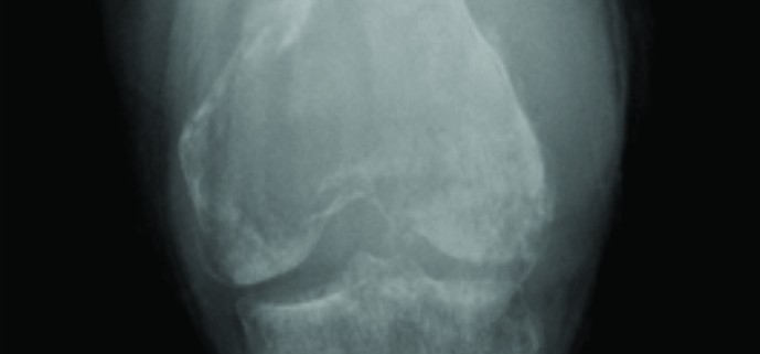
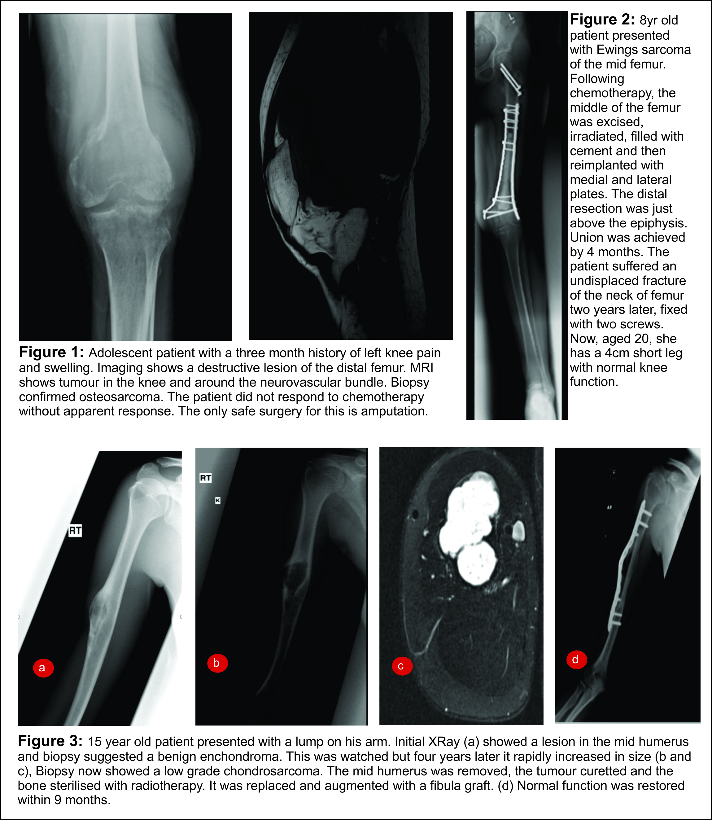
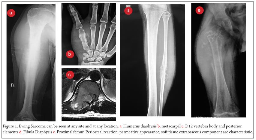
Clinical examination reveals a tender diffuse swelling in the affected area. The range of motion of the adjoining joint may be terminally restricted. The palpation may reveal local warmth. Though the most commonly affected site is the diaphysis in the bone, ES is known to affect the metaphyseal region as well [3]. ES may affect any bone in the body (Fig 1 a-e). Periosteal ES located on the surface of bone and soft tissue ES, though rare, are well-defined clinical entities [3].
‘The clinical and radiological features in an ES of bone may be akin to osteomyelitis or an eosinophilic granuloma and these differentials have to be borne in mind and ruled out by subsequent investigations.
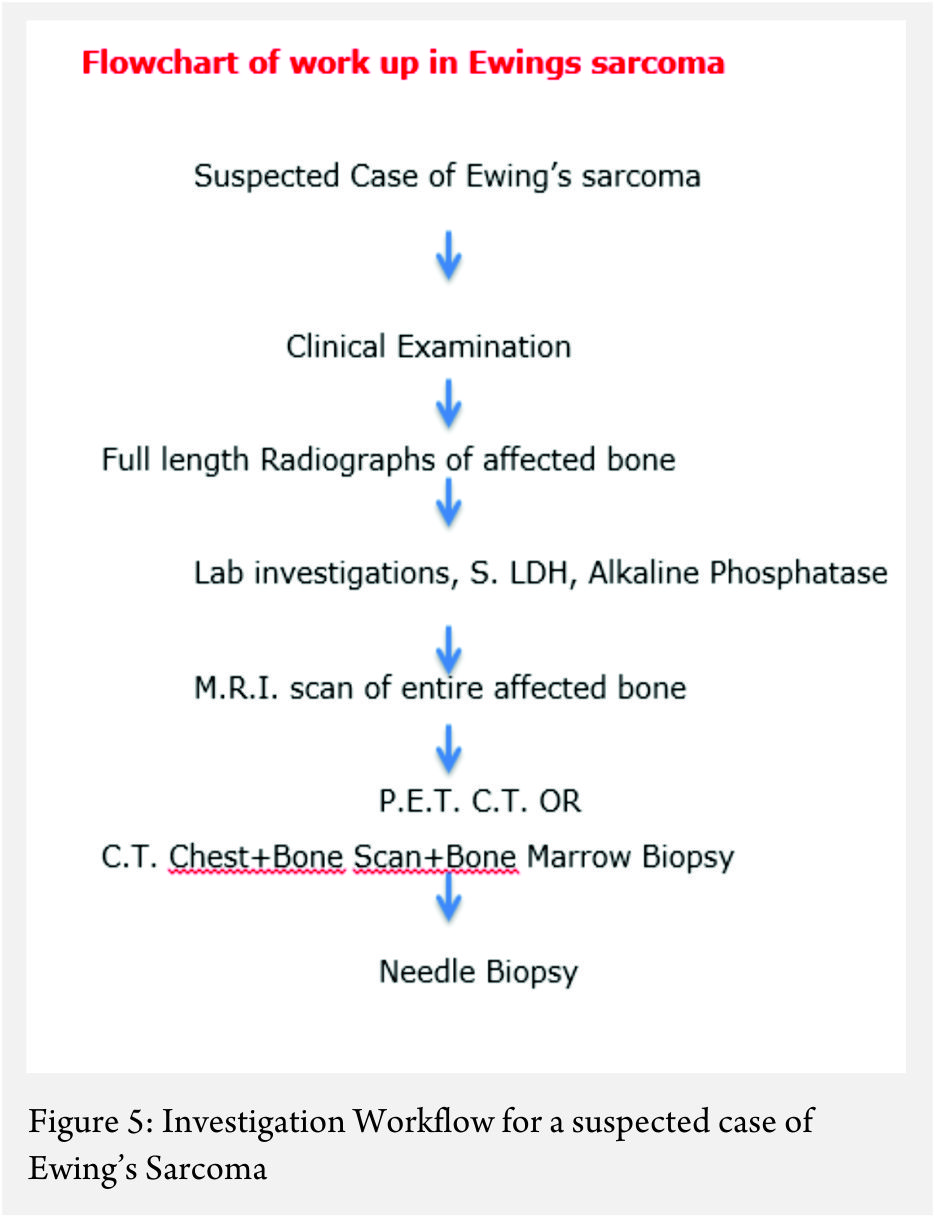
Figure 1
Work up:
The work up includes plain radiographs of the affected bone including the nearby joint, M.R.I. scan of the involved bone, and either a PET CT [7,8,9] or a CT Chest with Technetium Bone scan and a bone marrow aspiration biopsy [3]. The x ray ( Figure 1, a-e) usually shows a permeative, lytic lesion with lamellated periosteal reaction. In locally advance cases, an extra osseous soft tissue component is common [1,3,5]. A diaphyseal lesion may exhibit a characteristic “Onion peel” periosteal reaction. In some cases, “hair-on-end” or “sun-ray spicule” type of periosteal reaction may also be seen.
The laboratory investigations may reveal leukocytosis with elevated E.S.R. and C.R.P [3]. Serum levels of Lactate Dehydrogenase (S. LDH) are usually elevated and serve as a marker of disease activity and response to treatment [6].
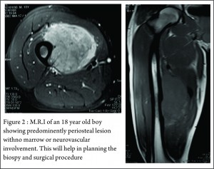
These clinico-radiological and laboratory parameters are akin to osteomyelitis and it requires a trained eye with high index of suspicion to pick the neoplastic nature early in order to avoid mistreating these patients. MRI scan (Fig 2 ) has emerged as one of the most important radiological investigation amongst the others, in the work-up of primary bone sarcomas. It helps immensely in delineating the marrow involvement, revealing skip lesions if any, understanding the extent of soft tissue component and its relationship with the neuro vascular bundle, joint involvement and to decide the ideal site for biopsy. M.R.I. can also be used to assess response to neo-adjuvant chemotherapy [3,5].

Staging
Staging in a case of ES is of paramount importance because of its bearing on the overall prognosis and treatment decisions [1,3]. The conventional staging investigations included a C.T. scan of the chest, a three phase technetium mendronate bone scan and a bone marrow aspiration biopsy [3,10]. However, with the advent of P.E.T. C.T., the bone marrow aspiration biopsy is being found unnecessary [8,9].

Biopsy
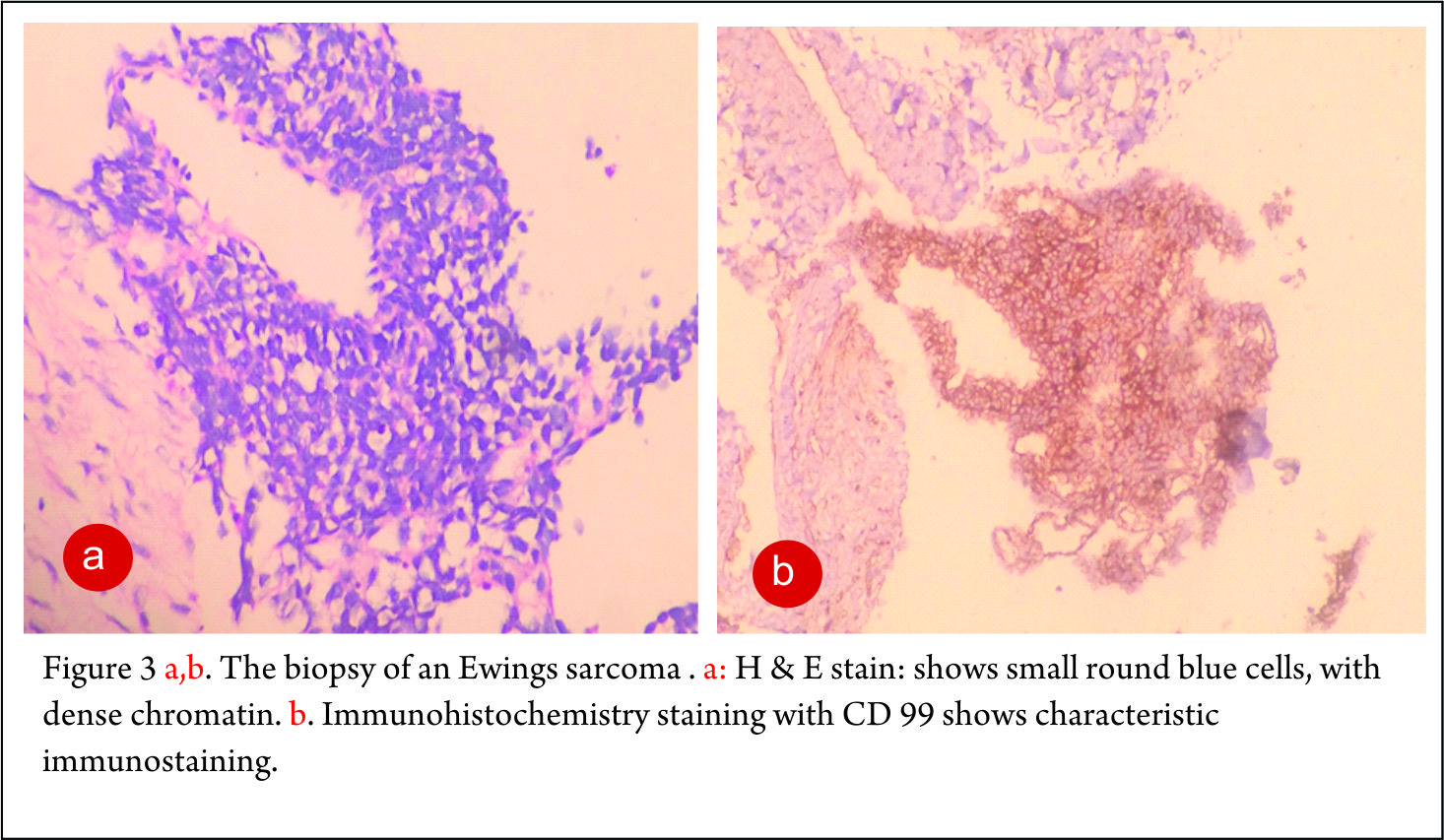
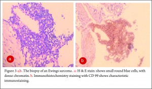
The clinico-radiological suspicion of Ewing sarcoma has to be corroborated by a biopsy and pathological examination before further treatment is commenced. The biopsy of such a lesion is to be done preferably by the orthopaedic oncologist who will be treating the case, at a multi disciplinary cancer centre [3, 11, 13, 14, 15, 16, 17]. Most of the lesions are accurately diagnosed by a needle biopsy. Under the microscope, the tumor is arranged in sheets, nests or clusters of small round blue cells invading the native bone [1]. (Fig 3 a,b). The cells show dense blue chromatin with scanty cytoplasm and the contained glycogen is evident by the P.A.S. (periodic acid-Schiff) stain positivity. Immunohistochemical markers as CD 99 (a mic-2 gene product) and Fli-1 are diagnostic of Ewing sarcoma and are used as confirmatory tools [1,3].

Figure 2 Figure 3

Treatment
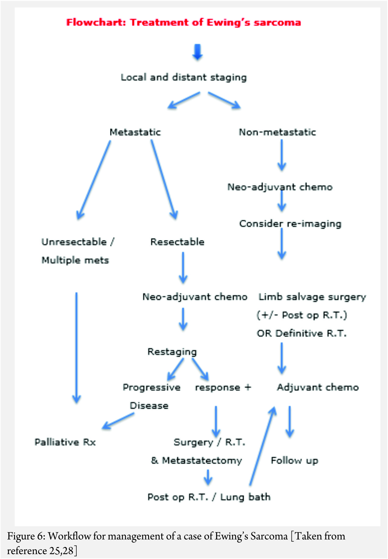
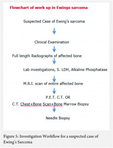
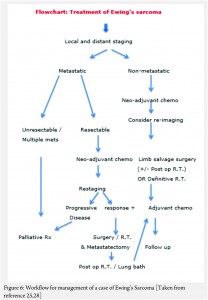
The treatment of ES is handled by a multi disciplinary team comprising of the orthopaedic oncologist, Medical oncologist, Radiation Oncologist, Pathologist and Radiologist [11, 12, 13 , 16, 17, 18]. The patient and the family need to be informed about the clinical results and the expected prognosis and have to participate in the decision making process. Flowcharts of both diagnostic work up and management protocol are provided in figures 5 and 6. The prognosis depends upon the metastatic status of the patient, with the non-metastatic patients having a better outcome [1,3].
The actual management of non-metastatic ES requires neo-adjuvant chemotherapy, followed by surgical resection (if feasible and indicated) followed by post op radiotherapy if necessary (OR definitive local radiotherapy) and then adjuvant (post operative) chemotherapy [1,3] .
In general, the local treatment outcome of an extremity ES is better with surgical wide resection than compared to definitive local radiotherapy alone [18]. In an axially located ES as in pelvis and spine, the decision regarding excision will have to be weighed against the morbidity of the surgery [19]. In a non-metastatic axially located ES, surgery or combined surgery and radiotherapy appears to have an edge over only radiotherapy; the latter being used only in unresectable tumors [20, 21, 22, 23].
The neo adjuvant chemotherapy helps in multiple ways. It is useful in downstaging the local disease, reducing the vascularity, controlling the micro-metastases, sterilizing the satellite lesions in the surrounding zone of hyperemia, helping formation of a thicker capsule, reducing the local edema, healing of pathological fractures and prognosticating outcome of the treatment based on the analysis of percentage necrosis in the tumor. All of these help in making the surgical excision easier and reduce the local recurrence rates [24,25,26,27,28,29,30,31].
The decision regarding limb salvage in a non-metastatic case of Ewings sarcoma is based upon the local extent of the disease. The status of the neuro-vascular bundle and amount of muscles involved by the soft tissue component, determine feasibility of a limb salvage surgery. The only absolute contra indication to a limb salvage surgery would be encasement of a major motor nerve in the extremity and inadequate muscles left after wide excision of the lesion, which would result in a non-functional extremity.
In a case that there are metastases at diagnosis, the decision regarding the approach is based on the number and type of metastases. In a widely metastatic case, only palliative treatment is offered. If there are few pulmonary metastases amenable to excision or are of doubtful significance, the patient is given neo adjuvant chemotherapy and re-staged. The decision regarding treatment is then based on the response to the chemotherapy. If there is progression despite the neo-adjuvant chemotherapy, palliative protocol is followed. If the metastatic lesions have responded to the chemotherapy then the local treatment decision can be taken accordingly with curative intent [25, 28].
The local control rates and the overall survival rates for patients of primary bone sarcomas treated with limb salvage and for those treated by amputation are comparable, with limb salvage surgery carrying better functional outcome [26,28,30,34]. In developing countries, it is worthwhile to offer limb salvage to patients who have a better prognosis, in whom the function of the salvaged extremity is going to be acceptable and for those who are willing to complete the necessary treatment and understand the complications involved.
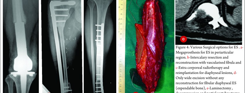
The exact modality of reconstruction after limb salvage is decided by the site of disease, the extent, the patients age [11,34] and expectations and in the developing world, by the socio-economic status of the patient (Fig 4). For periarticular ES, reconstruction can be done by using megaprosthesis [34] or allo-prosthesis composite. This restores the function in the operated extremity fast, shortens the rehabilitation time post operatively, enables early resumption of adjuvant treatment modalities, is a durable option with acceptable complication rate. Arthrodesis can be an alternative to megaprosthetic reconstruction. In cases with diaphyseal involvement, joint sparing inter-calary resections can be done and the defect reconstructed using allograft – live fibula composite or only live vascularised fibula or extra corporeal radiotherapy and reimplantation [35,41]. Rotationplasty is a viable alternative for very young children [36] and in cases of failed limb salvage surgery [37].
The post operative margins of the resected specimen and the percentage necrosis after chemotherapy decide the need for post operative radiotherapy. In cases where the margins are inadequate or the tumor is viable, radiation is used post operatively in order to achieve better control rates [3,21-27,29]. The adjuvant chemo continues in the post operative period. [3, 4, 22, 23, 24, 25, 26, 27, 30, 31, 34].
Patients treated thus need to undergo the prescribed rehabilitation program in order to attain the maximum functional outcome [38]. Functional outcomes in these patients are measured by the Musculo Skeletal Tumor society scoring system (MSTS) or the Toronto extremity salvage score (TESS) [39, 40]. These scores basically reflect the ability of the patient to carry out activities of daily living.

Figure 4

Follow up
The patients are advised to follow up every 3 monthly in the first two years, every six monthly for next three years and annually thereafter. At every visit, radiographs and appropriate staging investigations follow clinical examination [3,25]. Fuchs et al have reported long term complications in 59% percent of patients treated for ES over a average follow up of 25 years [46]. These complications comprised of metastases, local recurrence, secondary malignancies, pathologic fractures, and radiation-associated and chemotherapy-associated morbidities. Hence it is recommended to follow all these patients over a longer period.

Fig 5            Fig 6

Results
In various studies, the overall survival (at 3 or 5 year follow up) for non metastatic ES has been reported to be between 43.5% to 80% [1,23,42-48]. The local recurrence rates are reported to be around 10% to 12.5% [44,48]. In long term follow up of an average of 18 years, Bacci et al have reported overall survival at 5, 10, 15 and 20 years as 57.2%, 49.3%, 44.9% and 38.4% respectively [45]. The poor prognostic indicators in a case of ES are presence of metastases (especially bone and bone marrow metastases), age older than 10 years, a size larger than 200 ml, more central lesions (as in the pelvis or spine), and poor response to chemotherapy [3]. New pharmacological agents and radiotherapeutical modalities are being investigated as discussed in the earlier two articles in the symposium [49,50] and possibility of imporving the survival and quality of life of patients with ES looks promising.


Conclusion

ES is one of the common primary bone malignancies. Appropriate diagnosis, staging, biopsy and treatment at specialized centers is essential for a good outcome. Treatment is multi modal with neoadjuvant and adjuvant chemotherapy, surgery with appropriate margins and radiation in adjuvant or definitive setting; all playing important role in achieving good overall survival rates. Limb salvage surgery in non-metastatic ES is now a norm. The survivors are prone to many long-term complications and need to be followed up for a longer duration. .


References

1. Hameed M. Small Round Cell Tumors Of Bone. Arch Pathol Lab Med. 2007;131:192-204.
2. Rajwanshi A, Srinivas R, Upasana G. Malignant small round cell tumors.J Cytol. 2009;26(1): 1–10.
3. Iwamoto Y. Diagnosis and treatment of Ewing’s sarcoma. Jpn J Clin Oncol. 2007 Feb;37(2):79-89.
4. Bacci G, Ferrari S, Rosito P, Avella M, Barbieri E, Picci P, Battistini A, Brach del Prever A. Ewing’s sarcoma of the bone. Anatomoclinical study of 424 cases. Minerva Pediatr. 1992 Jul-Aug;44(7-8):345-59.
5. Eggli KD, Quiogue T, Moser RP Jr. Ewing’s sarcoma. Radiol Clin North Am. 1993 Mar;31(2):325-37.
6. Bacci G1, Avella M, McDonald D, Toni A, Orlandi M, Campanacci M. Serum lactate dehydrogenase (LDH) as a tumor marker in Ewing’s sarcoma. Tumori. 1988 Dec 31;74(6):649-55.
7. Quartuccio N1, Fox J, Kuk D, Wexler LH, Baldari S, Cistaro A, Schöder H. Pediatric bone sarcoma: diagnostic performance of ¹⁸F-FDG PET/CT versus conventional imaging for initial staging and follow-up. AJR Am J Roentgenol. 2015 Jan;204(1):153-60.
8. Anderson PM. Futility versus utility of marrow assessment in initial Ewing sarcoma staging workup. Pediatr Blood Cancer. 2015 Jan;62(1):1-2.
9. Gerth HU, Juergens KU, Dirksen U, Gerss J, Schober O, Franzius C. Significant benefit of multimodal imaging: PET/CT compared with PET alone in staging and follow-up of patients with Ewing tumors. J Nucl Med. 2007 Dec;48(12):1932-9.
10. Oberlin O1, Bayle C, Hartmann O, Terrier-Lacombe MJ, Lemerle J. Incidence of bone marrow involvement in Ewing’s sarcoma: value of extensive investigation of the bone marrow. Med Pediatr Oncol. 1995 Jun;24(6):343-6.
11. Meyers PA1, Levy AS. Ewing’s sarcoma. Curr Treat Options Oncol. 2000 Aug;1(3):247-57.
12. Exner GU, von Hochstetter AR.Technique and tactics of biopsy including puncture. Z Orthop Ihre Grenzgeb. 1992 Jul-Aug;130(4):272-5.
13. Mavrogenis AF, Angelini A, Vottis C, Palmerini E, Rimondi E, Rossi G, Papagelopoulos PJ, Ruggieri P. State-of-the-art approach for bone sarcomas. Eur J Orthop Surg Traumatol. 2015 Jan;25(1):5-15.
14. Burke NG, Moran CJ, Hurson B, Dudeney S, O’Toole GC. Musculoskeletal oncology training during residency. J Orthop Surg (Hong Kong). 2011 Dec;19(3):350-3.
15. Huang AJ, Kattapuram SV. Musculoskeletal neoplasms: biopsy and intervention. Radiol Clin North Am. 2011 Nov;49(6):1287-305.
16. Scarborough MT. The biopsy. Instr Course Lect. 2004;53:639-44.
17. Bickels J, Jelinek JS, Shmookler BM, Neff RS, Malawer MM. Biopsy of musculoskeletal tumors. Current concepts. Clin Orthop Relat Res. 1999 Nov;(368):212-9.
18. Sudanese A, Toni A, Ciaroni D, Avella M, Dallari D, Picci P, Bacci G, Barbieri E, Mancini A, Campanacci M, et al. The role of surgery in the treatment of localized Ewing’s sarcoma. Chir Organi Mov. 1990 Jul-Sep;75(3):217-30
19. Beadel GP, McLaughlin CE, Aljassir F, Turcotte RE, Isler MH, Ferguson P, Griffin AM, Bell RS, Wunder JS. Iliosacral resection for primary bone tumors: is pelvic reconstruction necessary? Clin Orthop Relat Res. 2005 Sep;438:22-9.
20. Scully SP, Temple HT, O’Keefe RJ, Scarborough MT, Mankin HJ, Gebhardt MC. Role of surgical resection in pelvic Ewing’s sarcoma. J Clin Oncol. 1995 Sep;13(9):2336-41.
21. Carrie C, Mascard E, Gomez F, Habrand JL, Alapetite C, Oberlin O, Moncho V, Hoffstetter S. Nonmetastatic pelvic Ewing sarcoma: report of the French society of pediatric oncology. Med Pediatr Oncol. 1999 Nov;33(5):444-9.
22. Donati D, Yin J, Di Bella C, Colangeli M, Bacci G, Ferrari S, Bertoni F, Barbieri E, Mercuri M. Local and distant control in non-metastatic pelvic Ewing’s sarcoma patients. J Surg Oncol. 2007 Jul 1;96(1):19-25.
23. Biswas B, Rastogi S, Khan SA, Mohanti BK, Sharma DN, Sharma MC, Mridha AR, Bakhshi S. Outcomes and prognostic factors for Ewing-family tumors of the extremities. J Bone Joint Surg Am. 2014 May 21;96(10):841-9.
24. Sciubba DM, Okuno SH, Dekutoski MB, Gokaslan ZL. Ewing and osteogenic sarcoma: evidence for multidisciplinary management. Spine (Phila Pa 1976). 2009 Oct 15;34(22 Suppl):S58-68.
25. ESMO Guidelines Working Group, Saeter G. Ewing’s sarcoma of bone: ESMO clinical recommendations for diagnosis, treatment and follow-up. Ann Oncol. 2007 Apr;18 Suppl 2:ii79-80.
26. Bacci G, Balladelli A, Forni C, Ferrari S, Longhi A, Bacchini P, Alberghini M, Fabbri N, Benassi M, Briccoli A, Picci P. Adjuvant and neoadjuvant chemotherapy for Ewing sarcoma family tumors in patients aged between 40 and 60: report of 35 cases and comparison of results with 586 younger patients treated with the same protocols in the same years. Cancer. 2007 Feb 15;109(4):780-6.
27. Sluga M, Windhager R, Lang S, Heinzl H, Krepler P, Mittermayer F, Dominkus M, Zoubek A, Kotz R. A long-term review of the treatment of patients with Ewing’s sarcoma in one institution. Eur J Surg Oncol. 2001 Sep;27(6):569-73.
28. Paulussen M, Ahrens S, Craft AW, Dunst J, Fröhlich B, Jabar S, Rübe C, Winkelmann W, Wissing S, Zoubek A, Jürgens H. Ewing’s tumors with primary lung metastases: survival analysis of 114 (European Intergroup) Cooperative Ewing’s Sarcoma Studies patients. J Clin Oncol. 1998 Sep;16(9):3044-52.
29. Picci P, Rougraff BT, Bacci G, Neff JR, Sangiorgi L, Cazzola A, Baldini N, Ferrari S, Mercuri M, Ruggieri P, et al. Prognostic significance of histopathologic response to chemotherapy in nonmetastatic Ewing’s sarcoma of the extremities. J Clin Oncol. 1993 Sep;11(9):1763-9.
30. Bacci G, Ferrari S, Avella M, Barbieri E, Picci P, Casadei R, Rosito P, Neri S, Capanna R, Battistini A, et al. Non-metastatic Ewing’s sarcoma: results in 98 patients treated with neoadjuvant chemotherapy. Ital J Orthop Traumatol. 1991 Dec;17(4):449-65.
31. Cagnano R, Avella M, Rosito P, Ciaroni D, Ferraro A, Di Scioscio M, Putti C, Bacci G. Neoadjuvant chemotherapy for localized Ewing’s sarcoma. Preliminary results of a new protocol which uses surgery alone or followed by radiotherapy for local control. J Chemother. 1989 Jul;1(4 Suppl):1248-9.
32. Sluga M, Windhager R, Lang S, Heinzl H, Krepler P, Mittermayer F, Dominkus M, Zoubek A, Kotz R.The role of surgery and resection margins in the treatment of Ewing’s sarcoma. Clin Orthop Relat Res. 2001 Nov;(392):394-9.
33. Hobusch GM, Lang N, Schuh R, Windhager R, Hofstaetter JG. Do patients with ewing’s sarcoma continue with sports activities after limb salvage surgery of the lower extremity? Clin Orthop Relat Res. 2015 Mar;473(3):839-46.
34. Dai X, Ma W, He X, Jha RK. Review of therapeutic strategies for osteosarcoma, chondrosarcoma, and Ewing’s sarcoma. Med Sci Monit. 2011 Aug;17(8):RA177-190.
35. Poffyn B, Sys G, Mulliez A, Van Maele G, Van Hoorebeke L, Forsyth R, Uyttendaele D. Extracorporeally irradiated autografts for the treatment of bone tumours: tips and tricks. Int Orthop. 2011 Jun;35(6):889-95.
36. Hardes J, Gosheger G, Vachtsevanos L, Hoffmann C, Ahrens H, Winkelmann W. Rotationplasty type BI versus type BIIIa in children under the age of ten years. Should the knee be preserved? J Bone Joint Surg Br. 2005 Mar;87(3):395-400.
37. Ramseier LE, Dumont CE, Exner GU. Rotationplasty (Borggreve/Van Nes and modifications) as an alternative to amputation in failed reconstructions after resection of tumours around the knee joint. Scand J Plast Reconstr Surg Hand Surg. 2008;42(4):199-201
38. Shehadeh A, El Dahleh M, Salem A, Sarhan Y, Sultan I, Henshaw RM, Aboulafia AJ. Standardization of rehabilitation after limb salvage surgery for sarcomas improves patients’ outcome. Hematol Oncol Stem Cell Ther. 2013 Sep-Dec;6(3-4):105-11
39. Cassidy RJ, Indelicato DJ, Gibbs CP, Scarborough MT, Morris CG, Zlotecki RA. Function Preservation After Conservative Resection and Radiotherapy for Soft-tissue Sarcoma of the Distal Extremity: Utility and Application of the Toronto Extremity Salvage Score (TESS). Am J Clin Oncol. 2014 Jul 17.
40. P. U. Tunn & D. Pomraenke & U. Goerling & P. Hohenberger. Functional outcome after endoprosthetic limb-salvage therapy of primary bone tumours—a comparative analysis using the MSTS score, the TESS and the RNL index. International Orthopaedics (SICOT) (2008) 32:619–625.
41. Puri A, Gulia A, Jambhekar N, Laskar S. The outcome of the treatment of diaphyseal primary bone sarcoma by resection, irradiation and re-implantation of the host bone: extracorporeal irradiation as an option for reconstruction in diaphyseal bone sarcomas. J Bone Joint Surg Br. 2012 Jul;94(7):982-8
42. Bacci G, Palmerini E, Staals EL, Longhi A, Barbieri E, Alberghini M, Ferrari S Ewing’s sarcoma family tumors of the humerus: outcome of patients treated with radiotherapy, surgery or surgery and adjuvant radiotherapy. Radiother Oncol. 2009 Nov;93(2):383-7.
43. La TH, Meyers PA, Wexler LH, Alektiar KM, Healey JH, Laquaglia MP, Boland PJ, Wolden SL. Radiation therapy for Ewing’s sarcoma: results from Memorial Sloan-Kettering in the modern era. Int J Radiat Oncol Biol Phys. 2006 Feb 1;64(2):544-50.
44. Krasin MJ1, Davidoff AM, Rodriguez-Galindo C, Billups CA, Fuller CE, Neel MD, Merchant TE. Definitive surgery and multiagent systemic therapy for patients with localized Ewing sarcoma family of tumors: local outcome and prognostic factors. Cancer. 2005 Jul 15;104(2):367-73.
45. Bacci G1, Forni C, Longhi A, Ferrari S, Donati D, De Paolis M, Barbieri E, Pignotti E, Rosito P, Versari M. Long-term outcome for patients with non-metastatic Ewing’s sarcoma treated with adjuvant and neoadjuvant chemotherapies. 402 patients treated at Rizzoli between 1972 and 1992. Eur J Cancer. 2004 Jan;40(1):73-83.
46. Fuchs B1, Valenzuela RG, Inwards C, Sim FH, Rock MG. Complications in long-term survivors of Ewing sarcoma. Cancer. 2003 Dec 15;98(12):2687-92.
47. Marcus Jr RB, Berrey BH, Graham-Pole J, Mendenhall NP, Scarborough MT. The treatment of Ewing’s sarcoma of bone at the University of Florida: 1969 to 1998. Clin Orthop Relat Res. 2002 Apr;(397):290-7.
48. Elomaa I1, Blomqvist CP, Saeter G, Akerman M, Stenwig E, Wiebe T, Björk O, Alvegård TA. Five-year results in Ewing’s sarcoma. The Scandinavian Sarcoma Group experience with the SSG IX protocol. Eur J Cancer. 2000 May;36(7):875-80.
49. Irukulla MM, Joseph DM. Management of Ewing Sarcoma:Current Management and the Role of Radiation Therapy. Journal of Bone and Soft Tissue Tumors May-Aug 2015; 1(1):4-6
50.Valvi S & Kellie SJ. Ewing Sarcoma: Focus on Medical Management. Journal of Bone and Soft Tissue Tumors May-Aug 2015; 1(1):4-6.


How to Cite this article: Panchwagh Y. Ewing Sarcoma: Focus on Surgical Management. Journal of  Bone and Soft Tissue Tumors May-Aug 2015;1(1):23-28.

Dr.Yogesh Panchwagh
Dr.Yogesh Panchwagh

(Abstract)      (Full Text HTML)      (Download PDF)