Tag Archive for: Ewing sarcoma

Resection and Reconstruction of Calcaneal Tumors – A Review

Original Article | Volume 6 | Issue 2 | JBST May-August 2020 | Page 2-4 | Subbiah Shanmugam, Sujay Susikar, Murali Kannan. DOI: 10.13107/jbst.2020.v06i02.22

Author: Subbiah Shanmugam[1], Sujay Susikar[1], Murali Kannan[1]

[1]Department of Surgical Oncology, Associate Professor, Centre for Oncology, Government Royapettah Hospital, Chennai, Tamil Nadu, India.

Address of Correspondence
Dr. Sujay Susikar,
Department of Oncology, Centre for Oncology, Government Royapettah Hospital, Chennai, Tamil Nadu, India.
E-mail: sujaysusikar@gmail.com


Abstract

Introduction: Calcaneum is a rare site for Ewing’s sarcoma. The treatment includes neoadjuvant systemic therapy followed by surgical resection and adjuvant systemic therapy. The reconstruction options in this era of limb salvage for bone tumors are not much established in calcaneal tumors in view of limited reporting. Various other reconstruction options available are allograft, iliac crest autograft, and custom-made prosthesis.
Case Report: We have reported a 13-year-old female patient with Ewing’s sarcoma of calcaneum diagnosed by imaging and biopsy. The patient underwent neoadjuvant chemotherapy and then had undergone total calcanectomy with allograft reconstruction. The post-operative outcomes are fair, and the patient is on adjuvant chemotherapy at present.
Conclusion: Biological reconstruction in the form of allograft is a reliable option with regard to the functional outcomes for calcaneal resections.
Keywords: Calcaneum, Ewing sarcoma, total calcanectomy, allograft.


Reference:
1. Sherif PA, Santa A. Ewing’s sarcoma of the calcaneum. Indian J Med Paediatr Oncol 2017;38:542-4.
2. Imanishi J, Choong PF. Three-dimensional printed calcaneal prosthesis following total calcanectomy. Int J Surg Case Rep 2015;10:83-7.
3. Ottolenghi CE, Petracchi LJ. Chondromyxosarcoma of the calcaneus; report of a case of total replacement of involved bone with a homogenous refrigerated calcaneus. J Bone Joint Surg 1953;35:211-4.
4. Scoccianti G, Campanacci DA, Innocenti M, Beltrami G, Capanna R. Total calcanectomy and reconstruction with vascularized iliac bone graft for osteoblastoma: A report of two cases. Foot Ankle Int 2009;30:716-20.
5. Kurvin LA, Volkering C, Kessler SB. Calcaneus replacement after total calcanectomy via vascularized pelvis bone. Foot Ankle Surg 2008;14:221-4.
6. Anacak Y, Sabah D, Demirci S, Kamer S. Intraoperative extracorporeal irradiation and re-implantation of involved bone for the treatment of musculoskeletal tumors. J Exp Clin Cancer Res 2007;26:571.
7. Li J, Guo Z, Pei GX, Wang Z, Chen GJ, Wu ZG. Limb salvage surgery for calcaneal malignancy. J Surg Oncol 2010;102:48-53.
8. Brenner P, Zwipp H, Rammelt S. Vascularized double barrel ribs combined with free serrate us anterior muscle transfer for homologous restoration of the hind foot after calcanectomy. J Trauma Acute Care Surg 2000;49:331-5.
9. Elsalanty ME, Genecov DG. Bone grafts in craniofacial surgery. Craniomaxillofac Trauma Reconstr 2009;2:125-34.
10. Chou LB, Malawer MM. Osteosarcoma of the calcaneus treated with prosthetic replacement with twelve years of follow up: A case report. Foot Ankle Int 2007;28:841-4.


How to Cite this article: Shanmugam S, Susikar S, Kannan M | Resection and Reconstruction of Calcaneal Tumors – A Review | Journal of Bone and Soft Tissue Tumors | May-August 2020; 6(2): 2-4.

[Full Text HTML] [Full Text PDF] [XML]


Ewing Sarcoma: What’s in a name?

Vol 1 | Issue 1 | May – August 2015 | page:6-7 | Ashok K Shyam[1,2*]


Author: Ashok K Shyam[1,2*].

[1]Department of Orthopaedics, Sancheti Institute for Orthopaedics and Rehabilitation, Pune India.
[2]Indian Orthopaedic Research Group, Thane, India.

Address of Correspondence
Dr. Ashok K Shyam MS Orth.
Department of Orthopaedics, Sancheti Institute for Orthopaedicsand Rehabilitation, Pune India.
Email: drashokshyam@yahoo.co.uk


Abstract

There appears to be some ambiguity surrounding interpretation of collaborating disciplines on how ‘Ewing sarcoma’ is named, written or spoken. And they mostly are pretty sure about their version (or probably unaware of any other version!). The article will focus on the issue of how Ewing sarcoma is quoted in literature and shed some light on origin of the term. This will also serve as an introduction to our symposium on Management of Ewing Sarcoma.
Keywords: Ewing Sarcoma, Name.


History

Ewing sarcoma was first described by Dr James Ewing in 1921 in his paper read at New York Pathological society proceedings [1]. In his paper he described a 14 year old girl with a bone tumor arising from radius shaft which was diagnosed to be osteosarcoma. Although osteosarcomas were known to be radioresistent, this particular patient underwent radiotherapy and to everyone’s surprise had a ‘miraculous’ response both clinically and radiographically. The tumor did recur but biopsy done this time revealed it to be distinct form osteosarcoma. Ewing described the cells as round using the loose term ’round cell sarcoma’. To him the cells appeared similar to cells of endothelium of the blood vessels of the bone and thus he described the tumor as ‘Endothelioma of the bone’. He further described 6 such similar patients. This tumor was named as ‘Ewing sarcoma’ few years later by Ernest Codman [Ewing sarcoma is the most common differential of Codman Triangle] [2]. Although Dr James Ewing has contributed a lot to medicine his contributions in estabilishing oncology as an independent science is noted by naming him as ‘The Cancer Man’ or ‘Mr Cancer’ by his colleagues and media. He is still best known to every student of medicine by the eponym of ‘Ewing Sarcoma’.

What’s in the Name?

Ewing sarcoma has been reported to be written as ‘Ewing sarcoma’, Ewing’s Sarcoma’, ‘Ewings’ Sarcoma’ or simply ‘Ewings sarcoma’. Which is the correct version? I came across this issue rather accidently on preparing and reviewing for this particular symposium. We have three articles in the symposium [3,4,5], one on radiotherapy, one on medical management and one on surgical aspect. There was difference in how these three manuscripts spelled Ewing sarcoma. The non surgical articles insisted the name to be ‘Ewing Sarcoma’ while the surgical article insisted on ‘Ewing’s Sarcoma’. So what is the correct nomenclature? On sarcomahelp.com site I found this description “The tumor which bears his name is generally referred to as Ewing’s sarcoma when spoken and either Ewing’s sarcoma or Ewing sarcoma when written [6].” I reviewed the policies about the eponymus words and found an interesting fact about them. A cold war is been fought between US and Europe with US wishing to phase out use of eponyms and is against creating any new ones [7]. The argument is that the disease is not ‘Owned’ by a person. They called the eponymus use with an apostrophe as a possessive case of the word that indicates that the person either had the disease or owned the disease. By that example Ewing’s sarcoma indicates that Ewing had the sarcoma! They advocated a non possessive use of the eponyms like ‘Ewing sarcoma’. Following the rule the AMA Manual of Style: a guide to authors and editors recommends use of non-possessive case for writing eponyms. Even if we ask word nerds in true grammatical sense an apostrophe does indicate possessive nature of the noun to which it is attached. On the other hand the European literature holds the eponyms in high regards as historical testaments to physicians who first described the diseases and advice to write eponyms with an apostrophe as in Ewing’s sarcoma. To support this there remains arguments regarding using non possessive case in certain diseases like replacing ‘Down’s Syndrome’ with ‘Down syndrome’ which will then imply that there is an ‘Up Syndrome’! The controversy still rages and anyone interested should read these two articles published in British Medical Journal [8,9].
But what about our question about Ewing Sarcoma? Searching pubmed, I could find 1021 article with Ewing sarcoma , 2147 articles with Ewing’s Sarcoma , 17 article with Ewings sarcoma and 9 articles with Ewings’ sarcoma respectively in their titles (Fig 1 a-d). Thus most of them were divided into either possessive case or non-possessive case and possibly it depends on journal policy and geographical preference. Search of MESH (Medical Subject Heading) in pubmed identifies Ewing sarcoma as Mesh Major Subject Heading. I too would personally agree with non-possessive case [also as per AMA Style and Pubmed MESH term] in form of ‘Ewing Sarcoma’ although authors may choose other versions too. However when one of the version is used, authors should use the same version consistently throughout the manuscript. In this symposium we have used the non-possessive version in medical management and radiotherapy articles while a possessive version is used by the surgical focussed article. We hope the symposium is informative and any queries that remain in readers mind are welcomed as ‘Letter to Editor’ and will be answered by the respective authors.

Figure 1


References

1. Ewing J: Diffuse endothelioma of bone, Proc NY Pathol Soc 1921;21:17.
2. Timothy P. Cripe, “Ewing Sarcoma: An Eponym Window to History,” Sarcoma, vol. 2011, Article ID 457532
3. Valvi S & Kellie SJ. Ewing Sarcoma: Focus on Medical Management. Journal of Bone and Soft Tissue Tumors May-Aug 2015; 1(1):4-6
4. Irukulla MM, Joseph DM. Management of Ewing Sarcoma: Current Management and the Role of Radiation Therapy. Journal of Bone and Soft Tissue Tumors May-Aug 2015; 1(1):4-6
5. Panchwagh Y. Ewing Sarcoma: Focus on Surgical Management. Journal of Bone and Soft Tissue Tumors May-Aug 2015;1(1):4-6
6. http://sarcomahelp.org/ewings-sarcoma.html#tpm1_1
7. Jana N, Barik S, Arora N. Current use of medical eponyms–a need for global uniformity in scientific publications. BMC Med Res Methodol. 2009 Mar 9;9:18.
8. Woywodt A, Matteson E. Should eponyms be abandoned? Yes. BMJ. 2007;335:424
9. Whitworth JA. Should eponyms be abandoned? No. BMJ. 2007;335:425.


How to Cite this article: Shyam AK. Ewing Sarcoma: What’s in a name? Journal of  Bone and Soft Tissue Tumors May-Aug 2015;1(1):6-7.

Dr.Ashok Shyam

Dr.Ashok Shyam


(Abstract)      (Full Text HTML)      (Download PDF)


Management of Ewing Sarcoma: Current Management and the Role of Radiation Therapy

Vol 1 | Issue 1 | May – August 2015 | page:18-22 | Monica Malik Irukulla[1,*], Deepa M Joseph[1].


Author: Monica Malik Irukulla[1,*], Deepa M Joseph[1].

[1]Department of Radiation Oncology, Nizam’s Institute of Medical Sciences, Hyderabad. India

Address of Correspondence
Dr. Monica Malik Irukulla, MD.
Department of Radiation Oncology, Nizam’s Institute of Medical Sciences, Hyderabad. India.
Email: dr_monica11@yahoo.com


Abstract

The management of Ewing sarcoma has evolved over the last few decades with successive improvement in survival rates. Multidisciplinary management is the key to successful outcomes. Dose intensity of chemotherapy is of vital importance. Local control can be effectively achieved with surgery, radiation therapy or a combination of the two. The choice of appropriate local therapy should be individualized and depends on various factors such as site, size, resectability, expected morbidity, long term effects etc. Metastatic disease remains a significant challenge and optimal therapeutic strategies still need to be defined. Current management and the role of radiation therapy in Ewing sarcoma are reviewed.
Keywords: Ewing sarcoma, radiation therapy, management.


Introduction

Ewing sarcoma family of tumors (ESFT) are a group of small round cell tumors showing varying degrees of neuroectodermal differentiation with Ewing sarcoma being the least differentiated. Primitive neuroectodermal tumors (PNET) show neuroectodermal differentiation by light microscopy, immune histochemistry (IHC) or electron microscopy [1]. According to WHO classification of bone and soft tissue tumors, Ewings sarcoma/PNET is synonymous with Ewing tumor, peripheral neuroepithelioma, peripheral neuroblastoma and Askin tumor [1]. In most of the patients, a chromosomal translocation leads to the expression of the EWS-FLI1 chimeric transcription factor which is the major oncogene in this pathology [2].

Epidemiology
Ewing sarcoma is the second most common primary bone tumor of childhood and it most commonly occurs in the second decade of life with a slight male preponderance. The incidence of Ewing sarcoma has been reported to be low in Asian population as compared to Caucasians[3]. Data from Indian population show that it is not so uncommon[4]. The common sites of primary Ewing sarcoma are the long bones of the lower extremities (41%), pelvic bones (26%), and bones of the chest wall (16%)[5]. Extraosseous Ewing sarcoma is more commonly axial in location involving the trunk (32%), extremities (26%), head and neck (18%), the retroperitoneum (16%) etc[6]. Approximately 20-25% of patients present with metastasis at diagnosis. Common sites of metastases include lungs, bones and bone marrow.

Diagnostic Evaluation
Typical presenting symptoms include pain and swelling with occasional constitutional symptoms like fever, fatigue and loss of weight. Patients should be evaluated and managed by a multidisciplinary team of experts including pediatric oncologists, orthopedic surgeons, radiologists, pathologists and rehabilitation specialists. A biopsy should be performed in a way such that the track and scar can be included in the subsequent resection or radiation portal. Biopsy should be from soft tissue as often as possible to avoid increasing the risk of fracture and should be through rather than between muscle compartments avoiding the neurovascular bundles. A skilled pathologist should be available onsite to confirm adequacy of the material and review the frozen sections. A needle biopsy may be adequate if sufficient tissue can be obtained for histological, cytogenetic and molecular studies. The risk of diagnostic errors and complications increases by as much as 12-fold when the biopsy is improperly done [7]. Ewing sarcoma/PNETs usually strongly express the cell surface glycoprotein MIC2 (CD99) and this can be helpful in diagnosis of small blue round cell tumors. CD99 is however not exclusively specific for ES/PNET and is found in other tumors such as synovial sarcoma, NHL, GIST etc [8]. Approximately 85% patients have expression of EWS-FLI1 chimeric transcription factor resulting from translocation between EWS and FLI-1 gene t(11;22)(q24;q12) [9]. In most of the remaining patients, alternative translocations between EWS and another ETS- family member (ERG, FEV, ETV1, E1AF) are detected [2]. Molecular analysis for EWS-FLI 1 should be considered. The prognostic value of the same remains inconclusive until now[10]. This is being evaluated as potential therapeutic target [11]. Local imaging with MRI with or without CT scan is recommended. Conventional staging evaluation includes bilateral bone marrow aspiration and biopsy or MRI of spine and pelvis, bone scan and CT scan of the chest. Serum LDH is an important prognostic marker. Positron emission tomography (PET) combined with conventional imaging is a valuable tool in staging and restaging ESFT with a sensitivity of 96% and specificity of 92% [12]. FDG-PET can also serve as a non-invasive method to predict response to chemotherapy which is a useful prognostic marker.13 Whole-body MRI can be a useful radiation-free modality to detect metastatic lesions with a higher sensitivity than bone scintigraphy [14,15]. Fertility consultation should be done for patients desiring future child bearing before starting therapy.

Prognostic factors
5-year event free survival approaches around 70% with standard multimodality approach in localized disease [16]. Favorable prognostic factors include extremity tumors, tumor volume <100ml, normal LDH and absence of metastases at presentation. Common adverse prognostic factors include metastatic disease at presentation, extra skeletal presentation, pelvis as the primary site and poor response to induction chemotherapy. Metastatic disease is the most significant adverse prognostic factor. Those with isolated pulmonary metastasis have a slightly better outcome than those with bone or bone marrow metastasis [17]. Survival depends on the site and number of metastases and the tumor burden with 5 year survival rates ranging from approximately 30% with isolated lung metastasis to less than 20% with multiple bone metastases. Older patients do worse than patients younger than 15 years.18 Poor histologic response to chemotherapy is associated with worse outcomes in patients with localized disease [19].

Treatment
Treatment of Ewing sarcoma has evolved following evidence from large multinational trials over the past few decades with successive improvement in outcomes. Multimodality approach is the key in the management of nonmetastatic Ewing sarcoma.

Chemotherapy
The prognosis in Ewing sarcoma remained very poor until 1960s in spite of good initial response to local treatment. The introduction of chemotherapy into the treatment regimen dramatically improved the response rates and thus the cure rates. Patients are started on induction chemotherapy for 3-4 cycles followed by local therapy at 12weeks. Restaging should be done with a chest imaging and MRI of the local part before local therapy. Further adjuvant chemotherapy is continued for total treatment duration of about 10-12 months. Chemotherapy with Vincristine, Adriamycin/Actinomycin D, cyclophosphamide, (VAC) alternating with Ifosfamide and Etoposide (IE) administered at a three weekly fashion is the standard regimen. Maintaining adequate dose intensity of chemotherapy is of utmost importance. Interval compressed or dose dense chemotherapy improves DFS and has the potential to improve overall survival [20].

Local therapy
Local therapy is delivered at the completion of 3-4 cycles of chemotherapy at 12 weeks and comprises of surgery or radiotherapy or both. There are no randomized trials comparing the two modalities. The choice of local therapy depends on the site of the disease, age of the patient, expected functional outcomes and concern over the late morbidities. Although retrospective institutional series suggest superior local control and survival with surgery rather than radiation therapy, most of these studies are compromised by selection bias. A North American intergroup trial showed no difference in local control or survival based on local treatment modality – surgery, radiation therapy, or both [21]. In patients with localized Ewing sarcoma treated in cooperative intergroup studies there was no significant effect of local control modality (surgery, RT, or surgery plus RT) on OS or EFS rates. In the CESS 86 trial, although radical surgery and resection plus RT resulted in better local control rates (100% and 95%, respectively) than definitive RT (86%), there was no improvement in relapse free survival and overall survival [22]. Preoperative radiation therapy can achieve tumor shrinkage and surgical resection with negative margins in cases with borderline resectability and can potentially allow smaller fields and lower radiation doses [23].

Definitive Radiotherapy
Ewing sarcoma was described by James Ewing in 1921 as “diffuse endothelioma of bone”, a distinct entity from osteosarcoma due to its high response to radiation therapy. In the current scenario, definitive radiotherapy remains an effective local therapy strategy for patients with tumors in sites not amenable for surgical resection and in cases where resection is likely to result in unacceptable morbidity. Advances in imaging, tumor delineation, treatment planning and delivery is now allowing greater precision and sparing of normal tissues. Historically, patients were treated with whole bone irradiation. With the POG 8346 trial, adequate involved field RT with MRI based planning became the standard. Current guidelines recommend 1.5 to 2 cm margin from the gross tumor volume. A randomized study of 40 patients with Ewing sarcoma using 55.8 Gy to the prechemotherapy tumor extent with a 2 cm margin compared with the same total-tumor dose after 39.6 Gy to the entire bone showed no difference in local control or EFS [24]. Initial treatment volume include the pre-chemotherapy volume with margin up to a dose of 45Gy, further boost is delivered to the post chemotherapy volume upto a total dose of 55.8Gy to 60Gy. Tumor size and RT dose have been shown to be predictive of local control rates in patients with non-metastatic Ewing sarcoma treated with chemotherapy and definitive RT [25]. Role of hyperfractionated radiotherapy in management of Ewing sarcoma has been evaluated in the CESS 86 trial [22]. No significant advantage has been demonstrated over the standard fractionation and dose. Recent reports suggest that Proton beam therapy can potentially spare more amount of normal tissue but longer follow up is needed to determine its impact on morbidity and cure rates [26]. Radiation therapy is associated with the development of second malignant neoplasms. In a retrospective analysis, the incidence of second malignancy was 20% in patients who received doses of 60 Gy or more and 5% in those who received 48 Gy to 60 Gy. Those who received < 48 Gy did not develop a second malignancy [27]

Postoperative RT
Postoperative radiation (PORT) is recommended in cases of intralesional or marginal resection, intraoperative spill and poor pathological response to chemotherapy and is usually initiated at 6-8 weeks following surgery. Current Children’s Oncology Group (COG) protocols have more specifically defined adequate margin status. Complete resection is defined as a minimum of 1 cm margin and ideally 2–5 cm around the involved bone. The minimum soft tissue margin for fat or muscle planes is at least 5 mm and for fascial planes at least 2 mm. The Intergroup Ewing Sarcoma Study (INT-0091) recommends 45 Gy to the original disease site plus a 10.8 Gy boost for patients with gross residual disease and 45 Gy plus a 5.4 Gy boost for patients with microscopic residual disease. In the absence of gross residual disease there seems to be no clear benefit to doses over 45 Gy [28]. No radiation therapy is recommended for those who have no evidence of microscopic residual disease following surgical resection. Although not statistically significant, local relapse was least in the combined arm (10.5%) compared with 25% for either surgery or radiotherapy alone [21]. EICESS 92 evaluated the role of postoperative RT in patients with poor pathological response to induction chemotherapy (<90% necrosis). In their analysis there was reduction in local failures (5% vs.12%) in the poor responders if they received PORT [29 In the CESS and EICESS trials, the local failure rate for central primaries was reduced by 50% with PORT. However the role of adjuvant radiotherapy in poor responders and central tumors needs to be clearly defined and the benefits need to be balanced against potential risks of long term effects and second malignancies. For extraskeletal ES, PORT is generally recommended except in good prognosis superficial tumors [30].

Management of metastatic disease
Standard treatment guidelines for metastatic Ewing sarcoma recommend treatment similar to localized disease [30]. Different chemotherapy agents used are Vincristine, Adriamycin, Cyclophosphamide, Ifosphamide and Etoposide. Addition of IE to VAC does not seem to have additional benefit in this subset of patients [31]. Dose-intense treatment approach with high dose chemotherapy and autologous stem cell transplantation (HDT/SCT) was evaluated in the nonrandomized Euro-EWING 99 R3 study [32]. Even though this may have a potential to improve outcome, it has not become the standard of therapy.
Following induction chemotherapy, patients are reassessed with local and chest imaging and previously abnormal investigations are repeated. A progressive disease is treated with palliative intent and the good responders are managed with treatment of primary disease and metastatic sites. Timing of local therapy for both primary site and metastatic sites remain unclear.

Radiotherapy in metastatic disease
Whole lung irradiation (WLI) in patients with lung-only metastases has shown improved disease free and overall survival in various trials. Patients with lung metastasis should be considered for whole lung irradiation even after complete resolution following chemotherapy [33] Doses of 12 to 21 Gy have been used and are usually well tolerated [34]. Hemithoracic irradiation is recommended in patients with chest wall tumor with pleural nodules, pleural effusion or positive pleural cytology.
Bone metastases in Ewing sarcoma should be treated with similar doses as the primary site. They may be treated simultaneously or following completion of chemotherapy depending on the risk of marrow suppression. In the phase II POG/CCG trial which evaluated the role of intensive chemotherapy, local treatment for primary disease was done after completion of 21weeks of chemotherapy and that of metastatic disease was done after week 39 chemotherapy [35]. With the emergence of stereotactic body radiotherapy (SBRT), it is now possible to deliver ablative doses to sites of bone metastases with excellent sparing of normal tissues. SBRT delivered in one to five fractions can also minimize interruptions of systemic therapy.

Treatment of relapse
Outlook of patients who relapse remain unfavorable. Late onset relapse (>2years) and strictly localized disease has a favorable outcome [36]. Chemotherapy regimens in relapse settings are not standard. Two phase II studies have demonstrated upto 33% partial responses in relapsed refractory Ewing’s sarcoma with the combination of Topotecan and Cyclophosphamide [37]. The combination of Irinotecan and Temozolomide has demonstrated clinical responses.38 Gemcitabine in combination with Docetaxel has shown modest activity [39]. Newer drugs and targeted therapies are being evaluated. Radiotherapy and/or surgery may play a role in improving control rates.

Indian Data
There is paucity of data from Indian population. In a retrospective analysis, symptom duration >4 months, tumor diameter >8cm and baseline WBC >11×10(9)/L were predictive of poorer outcomes [40]. Optimal surgical margin in extra skeletal Ewing sarcoma in children was evaluated by Laskar et al who concluded that clear margins of resection correlated with local control irrespective of margin size[41] Survival rates in India remain dismal in spite of the advancement seen in the western world [42]. Patients tend to present with advanced stage disease and often default treatment due to socioeconomic factors.


Conclusion

The management of Ewing sarcoma has significantly evolved over the last few decades with consequent improvements in survival and functional outcomes. Treatment mandates multidisciplinary co-ordination involving Medical, Surgical and Radiation Oncologists, Orthopedic surgeons, Rehabilitation specialists, Pediatricians and others. Dose intensity of chemotherapy and optimal timing and modality of local therapy appear to significantly influence outcomes and survival rates. Metastatic disease represents a major challenge and optimal treatment strategies still need to be defined.


References

1.Fletcher CDM, World Health Organization., International Agency for Research on Cancer. WHO classification of tumours of soft tissue and bone. 4th ed. Lyon: IARC Press; 2013.
2.Stoll G, Surdez D, Tirode F, et al. Systems biology of Ewing sarcoma: a network model of EWS-FLI1 effect on proliferation and apoptosis. Nucleic acids research 2013;41:8853-71.
3.Parkin DM, Stiller CA, Nectoux J. International variations in the incidence of childhood bone tumours. International journal of cancer Journal international du cancer 1993;53:371-6.
4.Rao VS, Pai MR, Rao RC, Adhikary MM. Incidence of primary bone tumours and tumour like lesions in and around Dakshina Kannada district of Karnataka. Journal of the Indian Medical Association 1996;94:103-4, 21.
5.Bernstein M, Kovar H, Paulussen M, et al. Ewing’s sarcoma family of tumors: current management. The oncologist 2006;11:503-19.
6.Applebaum MA, Worch J, Matthay KK, et al. Clinical features and outcomes in patients with extraskeletal Ewing sarcoma. Cancer 2011;117:3027-32.
7. Mankin HJ, Mankin CJ, Simon MA. The hazards of the biopsy, revisited. Members of the Musculoskeletal Tumor Society. The Journal of bone and joint surgery American volume 1996;78:656-63.
8. Perlman EJ, Dickman PS, Askin FB, Grier HE, Miser JS, Link MP. Ewing’s sarcoma–routine diagnostic utilization of MIC2 analysis: a Pediatric Oncology Group/Children’s Cancer Group Intergroup Study. Human pathology 1994;25:304-7.
9. Delattre O, Zucman J, Plougastel B, et al. Gene fusion with an ETS DNA-binding domain caused by chromosome translocation in human tumours. Nature 1992;359:162-5.
10. Le Deley MC, Delattre O, Schaefer KL, et al. Impact of EWS-ETS fusion type on disease progression in Ewing’s sarcoma/peripheral primitive neuroectodermal tumor: prospective results from the cooperative Euro-E.W.I.N.G. 99 trial. Journal of clinical oncology : official journal of the American Society of Clinical Oncology 2010;28:1982-8.
11. Herrero-Martin D, Fourtouna A, Niedan S, Riedmann LT, Schwentner R, Aryee DN. Factors Affecting EWS-FLI1 Activity in Ewing’s Sarcoma. Sarcoma 2011;2011:352580.
12. Volker T, Denecke T, Steffen I, et al. Positron emission tomography for staging of pediatric sarcoma patients: results of a prospective multicenter trial. Journal of clinical oncology : official journal of the American Society of Clinical Oncology 2007;25:5435-41.
13. Dimitrakopoulou-Strauss A, Strauss LG, Egerer G, et al. Impact of dynamic 18F-FDG PET on the early prediction of therapy outcome in patients with high-risk soft-tissue sarcomas after neoadjuvant chemotherapy: a feasibility study. Journal of nuclear medicine : official publication, Society of Nuclear Medicine 2010;51:551-8.
14. Mentzel HJ, Kentouche K, Sauner D, et al. Comparison of whole-body STIR-MRI and 99mTc-methylene-diphosphonate scintigraphy in children with suspected multifocal bone lesions. European radiology 2004;14:2297-302.
15. Burdach S, Thiel U, Schoniger M, et al. Total body MRI-governed involved compartment irradiation combined with high-dose chemotherapy and stem cell rescue improves long-term survival in Ewing tumor patients with multiple primary bone metastases. Bone marrow transplantation 2010;45:483-9.
16. Grier HE, Krailo MD, Tarbell NJ, et al. Addition of Ifosfamide and Etoposide to Standard Chemotherapy for Ewing’s Sarcoma and Primitive Neuroectodermal Tumor of Bone. New England Journal of Medicine 2003;348:694-701.
17. Cotterill SJ, Ahrens S, Paulussen M, et al. Prognostic factors in Ewing’s tumor of bone: analysis of 975 patients from the European Intergroup Cooperative Ewing’s Sarcoma Study Group. Journal of clinical oncology : official journal of the American Society of Clinical Oncology 2000;18:3108-14.
18. Ladenstein R, Potschger U, Le Deley MC, et al. Primary disseminated multifocal Ewing sarcoma: results of the Euro-EWING 99 trial. Journal of clinical oncology : official journal of the American Society of Clinical Oncology 2010;28:3284-91.
19. Paulussen M, Ahrens S, Dunst J, et al. Localized Ewing tumor of bone: final results of the cooperative Ewing’s Sarcoma Study CESS 86. Journal of clinical oncology : official journal of the American Society of Clinical Oncology 2001;19:1818-29.
20. Womer RB, West DC, Krailo MD, et al. Randomized controlled trial of interval-compressed chemotherapy for the treatment of localized Ewing sarcoma: a report from the Children’s Oncology Group. Journal of clinical oncology : official journal of the American Society of Clinical Oncology 2012;30:4148-54.
21. Yock TI, Krailo M, Fryer CJ, et al. Local control in pelvic Ewing sarcoma: analysis from INT-0091–a report from the Children’s Oncology Group. Journal of clinical oncology : official journal of the American Society of Clinical Oncology 2006;24:3838-43.
22. Dunst J, Jurgens H, Sauer R, et al. Radiation therapy in Ewing’s sarcoma: an update of the CESS 86 trial. International journal of radiation oncology, biology, physics 1995;32:919-30.
23. Wagner TD, Kobayashi W, Dean S, et al. Combination short-course preoperative irradiation, surgical resection, and reduced-field high-dose postoperative irradiation in the treatment of tumors involving the bone. International journal of radiation oncology, biology, physics 2009;73:259-66.
24. Craft A, Cotterill S, Malcolm A, et al. Ifosfamide-containing chemotherapy in Ewing’s sarcoma: The Second United Kingdom Children’s Cancer Study Group and the Medical Research Council Ewing’s Tumor Study. Journal of clinical oncology : official journal of the American Society of Clinical Oncology 1998;16:3628-33.
25. Krasin MJ, Rodriguez-Galindo C, Billups CA, et al. Definitive irradiation in multidisciplinary management of localized Ewing sarcoma family of tumors in pediatric patients: outcome and prognostic factors. International journal of radiation oncology, biology, physics 2004;60:830-8.
26. Rombi B, DeLaney TF, MacDonald SM, et al. Proton radiotherapy for pediatric Ewing’s sarcoma: initial clinical outcomes. International journal of radiation oncology, biology, physics 2012;82:1142-8.
27. Kuttesch JF, Jr., Wexler LH, Marcus RB, et al. Second malignancies after Ewing’s sarcoma: radiation dose-dependency of secondary sarcomas. Journal of clinical oncology : official journal of the American Society of Clinical Oncology 1996;14:2818-25.
28. Donaldson SS. Ewing sarcoma: radiation dose and target volume. Pediatric blood & cancer 2004;42:471-6.
29. Schuck A, Ahrens S, Paulussen M, et al. Local therapy in localized Ewing tumors: results of 1058 patients treated in the CESS 81, CESS 86, and EICESS 92 trials. International journal of radiation oncology, biology, physics 2003;55:168-77.
30. Group TEESNW. Bone sarcomas: ESMO Clinical Practice Guidelines for diagnosis, treatment and follow-up. Annals of Oncology 2014;25:iii113-iii23.
31. Grier HE, Krailo MD, Tarbell NJ, et al. Addition of ifosfamide and etoposide to standard chemotherapy for Ewing’s sarcoma and primitive neuroectodermal tumor of bone. The New England journal of medicine 2003;348:694-701.
32. Ladenstein R, Pötschger U, Le Deley MC, et al. Primary Disseminated Multifocal Ewing Sarcoma: Results of the Euro-EWING 99 Trial. Journal of Clinical Oncology 2010;28:3284-91.
33. Paulussen M, Ahrens S, Burdach S, et al. Primary metastatic (stage IV) Ewing tumor: survival analysis of 171 patients from the EICESS studies. European Intergroup Cooperative Ewing Sarcoma Studies. Annals of oncology : official journal of the European Society for Medical Oncology / ESMO 1998;9:275-81.
34. Bolling T, Schuck A, Paulussen M, et al. Whole lung irradiation in patients with exclusively pulmonary metastases of Ewing tumors. Toxicity analysis and treatment results of the EICESS-92 trial. Strahlentherapie und Onkologie : Organ der Deutschen Rontgengesellschaft [et al] 2008;184:193-7.
35. Bernstein ML, Devidas M, Lafreniere D, et al. Intensive therapy with growth factor support for patients with Ewing tumor metastatic at diagnosis: Pediatric Oncology Group/Children’s Cancer Group Phase II Study 9457–a report from the Children’s Oncology Group. Journal of clinical oncology : official journal of the American Society of Clinical Oncology 2006;24:152-9.
36. Stahl M, Ranft A, Paulussen M, et al. Risk of recurrence and survival after relapse in patients with Ewing sarcoma. Pediatric blood & cancer 2011;57:549-53.
37. Saylors RL, 3rd, Stine KC, Sullivan J, et al. Cyclophosphamide plus topotecan in children with recurrent or refractory solid tumors: a Pediatric Oncology Group phase II study. Journal of clinical oncology : official journal of the American Society of Clinical Oncology 2001;19:3463-9.
38. Wagner LM, McAllister N, Goldsby RE, et al. Temozolomide and intravenous irinotecan for treatment of advanced Ewing sarcoma. Pediatric blood & cancer 2007;48:132-9.
39. Mora J, Cruz CO, Parareda A, de Torres C. Treatment of relapsed/refractory pediatric sarcomas with gemcitabine and docetaxel. Journal of pediatric hematology/oncology 2009;31:723-9.
40. Biswas B, Rastogi S, Khan SA, et al. Developing a prognostic model for localized Ewing sarcoma family of tumors: A single institutional experience of 224 cases treated with uniform chemotherapy protocol. Journal of surgical oncology 2015;111:683-9.
41. Qureshi S, Laskar S, Kembhavi S, et al. Extraskeletal Ewing sarcoma in children and adolescents: impact of narrow but negative surgical margin. Pediatr Surg Int 2013;29:1303-9.
42. Tiwari A, Gupta H, Jain S, Kapoor G. Outcome of multimodality treatment of Ewing’s sarcoma of the extremities. Indian journal of orthopaedics 2010;44:378-83.


How to Cite this article: Irukulla MM, Joseph DM. Management of Ewing Sarcoma: Current Management and the Role of Radiation Therapy. Journal of  Bone and Soft Tissue Tumors May-Aug 2015;1(1):18-22.

Dr. Monica M. Irukulla
Dr. Deepa M Joseph
Dr. Deepa M Joseph

(Abstract)      (Full Text HTML)      (Download PDF)


Ewing Sarcoma: Focus on Medical Management

Vol 1 | Issue 1 | May – August 2015 | page:1-2 | Santosh Valvi, Stewart J Kellie


Author: Santosh Valvi [1,2*], Stewart J Kellie [3,4]

[1]Kids Cancer Centre, Sydney Children’s Hospital, Randwick 2031, New South Wales, Australia
[2] Children’s Cancer Institute Australia, Lowy Cancer Research Centre, University of New South Wales, Randwick 2031, New South Wales, Australia
[3] Oncology Unit, The Children’s Hospital at Westmead, Westmead 2145, New South Wales, Australia
[4] Discipline of Paediatrics and Child Health, Faculty of Medicine, University of Sydney, Westmead 2145, New South Wales, Australia

Address of Correspondence
Dr. Santosh Valvi FRACP
Kids Cancer Centre, Sydney Children’s Hospital, Randwick 2031, New South Wales, Australia
Email: santosh.valvi@health.nsw.gov.au


Abstract

The management of Ewing sarcoma has evolved over the last few decades with successive improvement in survival rates. Multidisciplinary management is the key to successful outcomes. Dose intensity of chemotherapy is of vital importance. Local control can be effectively achieved with surgery, radiation therapy or a combination of the two. The choice of appropriate local therapy should be individualized and depends on various factors such as site, size, respectability, expected morbidity, long term effects etc. Metastatic disease remains a significant challenge and optimal therapeutic strategies still need to be defined. Current management and the role of radiation therapy in Ewing sarcoma are reviewed.
Keywords: Ewing sarcoma, radiation therapy, management


Introduction
In 1921, James Ewing reported a group of primary radiosensitive tumors as diffuse endothelioma of bone, believing they arose from the blood vessels of bone tissue [1]. A few years later the noted Boston surgeon, Ernest Codman, referred to this new entity as Ewing sarcoma (EWS) [2]. EWS, a rare malignancy with a strong pediatric predilection, typically presents as a bone tumor [3]. It is the second most common primary malignant bone tumor in children and young adults, following osteosarcoma and accounts for approximately 3% of all childhood malignancies [4].

Epidemiology
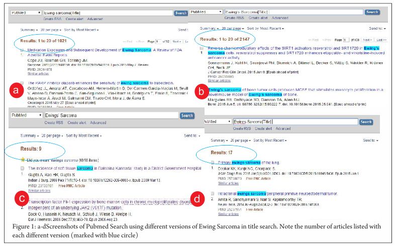
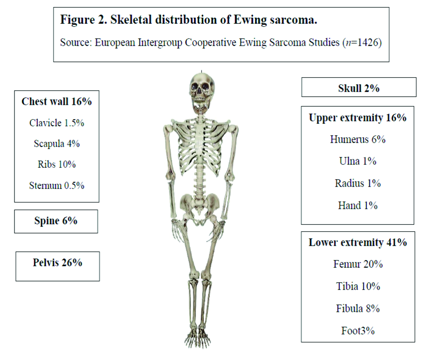
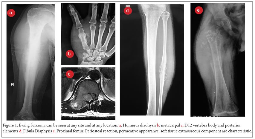
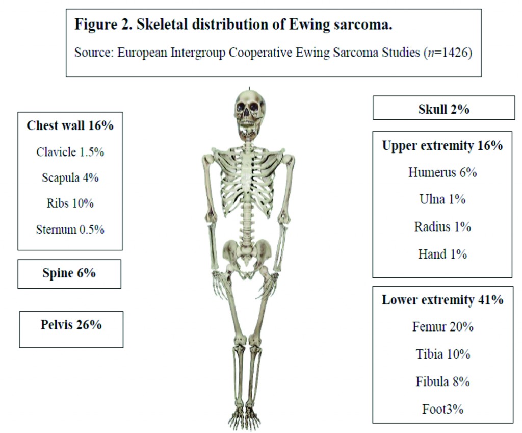
Over the last 30 years, the incidence of EWS has remained unchanged at around 3 cases per million per year [5]. With a median age of 15 years, it most commonly occurs in the second decade of life (Fig 1) [6]. There is a slight male predilection (male: female 1.2:1) and Caucasians are much more frequently affected than Asians and Africans [7,8]. Lower extremities are the most common site of bone disease (43%) while extraosseous primary tumors mostly occur in the trunk (32%) (Fig 2). Metastatic disease is present at diagnosis in about 20-25% of patients and affects the lungs, other bones or multiple systems [5,9].

Biology & Pathology
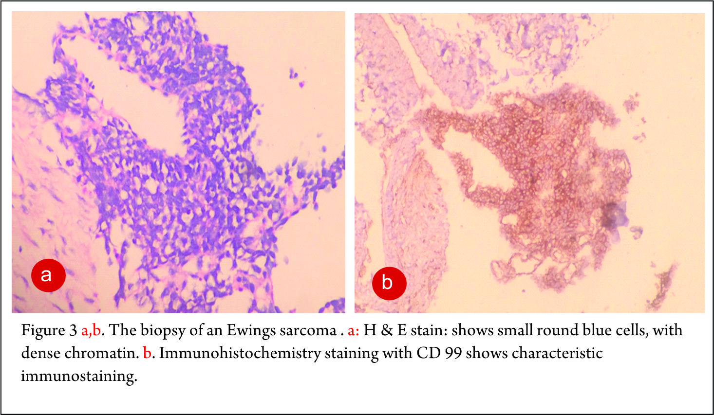
The World Health Organisation (WHO) classification uses EWS/primitive neuroectodermal tumor (PNET) as an inclusive term which encompasses classic EWS, Askin tumor of the thoracic wall, Ewing tumor, peripheral neuroepithelioma, peripheral neuroblastoma, Ewing family of tumors and Ewing sarcoma family of tumors [10]. EWS is derived from a primordial bone marrow-derived mesenchymal stem cell [11,12]. Histologically, EWS is characterised by a monotonous population of small round blue cells with a low mitotic activity of 15-20%. Cytoplasmic glycogen is abundant which gives periodic acid-Schiff (PAS) positivity [13]. The MIC2 gene product, CD99, a surface membrane glycoprotein is overexpressed [14] but it is not specific for EWS. Neural differentiation is evident in the form of positive vimentin in approximately one third of cases.
A reciprocal chromosomal translocation involving the EWSR1 gene on chromosome 22 band q12 combined with any of a number of partner chromosomes is pathognomonic of the diagnosis of EWS. The breakpoint was first cloned in the 1990s [12,15]. Although abnormalities of chromosome 11 are involved in 95% of cases [16], the translocation may involve chromosomes 21, 7 and 17 uncommonly [17,18]. The fusion protein resulting from this chromosomal rearrangement is a potent transcriptional factor which inappropriately activates the target genes, thereby exerting the oncogenic activity.
Other numerical and structural alterations seen in EWS are gains of chromosomes 2, 5, 8, 9, 12, and 15; deletions on the short arm of chromosome 6; the nonreciprocal translocation t(1;16)(q12;q11.2); and trisomy 20 [19,20].

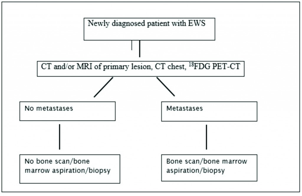
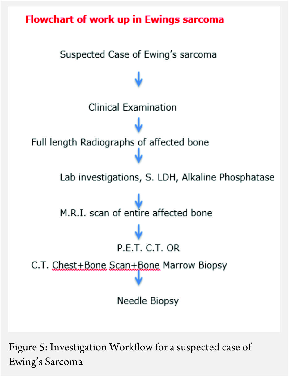
Figure 1: Investigation Workflow for a newly diagnosed Patient with EWS

Staging
EWS is defined by clinical and imaging techniques as localized when there is no spread beyond the primary site or metastatic when the tumor has disseminated to distant organs. Of all imaging modalities, 18FDG PET-CT has the highest specificity (96%) and sensitivity (92%) [21] and is superior to the traditionally used 99mTc-MDP bone scan for detection of bone metastases except for skull lesions [22]. Current recommendations for staging work-up include CT and/or MRI of the primary tumor, chest CT to detect lung metastases and 18FDG PET-CT for identification of distant metastases [23]. As bone marrow involvement is an independent risk factor [24], marrow biopsy has been an integral part of the initial work-up and is still recommended in ongoing clinical trials [25] (26). But recent studies have questioned the utility of bone marrow biopsy in localized [22,27] and metastatic disease [23].

fig 2

Prognosis
The 5 year survival rate for EWS was less than 10% before the advent of modern chemotherapy [28,29]. Currently, the survival rates are 70% for the patients with localized disease [30] and 30% for the patients with metastatic disease [9]. Among patients with refractory or recurrent disease, fewer than 20% of patients can expect to be long term survivors [31,32].
The presence of metastatic disease at diagnosis remains the most important adverse prognostic factor in EWS [33,34,35,36]. In patients with metastatic disease the site(s) of metastases can have an impact on the outcome. Patients with only lung metastases fare better (event free survival, EFS 29% to 52%) than patients with bone and/or bone marrow involvement (EFS 19%) [37,38] or combined bones and lungs involvement (EFS 8%) (34). Unilateral lung involvement has a better outcome compared with bilateral lung lesions [39].
Younger age (<15 years old) [5,40,41], female gender [42], tumor site (distal extremity better than proximal extremity and pelvis) [9], tumor size (volume less than 200 ml and single dimension less than 8 cm) [43], normal serum lactate dehydrogenase (LDH) levels at diagnosis [44], and decreased metabolic activity on 18FDG PET scan after presurgical chemotherapy [45,46] are associated with a more favourable prognosis.
Complex karyotypic abnormalities or chromosome number less than 50 in tumor cells at diagnosis [19], detection of fusion transcripts by polymerase chain reaction (PCR) in morphologically normal bone marrow [47], p53 protein overexpression, Ki67 expression, loss of 16q [48,49], overexpression of microsomal glutathione S-transferase (associated with doxorubicin resistance [50] may be associated with inferior outcome. Patients with secondary Ewing sarcoma [51] or with a poor response to presurgical chemotherapy [52,53] and patients relapsing less than two years after diagnosis (early) have a poorer prognosis [54].

9

Treatment options
Chemotherapy for a total of 10-12 months before and after local control is common practice [33,55]. Initial chemotherapy aims to shrink the tumor to increase to probability of effective local control. Alkylating agents, mainly ifosfamide and cyclophosphamide and anthracyclines form the chemotherapeutic backbone Etoposide, vincristine and actinomycin-D make up the remainder of the four-to five-drug combination chemotherapy.

Chemotherapy for newly diagnosed patients:
Clinical trials in the early years (pre-1990)
Before 1960s, radiation therapy and surgery were used for the treatment of EWS which provided adequate control of the primary disease but patients invariably died of metastatic disease [56]. Chemotherapy was added based on the hypothesis that, in most cases of apparently localized disease, tumor cells were already disseminated without clinical manifestations. Single chemotherapy agents including cyclophosphamide [57,58,59], vincristine [60], daunorubicin [61] and actinomycin-D [62] were trialled in 1960s with promising results.
From two- to as many as six-drug combinations have been used in various randomized and non-randomized trials for the treatment of EWS. Hustu et al [63] used a first ever combination with vincristine and cyclophosphamide with 80% overall survival. In Europe, the French Society of Pediatric Oncology (SFOP) [64,65,66], the United Kingdom Children’s Cancer Study Group (UKCCSG) [35,67], the Scandinavian Study Group (SSG) [68, 69] and the German/Austrian Cooperative Ewing Sarcoma Study Group (CESS) [70,71] performed early clinical trials. Subsequently, the European Intergroup Cooperative Ewing Sarcoma Study group (EICESS) and the European Ewing Tumor Working Initiative of National Groups (EURO-EWING) continued the trials. In the United States, initially the Intergroup Ewing Sarcoma Study (IESS) group [72,73,74], the Children’s Cancer Group (CCG), the Pediatric Oncology Group (POG) and subsequently the Children’s Oncology Group (COG) conducted trials for EWS.
Four-drug combination chemotherapy including vincristine, actinomycin-D, cyclophosphamide and doxorubicin was universally accepted for the treatment by the early 1980s [75] with survival rates between 36-60%. Ifosfamide and etoposide were identified as effective single agents [76,77] and subsequent studies established a survival benefit of their addition to VACD [78]. National Cancer Institute protocol INT0091 was a randomized trial conducted by the Children’s Cancer Group (CCG) and Pediatric Oncology Group (POG) from 1988 through 1992. Patients were assigned to receive VACD or VACD plus ifosfamide and etoposide (VACD-IE). In patients without metastatic disease, the five-year EFS for the VACD group was 54% while the same for the VACD-IE group was 69%. These results established VACD-IE as the gold standard for the treatment of localised Ewing sarcoma [30].
Clinical trials for standard risk (SR) and high risk (HR) EWS since 1990
The disease risk stratification into SR and HR has varied depending on the trial but in general SR means localized small tumors (<200 mL), or tumors with a good histological response to preoperative chemotherapy (<10% cells). HR tumors include metastatic tumors, or large localized tumors (>200 mL).
The trials for SR EWS have tried to address the important questions like the superiority of one alkylating agent over the other (cyclophosphamide and ifosfamide) and survival advantage by dose intensification or addition newer chemotherapy agents.

t2

Cyclophosphamide vs Ifosfamide
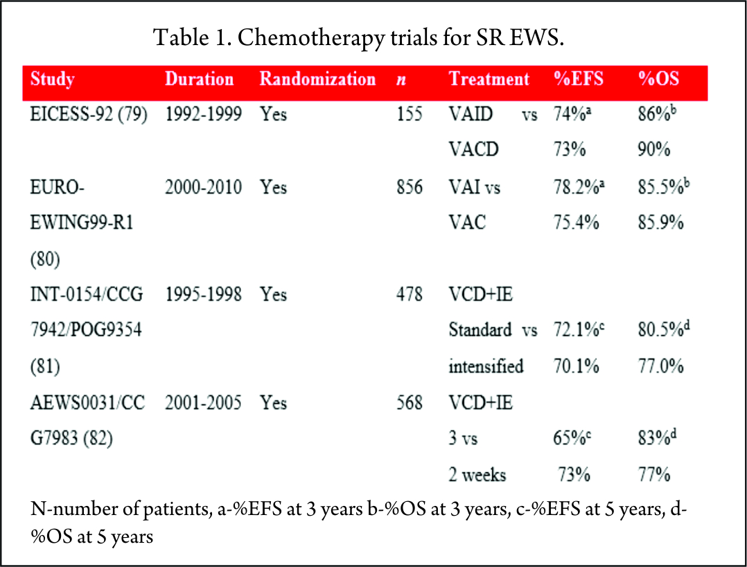
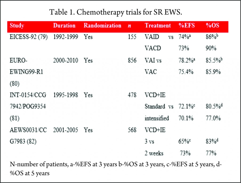
Historically, cyclophosphamide was used for the treatment of EWS. Promising results were seen with ifosfamide in relapsed patients who did not respond to cyclophosphamide [83]. It was postulated that 9 g/m2 of ifosfamide was equimyelotoxic to 2.1 g/m2 of cyclophosphamide [84]. With the potential for less myelotoxicity and high-dose administration, cyclophosphamide was replaced with ifosfamide in the 1980s. But the results of these non-randomized, single-arm studies were mixed, with one study showing no benefit [66] while others proving superiority of ifosfamide over cyclophosphamide [71,67,69]. With this uncertainty of greater efficacy and long-term renal tubular damage with the cumulative dose of ifosfamide [85], its role in the consolidation treatment of EWS was debated. Two large randomized trials, EICESS-92 [79] and its successor Euro-Ewing99-R1 [80] investigated if cyclophosphamide can replace ifosfamide in the consolidation treatment of standard-risk EWS. The results of these studies confirmed that both the drugs had similar efficacy and though cyclophosphamide was associated with more haematological toxicity, the incidence of renal toxicity was much less as compared to ifosfamide. But the question of superiority of one drug over the other is far from resolved and needs further investigation in light of their efficacy to improve the survival [75].

Standard dose vs dose intensification
To improve the outcome, intensification of chemotherapy drug doses was investigated. One way of achieving dose intensification is by escalating the doses of chemotherapy agents while keeping the interval stable. National Cancer Institute protocol INT0154 used VDC+IE chemotherapy and randomized patients to standard (17 cycles over 48 weeks) or intensified (11 cycles over 30 weeks) arms. This study showed no improvement in the outcome of patients with nonmetastatic disease by dose escalation of alkylating agents (81) which was in contrast to an earlier similar study, IESS-II [74].
AEWS0031 trial investigated the feasibility of dose intensification by interval compression (increased dose density) in patients with localized disease [82]. Patients treated every two weeks (intensified arm) had an improved five-year EFS (73%) compared with the standard arm group receiving chemotherapy every 3 weeks (65%) with no increase in toxicity. Due to its superiority, interval compression is used in many ongoing trials.
The Children’s Oncology Group is currently conducting a phase III randomized trial of adding vincristine, topotecan and cyclophosphamide to standard chemotherapy for patients with localized EWS in an attempt to improve the outcome further [25].
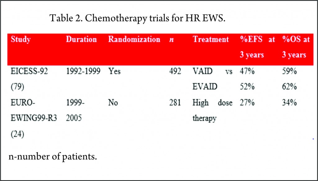
The EICESS-92 study recruited 492 high risk patients of which 157 had metastatic disease at diagnosis. These patients were randomized to receive either VAID or etoposide in addition to VAID (EVAID). Although there was evidence that etoposide had a more pronounced effect in localized HR group, there was no benefit for the patients with metastatic disease with a three-year EFS of 30% [79].
The EURO-EWING99-R3 study enrolled 281 patients with primary disseminated multifocal EWS. 169 patients received the high dose therapy (HDT)/stem cell transplant (SCT) post completion of chemotherapy and local therapy. 3-year EFS for whole cohort was 27% and for patients receiving HDT was 37% [24].

Local therapy
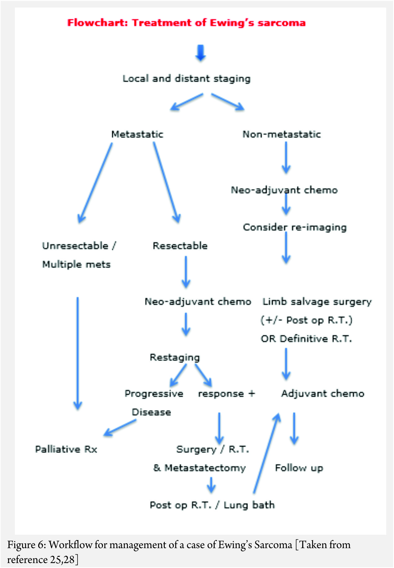
The goal of local therapy is to maximize the local control with minimal morbidity. Surgery and radiation therapy are the two local control modalities employed for EWS. No randomized trials have compared these and as such their relative roles remain controversial [13].
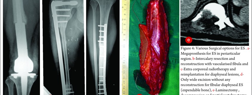
Surgical resection provides information about the amount of tumor necrosis and may be less morbid in the younger patients. Radiation therapy is also associated with the development of second malignant neoplasms in a dose and time dependent manner [86]. A retrospective analysis of patients treated on three consecutive clinical trials for localized EWS showed that the risk of local failure was greater for patients receiving definitive radiotherapy but the EFS and OS were comparable for both surgery and radiation as local control modalities [87]. Microscopically complete surgical resection of localised disease remains the goal of neoadjuvant (or upfront) chemotherapy. Large bone defects after the surgery may be reconstructed using autogenous or allogenic bone grafts and endoprosthetic replacements [13]. Radiation therapy may be used as the main modality of primary disease control in patients with axial or unresectable primary disease. Careful consideration about the use of radiation, dose and volume is required, particularly in younger patients.
In patients with lung metastases, upfront whole-lung radiation may be used irrespective of the radiographic response following chemotherapy [88]. The results of the recently concluded Euro-EWING99 R2 pulmonary (AEWS0331) study which compared the HDT and peripheral blood stem cell (PBSC) rescue with the standard chemotherapy and whole lung irradiation are awaited. A multivariate analysis of the R3 arm of this trial including patients with metastatic disease emphasized the importance of aggressive local control of primary and metastatic sites. The EFS was higher with combined surgery and radiation compared to either modality alone or no local control [89].

High-dose therapy (HDT) and stem cell transplantation (SCT)
Despite advances in multimodal therapy of EWS, there remains a group of patients with high risk of treatment failure. These are primarily the patients with metastatic disease or with extensive unresectable localized disease and patients with a poor response to chemotherapy. This group has a poor 20%-30% disease free survival (DFS) [90,91]. Although conventional chemotherapy regimens induce remission, patients with metastatic disease relapse after a median of one to two years after completion of therapy owing to minimal residual or metastatic disease (MRD/MMD). In the 1980s trials investigating the role of SCT to consolidate remission by reduction of MRD/MMD began. The results of the initial National Cancer Institute (NCI) studies investigating total body irradiation (TBI) with autologous bone marrow transplant (ABMT) showed no improvement in survival [92]. Since then multiple reports have been published of consolidation using HDT followed by SCT but its role in the treatment of EWS has yet to be conclusively determined [93].

Melphalan vs busulfan-based conditioning regimens
Response to melphalan-based HDT has been variable. Some studies showed no additional benefit with poor survival rates between 5%-27% [34,90,94,95] while others [96,97,98] reported improved survival rates of 45%-50%. As use of high-dose busulfan combined with melphalan or other agents has shown promising results with survival rates between 36%-60% [99,100,101,102,103,104], these regimens have been widely used in high-risk patients.

Role of total-body irradiation (TBI)
Use of TBI during the consolidation phase had no survival advantage but increased the incidence of toxicity [92,94]. Two Meta European Intergroup Cooperative Ewing Sarcoma Studies (MetaEICESS) assessed the role of TBI in consolidation treatment. Patients received systemic consolidation in the form of hyperfractionated TBI with melphalan/etoposide in the first HyperME study or two times the melphalan/etoposide in the second TandemME study. EFS were similar in both studies while TBI containing regimen was associated with a higher incidence of toxicity [105]. In conclusion, although EWS is a radiosensitive tumor, there is limited role of TBI in its treatment because of poor efficacy and increased toxicity.

Autologous vs allogenic BMT
Allogeneic transplant may overcome the concerns with tumor cell contamination of stem cell products during autologous transplant [106] and have a potential of graft-versus-tumor (GVT) effect with improved survival. A retrospective analysis of the MetaEICESS study data showed that the EFS was 25% after autologous and 20% after allogeneic transplant [54]. As there was increased incidence of toxicity and no evidence of GVT effect after allogeneic transplant, there seems to be no advantage of allogeneic over autologous transplant.

Chemotherapy for recurrent EWS
Although around 80% of relapses occur within 2 years of initial diagnosis [107], late relapses occurring more than five years from the initial diagnosis are more common in EWS than any other pediatric solid tumors. The Childhood Cancer Survivor Study (108) retrospectively assessed more than 12,700 childhood cancer survivors and concluded that survivors with EWS were at a higher risk of late recurrence, 5-20 years after the diagnosis, than survivors with acute lymphoblastic leukemia. Time to relapse is an important prognostic factor with recurrences occurring within two years of initial diagnosis having worse five-year survival of 7% compared to 30% for patients relapsing after two years [32,107]. Number of recurrences also impacts the outcome with multiple metastatic recurrences having worse prognosis than isolated local or metastatic recurrence [107]. There is no established treatment for these patients and the preferred approach is to combine multi-agent chemotherapy with local modality of surgery and/or radiotherapy [109,110].
High dose Ifosfamide alone [111] or with carboplatin and etoposide (ICE) has been commonly used with survival rates between 29%-33% [112,113]. Cyclophosphamide and topotecan combination achieved response rates of 23%-44% with low toxicity and an added advantage of outpatient administration [114,115] but with a small median duration of response of 8 months [116]. Response rates of 29% to 68% and median time to progression of 3 to 8.5 months were seen with irinotecan and temozolomide [117,118,119,120]. Diarrhea was a troublesome complication which was managed effectively with oral cephalosporins. The combination was otherwise well tolerated. Although gemcitabine and docetaxel showed activity in one study [121], the results were not confirmed by subsequent studies. [122].
In case of recurrent EWS, the addition of HDT to salvage regimens is controversial. Some studies showed a good response in specific groups of patients who responded to relapse therapy and underwent HDT with OS rates of 53 to 66% [123,124], but most of the reports indicate HDT does not improve prognosis [54,125,126].

Targeted therapy for EWS
Tyrosine kinase (TK) inhibitors
TKs are important modulators of growth factor signaling and play a critical role in tumor growth. TK inhibitors are used alone or in combination with conventional chemotherapy agents in treatment of various cancers (127). A number of TK inhibitors have been tried in EWS with variable response.

Insulin-like growth factor 1 receptor (IGF1R) inhibitors
IGF1R is necessary for growth and development of normal as well as cancer cells [128]. With promising pre-clinical results showing IGF1R inhibition in EWS cell lines and xenografts [129], more than 25 agents inhibiting IGF1R are currently under investigation [130].
IGF1R monoclonal antibodies including R1507 (131), figitumumab [132], ganitumab (AMG479) [133], cixutumumab [134,135], and robatumumab (SCH-717454) [136] have shown activity in early phase clinical trials with response rates ranging from 6-14% and a favourable safety profile. But the results of the phase II studies were less impressive compared with the promising preclinical and early clinical data [137]. Small-molecule inhibitors of IGF1R such as GSK1838705A [138], GSK1904529A [139], BMS-754807 [140], and INSM-18 [141] are also in preclinical and clinical development.
Phase II clinical trials of imatinib, a TK inhibitor of the BCR-ABL fusion protein [142,143,144] and dasatinib, a multitargeted TK inhibitor [145] showed no efficacy in EWS.

Biologic agents
Angiogenesis inhibitors
Neovascularization plays a critical role in the pathogenesis of EWS [146] and targeting vascular endothelial growth factor (VEGF) may interfere with vasculogenesis, providing a novel therapeutic approach [147]. A phase I study [148] and a randomized phase II trial [149] conducted by the Children’s Oncology Group have shown the feasibility and tolerability of bevacizumab in EWS patients. Another phase II study investigated the role of vinblastine and celecoxib as angiogenesis inhibitors in combination with the standard chemotherapy (150). Although the feasiblity of this combination was established, there were significant pulmonary and bladder toxicities.

Histone deacetylase (HDAC) inhibitors
HDAC inhibition suppresses EWS-FLI1 expression and may represent a novel therapeutic target for EWS (151).

Mammalian target of rapamycin (mTOR) inhibitors
MTOR is a serine/threonine kinase with critical role in protein synthesis, cell growth and proliferation regulation. mTOR inhibitors have shown activity in preclinical models. A phase I study of temsirolimus, irinotecan and temozolomide demonstrated efficacy and tolerability [152]. But another phase II study of temsirolimus with cixutumumab did not show any objective response despite the encouraging preclinical data [153]. Ridaforolimus was associated with a statistically significant but clinically small benefit on PFS [154].

Aurora A kinase inhibitors
Although alisertib (MLN8237), an Aurora A kinase inhibitor produced promising results in the Pediatric Preclinical Testing Program [155], a recently concluded Children’s Oncology Group phase II trial failed to establish its efficacy in EWS [156].

Hedgehog pathway modulation
Arsenic trioxide was effective in inhibiting EWS growth in preclinical cell culture models by targeting p38(MAPK) and c-Jun N-terminal kinase [157]. These observations warrant further investigation.

Bisphosphonates
Zoledronic acid acts by inducing apoptosis by upregulating osteoprotegerin which was the basis of activity seen in EWS pre-clinical models [158,159]. However, confirmatory clinical trials have not been performed.

Immune therapy
Interleukin-15-activated natural killer (NK) cells combined with HDAC inhibitors improve immune recognition of therapy-sensitive and –resistant EWS and sensitize for NK cell cytotoxicity [160]. Allogenic NK cells have shown activity against EWS cells on their own [161].

EWS-FLI1 targeting
Targeting the EWS-FLI1 fusion protein or its key signalling pathway is another attractive approach [162]. YK-4279, a small molecule inhibitor of EWS-FLI1 protein activity [163,164], mithramycin, a chemotherapy drug [165] and midostaurin (PKC412), a multikinase inhibitor [166] have shown activity in preclinical models.


 Conclusion 

Many advances have been made in the management of EWS since its first description almost 100 years ago. Molecular and imaging techniques are progressing at a rapid pace allowing for newer insights into the biology of this disease. From radiation therapy alone, the treatment has evolved to include multiple modalities. The outcome for localized disease has improved dramatically but more needs to be done for patients with metastatic or recurrent EWS. Targeted therapies may offer some hope for the latter group.


References

1. Diffuse endothelioma of bone. Ewing, JR. 1921, Proceedings of the New York Pathological Society, Vol. 21, pp. 17-24.
2. Ewing sarcoma: an eponym window to history. Cripe, TP. 2011, Sarcoma, Vol. 2011, pp. 1-4.
3. Ewing’s sarcoma. Karosas, AO. 2010, American Journal of Helath-System Pharmacy, Vol. 67, pp. 1599-1605.
4. Hawkins, DS, et al. Ewing sarcoma. [ed.] DG Poplack PA Pizzo. Principles and practice of pediatric oncology. 6th. Philadelphia : Lippincott, Williams and Wilkins, 2011, pp. 987-1014.
5. Changes in incidence and survival of Ewing sarcoma patients over the past 3 decades: Surveillance Epidemiology and End Results data. Esiashvili, N, Goodman, M and Marcus, Jr, RB. 2008, Journal of Pediatric Hematology/Oncology, Vol. 30, pp. 425-430.
6. Patterns of care and survival for patients aged under 40 years with bone sarcoma in Britain, 1980-1994. Stiller, CA, et al. 2006, British Journal of Cancer, Vol. 94, pp. 22-29.
7. Rarity of Ewing’s sarcoma among U.S. Negro children. Fraumeni, Jr, JF and Glass, AG. 1970, Lancet, Vol. 1, pp. 366-367.
8. Rarity of Ewing’s sarcoma in China. Li, FP, et al. 1980, Lancet, Vol. 1, p. 1255.
9. Prgnostic factors in Ewing’s tumor of bone: analysis of 975 patients from the European Intergroup Cooperative Ewing’s Sarcoma Study Group. Cotterill, SJ, et al. 2000, Journal of Clinical Oncology, Vol. 18, pp. 3108-3114.
10. Ushigome, S, Machinami, R and Sorensen, PH. Ewing sarcoma/primitive neuroectodermal tumor (PNET). [ed.] KK Unni, F Martens, eds CDM Fletcher. World Health Organization classification of tumours. Pathology and genetics. Tumours of soft tissues and bone. Lyon, France : IARC Press, 2002.
11. Identification of cancer stem cells in Ewing’s sarcoma. Suva, ML, et al. 2009, Cancer Research, Vol. 69, pp. 1776-1781.
12. The Ewing family of tumors-a subgroup of small-round-cell tumors defined by specific chimeric transcripts. Delattre, O, et al. 1994, The New England Journal of Medicine, Vol. 331, pp. 294-299.
13. Diagnosis and treatment of Ewing’s sarcoma. Iwamoto, Y. 2007, Japanese Journal of Clinical Oncology, Vol. 37, pp. 79-89.
14. Neuroectodermal differentiation in Ewing’s sarcoma family of tumors does not predict tumor behavior. Parham, DM, et al. 1999, Human Pathology, Vol. 30, pp. 911-918.
15. Cloning and characterization of Ewing’s sarcoma and peripheral neuroepithelioma t(11;22) translocation breakpoints. Zucman, J, et al. 1992, Genes Chromosomes and Cancer, Vol. 5, pp. 271-277.
16. Diagnositc value of the molecular genetic detection of the t(11;22) translocation in Ewing’s tumors. Dockhorn-Dworniczak, B, et al. 1994, Virchows Archives, Vol. 425, pp. 107-112.
17. Molecular analysis of Ewing’s sarcoma: another fusion gene, EWS-E1AF, available for diagnosis. Urano, F, et al. 1998, Japanese Journal of Cancer Research, Vol. 89, pp. 703-711.
18. Promiscuous partnerships in Ewing’s sarcoma. Sankar, S and Lessnick, SL. 2011, Cancer Genetics, Vol. 204, pp. 351-365.
19. Ploidy and karyotype complexity are powerful prognostic indicatiors in the Ewing’s sarcoma family of tumors: a study by the United Kingdom Cancer Cytogenetics and the Children’s Cancer and Leukemia Group. Roberts, P, et al. 2008, Genes, Chromosomes and Cancer, Vol. 47, pp. 207-220.
20. Prognostic impact of deletions at 1p36 and numerical aberrations in Ewing tumors. Hattinger, CM, et al. 1999, Genes Chromosomes and Cancer, Vol. 24, pp. 243-254.
21. Diagnostic accuracy of 18F-FDG-PET and PET/CT in patients with Ewing sarcoma family tumors: a systematic review and a meta-analysis. Treglia, G, et al. 2012, Skeletal Radiology, Vol. 41, pp. 249-256.
22. An evaluation of [F-18]-fluorodeoxy-d-glucose positron emission tomography, bone scan, and bone marrow aspiration/biopsy as staging investigations in Ewing sarcoma. Newman, EN, et al. 2013, Pediatric Blood and Cancer, Vol. 60, pp. 1113-1117.
23. Futility versus utility of marrow assessment in initial Ewing sarcoma staging workup. Anderson, P. 2015, Pediatric Blood and Cancer, Vol. 62, pp. 1-2.
24. Primary disseminated multifocal Ewing sarcoma: results of the Euro-EWING 99 trial. Landenstein, R, et al. 2010, Journal of Clinical Oncology, Vol. 28, pp. 3284-3291.
25. https://www.clinicaltrials.gov/ct2/show/NCT01231906. ClinicalTrials.gov. [Online] [Cited: 5 June 2015.]
26. https://www.clinicaltrials.gov/ct2/show/NCT02306161. ClinicalTrials.gov. [Online] [Cited: 5 June 2015.]
27. Utility of bone marrow aspiration and biopsy in initial staging of Ewing sarcoma. kopp, LM, et al. 2015, Pediatric Blood and Cancer, Vol. 62, pp. 12-15.
28. Ewing’s sarcoma: a study of treatment methods. Jenkin, RD. 1966, Clinical Radiology, Vol. 17, pp. 97-106.
29. The curability of Ewing’s endothelioma of bone in children. Phillips, RF and Higinbotham, NL. 1967, Journal of Pediatrics, Vol. 70, pp. 391-397.
30. Addition of ifosfamide and etoposide to standard chemotherapy for Ewing’s sarcoma and primitive neuroectodermal tumor of bone. Grier, HE, et al. 2003, New England Journal of Medicine, Vol. 348, pp. 694-701.
31. Long-term survival in patients with Ewing’s sarcoma relapsing after completing therapy. Hayes, FA, et al. 1987, Medical and Pediatric Oncology, Vol. 15, pp. 254-256.
32. Risk of recurrence and survival after relapse in patients with Ewing sarcoma. Stahl, M, et al. 2011, Pediatric Blood and Cancer, Vol. 57, pp. 549-553.
33. Ewing sarcoma treatment. Jurgens, H and Dirksen, U. 2011, European Journal of Cancer, Vol. 47 Suppl 3, pp. S366-S367.
34. Primary metastatic (stage IV) Ewing tumor: survival analysis of 171 patients from the EICESS studies-European Intergroup Cooperative Ewing Sarcoma Studies. Paulussen, M, et al. 1998, Annals of Oncology, Vol. 9, pp. 275-281.
35. Long-term results from the first UKCCSG Ewing’s tumor study (ET-1): United Kingdom Children’s Cancer Study Group (UKCCSG) and the Medical Research Council Bone Sarcoma Working Party. Craft, AW, et al. 1997, European Journal of Cancer, Vol. 33, pp. 1061-1069.
36. Long-term event-free survival after intensive chemotherapy for Ewing’s family of tumors in children and young adults. Kolb, EA, et al. 2003, Journal of Clinical Oncology, Vol. 21, pp. 3423-3430.
37. Analysis of prognostic factors in Ewing sarcoma family of tumors: review of St. Jude Children’s Research Hospital studies. Rodriguez-Galindo, C, et al. 2007, Cancer, Vol. 110, pp. 375-384.
38. Treatment of metastatic Ewing’s sarcoma or primitive neuroectodermal tumor of bone: evaluation of combination ifosfamide and etoposide-a Children’s Cancer Group and Pediatric Oncology Group study. Miser, JS, et al. 2004, Journal of Clinical Oncology, Vol. 22, pp. 2873-2876.
39. Ewing’s tumors with primary lung metastases: survival analysis of 114 (European Intergroup) Cooperative Ewing’s Sarcoma Studies patients. Paulussen, M, et al. 1998, Journal of Clinical Oncology, Vol. 16, pp. 3044-3052.
40. Treatment of nonmetastatic Ewing’s sarcoma family tumors of the spine and sacrum: the experience from a single institution. Bacci, G, et al. 2006, European Spine Journal, Vol. 18, pp. 1091-1095.
41. Ewing tumors in infants. van den Berg, H, et al. 2008, Pediatric Blood and Cancer, Vol. 50, pp. 761-764.
42. Ewing sarcoma demonstrates racial disparities in incidence-related and sex-related differences in outcome: an analysis of 1631 cases from the SEER database, 1973-2005. Jawad, MU, et al. 2009, Cancer, Vol. 115, pp. 3526-3536.
43. Evaluation of prognostic factors in a tumor volume-adapted treatment strategy for localized Ewing sarcoma of bone: the CESS 86 experience. Ahrens, S, et al. 1999, Medical and Pediatric Oncology, Vol. 32, pp. 186-195.
44. Prognostic factors in non-metastatic Ewing’s sarcoma tumor of bone: an analysis of 579 patients treated at a single institution with adjuvant or neoadjuvant chemotherapy between 1972 and 1998. Bacci, G, et al. 2006, Acta Oncologica, Vol. 45, pp. 469-475.
45. [18F]Fluorodeoxyglucose positron emission tomography predicts outcome for Ewing sarcoma family of tumors. Hawkins, DS, et al. 2005, Journal of Clinical Oncology, Vol. 23, pp. 8828-8834.
46. Assessment of histological response of pediatric bone sarcomas using FDG PET in comparison to morphological volume measurement and standardized MRI parameters. Denecke, T, et al. 2010, European Journal of Nuclear Medicine and Molecular Imaging, Vol. 37, pp. 1842-1853.
47. Increased risk of systematic relapse associated with bone marrow micrometastasis and circulating tumor cells in localized Ewing tumor. Schleiermacher, G, et al. 2003, Journal of Clinical Oncology, Vol. 21, pp. 85-91.
48. Overexpression of p53 protein in primary Ewing’s sarcoma of bone: relationship to tumor stage, response and prognosis. Abudu, A, et al. 1999, British Journal of Cancer, Vol. 79, pp. 1185-1189.
49. Genetic imbalances revealed by comparative genomic hybridization in Ewing tumors. Ozaki, T, et al. 2001, Genes Chromosomes and Cancer, Vol. 2001, pp. 164-171.
50. Overcoming resistance to conventional drugs in Ewing sarcoma and identification of molecular predictors of outcome. Scotlandi, K, et al. 2009, Journal of Clinical Oncology, Vol. 27, pp. 2209-2216.
51. Clinical features and outcomes in patients with secondary Ewing sarcoma. Applebaum, MA, et al. 2013, Pediatric Blood and Cancer, Vol. 60, pp. 611-615.
52. The histological response to chemotherapy as a predictor of the oncological outcome of operative treatment of Ewing sarcoma. Wunder, JS, et al. 1998, Journal of Bone and Joint Surgery, American volume, Vol. 80, pp. 1020-1033.
53. Chemotherapy response is an important predictor of local recurrence in Ewing sarcoma. Lin, PP, et al. 2007, Cancer, Vol. 109, pp. 603-611.
54. Allogeneic and autologous stem-cell transplantation in advanced Ewing tumors. An update after long-term follow-up from two centers of the European Intergroup study EICESS. . Burdach, S, et al. 2000, Annals of Oncology, Vol. 11, pp. 1451-1462.
55. Current treatment of Ewing’s sarcoma. Thacker, MM, et al. 2005, Expert Review of Anticancer Therapy, Vol. 5, pp. 319-331.
56. Ewing’s sarcoma-A critical analysis of 165 cases. Dahlin, DC, Coventry, MD and canlon, PW. 1961, Journal of bone and Joint Surgery, Vol. 43 (A), pp. 185-192.
57. Cyclophosphamide therapy in children with Ewin’g sarcoma. Sutow, WW and Sullivan, MP. 1962, Cancer Chemotherapy Reports, Vol. 23, pp. 55-60.
58. Cyclophosphamide in children with cancer. Pinkel, D. 1962, Cancer, Vol. 15, pp. 42-49.
59. Cyclophosphamide in the management of Ewing’s sarcoma. Samuels, ML and Howe, CD. 1967, Canver, Vol. 20, pp. 961-966.
60. Vincristine in children with malignant solid tumors. James Jr, DH and George, P. 1964, The Journal of Pediatrics, Vol. 64, pp. 534-541.
61. Daunomycin, an antitumor antibiotic, in the treatment of neoplastic disease. Clinical evaluation with special reference to childhood leukemia. Tan, C, et al. 1967, Cancer, Vol. 20, pp. 333-353.
62. Treatment of pulmonary metastatic disease with radiation therapy and adjuvant actinomycin D. Preliminary observations. Cupps, RE, Ahmann, DL and Soule, EH. 1969, Cancer, Vol. 24, pp. 719-723.
63. Treatment of clinically localized Ewing’s sarcoma with radiotherapy and combination chemotherapy. Hustu, HO, Pinkel, D and Pratt CB. 1972, Cancer, Vol. 30, pp. 1522-1527.
64. The response to initial chemotherapy as a prognostic factor in localized Ewing sarcoma. Oberlin, O, et al. 1985, European Journal of Clinical Oncology, Vol. 21, pp. 463-467.
65. Prognostic factors in localized Ewing tumors and peripheral neuroectodermal tumors: the third study of the French Society of Pediatric Oncology (EW88 study). Oberlin, O, et al. 2001, British Journal of Cancer, Vol. 85, pp. 1646-1654.
66. No benefit of ifosfamide in Ewing’s sarcoma: A nonrandomised study of the French Society of Pediatric Oncology. Oberlin, O, et al. 1992, Journal of Clinical Oncology, Vol. 10, pp. 1407-1412.
67. Ifosfamide-containing chemotherapy in Ewing’s sarcoma: the second United Kingdom Children’s Cancer Study Group and the Medical Research Council Ewing’s Tumor Study (ET-2). Craft, A, et al. 1998, Journal of Clinical Oncology, Vol. 16, pp. 3628-3633.
68. Ewing’s sarcoma treatment in Scandinavia 1984-1990: Ten-year results of the Scandinavian Sarcoma Group Protocol SSGIV. Nilbert, M, et al. 1998, Acta Oncologica, Vol. 37, pp. 375-378.
69. Five-year results in Ewing’s sarcoma. The Scandinavian Sarcoma Group experience with the SSG IX protocol. Elomaa, I, et al. 2000, European Journal of Cancer, Vol. 36, pp. 875-880.
70. Multidisciplinary treatment of primary Ewing’s sarcoma of bone. A 6-year experience of a European Cooperative Trial. Jurgens, H, et al. 1988, Cancer, Vol. 61, pp. 23-32.
71. Localized Ewing’s tumor of bone: final results of the Cooperative Ewing’s Sarcoma Study CESS 86. Paulussen, M, et al. 2001, Journal of Clinical Oncology, Vol. 19, pp. 1818-1829.
72. Multimodal therapy for the management of primary, nonmetastatic Ewing’s sarcoma of bone: a long-term follow-up of the First Intergroup Study. Nesbit Jr, ME, et al. 1990, Journal of Clinical Oncology, Vol. 8, pp. 1664-1674.
73. Multimodality therapy for the management of localized Ewing’s sarcoma of pelvic and sacral bomes: a report from the Second Intergroup Study. Evans, RG, et al. 1991, Journal of Clinical Oncology, Vol. 9, pp. 1173-1180.
74. Multimodal therapy for the management of nonpelvic, localized Ewing’s sarcoma of bone: Intergroup Study IESS-II. Burgert Jr, EO, et al. 1990, Journal of Clinical Oncology, Vol. 8 , pp. 1514-1524.
75. Progress in the treatment of Ewing sarcoma: are the rumors of the demise of cytotoxic chemotherapy premature? Rosen, G. 2015, Klinische Padiatrie, Vol. 227, pp. 105-107.
76. Phase II study of VP-16-213 in childhood malignant disease: a Children’s Cancer Study Group report. Chard Jr, RL, et al. 1979, Cancer Treatment Reports, Vol. 63, pp. 1755-1759.
77. A phase II study of ifosfamide in children with recurrent solid tumors. Pinkerton, CR, Rogers, H and James, C. 1985, Cancer Chemotherapy and Pharmacology, Vol. 15, pp. 258-262.
78. Ifosfamide plus etoposide in newly diagnosed Ewing’s sarcoma of bone. Meyer, WH, et al. 1992, Journal of Clinical Oncology, Vol. 10, pp. 1737-1742.
79. Results of the EICESS-92 study: Two randomised trials of Ewing’s sarcoma treatment-Cyclophosphamide compared with ifosfamide in standard-risk patients and assessment of benefit of etoposide added to standard treatment in high-risk patients. Paulussen, M, et al. 2008, Journal of Clinical Oncology, Vol. 26, pp. 4385-4393.
80. Cyclophosphamide compared with ifosfamide in consolidation treatment of standard-risk Ewing sarcoma: results of the randomized noninferiority Euro-Ewing99-R1 trial. Le Deley, M, et al. [ed.] 2448. 2014, Journal of Clinical Oncology, Vol. 32, p. 2440.
81. Dose-intensified compared with standard chemotherapy for nonmetastatic Ewing sarcoma family of tumors: A Children’s Oncology Group study. Granowetter, L, et al. 2009, Journal of Clinical Oncology, Vol. 27, p. 25362541.
82. Randomized controlled trial of interval-compressed chemotherapy for the treatment of localized Ewing sarcoma: a report from the Children’s Oncology Group. Womer, RB, et al. 2012, Journal of Clinical Oncology, Vol. 30, pp. 4148-4154.
83. Ifosfamide in combination chemotherapy for sarcomas and testicular carcinoma. Niederle, N, et al. 1985, Cancer Treatment Reviews, Vol. 10(Suppl 1), pp. 129-135.
84. Cyclophosphamide dose escalation in comparison with vincrstine and actinomycin-D (VAC) in gross residual sarcoma: A pilot study without hematopoietic growth factor support evaluating toxicity and response. Ruymann, FB, et al. 1995, Journal of Pediatric Hematology/Oncology, Vol. 17, pp. 331-337.
85. Long-term follow-up of ifosfamide renal toxicity in children treated for malignant mesenchymal tumors: An International Society of Pediatric Oncology report. Suarez, A, et al. 1991, Journal of Clinical Oncology, Vol. 9, pp. 2177-2182.
86. Second malignancies after Ewing’s sarcoma: radiation dose-dependency of secondary sarcomas. Kuttesch, JF Jr, et al. 1996, Journal of Clinical Oncology, Vol. 14, pp. 2818-2825.
87. Comparative evaluation of local control strategies in localized Ewing sarcoma of bone: a report from the Children’s Oncology Group . DuBois, SG, et al. 2015, Cancer, Vol. 121, pp. 467-475.
88. Current therapeutic approaches in metastatic and recurrent Ewing sarcoma. Huang , M and Lucas, K. 2011, Sarcoma.
89. The value of local treatment in patients with primary, disseminated, multifocal Ewing sarcoma (PDMES). Haeusler, J, et al. 2010, Cancer, Vol. 116, pp. 443-450.
90. High-risk Ewing’s sarcoma: end-intensification using autologous bone marrow transplantation. Marcus, RB, Jr, et al. 1988, International Journal of Radiation Oncology*Biology*Physics, Vol. 15, pp. 53-59.
91. Ewing’s sarcoma metastatic at diagnosis. Results and comparisons of two intergroup Ewing’s sarcoma studies. Cangir, A, et al. 1990, Cancer, Vol. 66, pp. 887-893.
92. Total-body irradiation and autologous bone marrow transplant in the treatment of high-risk Ewing’s sarcoma and rhabdomyosarcoma. Horowitz, ME, et al. 1993, Journal of Clinical Oncology, Vol. 11, pp. 1911-1918.
93. Therapy for metastatic ESFT: is it time to ask new questions? Snyder, KM and Mackall, CL. 2007, Pediatric Blood and Cancer, Vol. 49, pp. 115-116.
94. High-dose melphalan, etoposide, total-body irradiation, and autologous stem-cell reconstitution as consolidation therapy for high-risk Ewing’s sarcoma does not improve prognosis. Meyers, PA, et al. 2001, Journal of Clinical Oncology, Vol. 19, pp. 2812-2820.
95. How effective is dose-intensive/myeloablative therapy against Ewing’s sarcoma/primitive neuroectodermal tumor metastatic to bone or bone marrow?The Memorial Sloan-Kettering experience and a literature review. Kushner, B and Meyers, P. 2001, Journal of Clinical Oncology, Vol. 19, pp. 870-880.
96. Myeloablative radiochemotherapy and hematopoietic stem-cell rescue in poor-prognosis Ewing’s sarcoma. Burdach, S, et al. 1993, Journal of Clinical Oncology, Vol. 11, pp. 1482-1488.
97. Does consolidation with autologous stem cell transplantation improve the outcome of children with metastatic or relapsed Ewing sarcoma? Al-Faris, N, et al. 2007, Pediatric Blood and Cancer, Vol. 49, pp. 190-195.
98. High-dose chemotherapy and autologous peripheral blood stem cell transfusion for adult and adolescent patients with small round cell sarcomas. Yamada, K, et al. 2007, Bone Marrow Transplantation, Vol. 29, pp. 471-476.
99. High-dose busulfan/melphalan with autologous stem cell rescue in Ewing’s sarcoma. Atra, A, et al. 1997, Bone Marrow Transplantation, Vol. 20, pp. 843-846.
100. Impact of high-dose busulfan plus melphalan as consolidation in metastatic Ewing tumors: a study by the Scoiete Francaise des Cancers de l’Enfant. Oberlin, O, Rey, A and Desfachelles, AS. 2006, Journal of Clinical Oncology, Vol. 24, pp. 3997-4002.
101. Autologous stem cell trasplantation for high-risk Ewing’s sarcoma and other pediatric solid tumors. Fraser, CJ, et al. 2006, Bone Marrow Transplantation, Vol. 37, pp. 175-181.
102. High-dose therapy with hematopoietic stem cell rescue in patients with poor prognosis Ewing family tumors. Rosenthal, J, et al. 2008, Bone Marrow Transplantation, Vol. 42, pp. 311-318.
103. Risk adapted chemotherapy for localised Ewing’s sarcoma of bone: the French EW93 study. Gasper, N, et al. 2012, European Journal of Cancer, Vol. 48, pp. 1376-1385.
104. Consolidation of first-line therapy with busulfan and melphala, and autologous stem cell rescue in children with Ewing’s sarcoma. Drabko, K, et al. 2012, Bone Marrow Transplantation, Vol. 47′, pp. 1530-1534.
105. High-dose therapy for patients with primary multifocal and early relapsed Ewing’s tumors: results of two consecutive regimens assessing the role of total-body irradiation. Burdach, S, et al. 2003, Journal of Clinical Oncology, Vol. 21, pp. 3072-3078.
106. Gene-marking to trace origin of relapse after autologous bone-marrow transplantation. Brenner, MK, et al. 1993, Lancet, Vol. 341, pp. 85-86.
107. Prognostic factors for patients with Ewing sarcoma (EWS) at first recurrence following multi-modality therapy: a report from the Children’s Oncology Group. Leavey, PJ, et al. 2008, Pediatric Blood and Cancer, Vol. 51, pp. 334-338.
108. Late recurrence in pediatric cancer: a report from the Childhood Cancer Survivor Study. Wasilewski-Masker, K, et al. 2009, journal of National Cancer Institute, Vol. 101, pp. 1709-1720.
109. Ewing’s sarcoma family of tumors: current management. Bernstein, M, et al. 2006, Oncologist, Vol. 11, pp. 503-519.
110. Ewing’s sarcoma: standard and experimental treatment options. Subbiah, V, et al. 2009, Current Treatment Options in Oncology, Vol. 10, pp. 126-140.
111. Response to high dose ifosfamide in patients with advanced/recurrent Ewing’s tumors. Ferrari, S, et al. 2009, Pediatric Blood and Cancer, Vol. 52, pp. 581-584.
112. Survival after recurrence of Ewing tumors: The St. Jude Children’s Research Hospital experience, 1979-1999. Rodriguez-Galindo, C, et al. 2002, Cancer, Vol. 94, pp. 561-560.
113. Ifosfamide, carboplatin and etoposide (ICE) reinduction chemotherapy in a large cohort of children and adolescents with recurrent/refractory sarcoma: the Children’s Cancer Group (CCG) experience. Van Winkle, P, et al. 2005, Pediatric Blood and Cancer, Vol. 44, pp. 338-347.
114. Cyclophosphamide plus topotecan in children with recurrent or refractory solid tumors: a Pediatric Oncology Group phase II study. Saylors, RLIII, et al. 2001, Journal of Clinical Oncology, Vol. 19, pp. 3463-3469.
115. Topotecan and cyclophosphamide in patients with refractory or relapsed Ewing tumors. Hunold, A, et al. 2006, Pediatric Blood and Cancer, Vol. 47, pp. 795-800.
116. Cyclophosphamide and topotecan as first-line salvage therapy in patients with relapsed Ewing sarcoma at a single institution . Farhat, R, et al. 2013, Journal of Pediatric Hematology/Oncology, Vol. 35, pp. 356-360.
117. Temozolomide and intravenous irinotecan for treatment of advanced Ewing sarcoma. Wagner, LM, et al. 2007, Pediatric Blood and Cancer, Vol. 48, pp. 132-139.
118. Irinotecan and temozolomide for Ewing sarcoma. Casey, DA, et al. 2009, Pediatric Blood and Cancer, Vol. 2009, pp. 1029-1034.
119. Vincristine, irinotecan, and temozolomide in patients with relapsed and refractory Ewing sarcoma. Raciborska, A, et al. 2013, Pediatric Blood and Cancer, Vol. 60, pp. 1621-1625.
120. Irinotecan and Temozolomide treatment for relapsed Ewing sarcoma: a single center experience and review of the literature. Kurucu, N, Sari, N and Ilhan, IE. 2015, Pediatric Hematology and Oncology, Vol. 32, pp. 50-59.
121. Treatment of relapsed/refractory pediatric sarcomas with gemcitabine and docetaxel. Mora, J, et al. 2009, Journal of Pediatric Hematology/Oncology, Vol. 31, pp. 723-729.
122. Phase II study of sequential gemcitabine followed by docetaxel for recurrent Ewing sarcoma, osteosarcoma, or unresectable or locally recurrent chondrosarcoma: results of Sarcoma Alliance for Research Through Collaboration tudy 003. Fox, E, et al. 2012, Oncologist, Vol. 17, p. 321.
123. Survival after recurrence of Ewing’s sarcoma family of tumors. Barker, LM, et al. 2005, Journal of Clinical Oncology, Vol. 23, pp. 4354-4362.
124. The value of high-dose chemotherapy in patients with first relapsed Ewing sarcoma. Rasper, M, et al. 2014, Pediatric Blood and Cancer, Vol. 61, pp. 1382-1386.
125. High dose chemotherapy with bone marrow or peripheral stem cell rescue is an effective treatment option for patients with relapsed or progressive Ewing’s sarcoma family of tumors. McTiernan, A, et al. 2006, Annals of Oncology, Vol. 17, pp. 1301-1305.
126. Myeloablative therapy with autologous stem cell rescue for Ewing sarcoma. Gardner, SL, et al. 2008, Bone Marrow Transplantation, Vol. 41, pp. 867-872.
127. Role of tyrosine kinase inhibitors in cancer therapy. Arora, A and Scholar, EM. 2005, Journal of Pharmacology and Experimental Therapeutics, Vol. 315, pp. 971-979.
128. The role of IGF1R in pediatric malignancies. KIm, SY, et al. 2009, Oncologist, Vol. 14, pp. 83-91.
129. IGF-1R taargeted treatment of sarcoma. Toretsky, JA and Gorlick, R. 2010, The Lancet Oncology, Vol. 11, pp. 1015-106.
130. Targeted therapy for Ewing’s sarcoma. Subbiah, V and Anderson, P. 20011, Sarcoma.
131. R1507,a monoclonal antibody to the insulin-like grwoth factor 1 receptor,in patients with recurrent or refractory Ewing sarcoma family of tumors:results of a phase II Sarcoma Alliance for Research through Collaboration study. Pappo, AS, et al. 2011, Journal of Clinical Oncology, Vol. 29, pp. 4541-4547.
132. Preliminary efficacy of the anti-insulin-like growth factor type 1 receptor antibody figitumumab in patients with refractory Ewing sarcoma. Jurgens, H, et al. 2011, Journal of Clinical Oncology, Vol. 29, pp. 4534-4540.
133. Phase II study of ganitumab, a fully human anti-type-1 insulin-like growth factor receptor antibody, in patients with metastatic Ewing family tumors or desmoplastic small round cell tumors. Tap, WD, et al. 2012, Journal of clinical oncology, Vol. 30, pp. 1849-1856.
134. Phase I/II trial and pharmakokinetic study of cixutumumab in pediatric patients with refractory solid tumors and Ewing sarcoma: a report from the Children’s Oncology Group. Melampati, S, et al. 2012, Journal of Clinical Oncology, Vol. 30, pp. 256-262.
135. Insulin growth factor-receptor (IGF-1R) antibody cixutumumab combined with the mTOR inhibitor temsirolimus in patients with refractory Ewing’s sarcoma family tumors. Naing, A, et al. 2012, Clinical Cancer Research, Vol. 18, pp. 2625-2631.
136. Activity of SCH-717454 in subjects with relapsed osteosarcoma or Ewing’s sarcoma (study P04720). Anderson, P, et al. London, UK : s.n., 2007. Proceedings of the 14th Annual Meeting of the Connective Tissue Oncology Society (CTOS ’07). Abstract #35094.
137. Targeting the insulin-like growth factor 1 receptor in Ewing’s sarcoma: reality and expectations. Olmos, D, et al. 2011, Sarcoma.
138. GSK1838705A inhibits the insulin-like growth factor-1 receptor and anaplastic lymphoma kinase and shows antitumor activity in experimental models of human cancers. Sabbatini, P, et al. 2009, Molecular Cancer Therapeutics, Vol. 8, pp. 2811-2820.
139. Antitumor activity of GSK1904529A, a small-molecule inhibitor of the insulin-like growth factor-I receptor tyrosine kinase. Sabbatini, P, et al. 2009, Clinical Cancer Research, Vol. 15, pp. 3058-3067.
140. BMS-754807, a small molecule inhibotor of insulin-like growth factor-1R/IR. Carboni, JM, et al. 20009, Molecular Cancer Therapeutics, Vol. 8, pp. 3341-3349.
141. Biological rationale and current clinical experience with anti-insulin-like growth factor 1 receptor monoclonal antibodies in treating sarcoma: twenty years from the bench to the bedside. Olmos, D, et al. 2010, Cancer Journal, Vol. 16, pp. 183-194.
142. A phase II study of imatinib mesylate in children with refractory or relapsed solid tumors: a Children’s Oncology Group study. Bond, M, et al. 2008, Pediatric Blood and Cancer, Vol. 50, pp. 254-258.
143. Phase II multicentre trial of imatinib in 10 histologic subtypes of sarcoma using a Bayesian Hierarchical statistical model. Chugh, R, et al. 2009, Journal of Clinical Oncology, Vol. 27, pp. 3148-3153.
144. Phase II clinical trial of imatinib mesylate in therapy of KIT and/or PDGFRa-expressing Ewing sarcoma family of tumors and desmoplastic small round cell tumors. Chao, J, et al. 2010, Anticancer Research, Vol. 30, pp. 547-552.
145. Results of a Sarcoma Alliance for Research through Collaboration (SARC) phase II trial of dasatinib in previously treated, high-grade, advanced sarcoma. Schuetze, S, et al. 15s, 2010, Journal of Clinical Oncology, Vol. 28, p. abstract 10009.
146. Novel bone cancer drugs: investigational agents and control paradigms for primary bone sarcomas (Ewing’s sarcoma and osteosarcoma). Anderson, P, et al. 2008, Expert Opinion on Investigational Drugs, Vol. 17, pp. 1703-1715.
147. Suppression of Ewing’s sarcoma tumor growth, tumor vessel formation, and vasculogenesis following anti-vascular endothelial growth factor receptor-2 therapy. Zhou, Z, et al. 2007, Clinical Cancer Research, Vol. 13, pp. 4867-4873.
148. Phase I trial and pharmacokinetic study of bevacizumab in pediatric patients with refractory solid tumors: a Children’s Oncology Group study. Bender, JLG, et al. 2008, Journal of Clinical Oncology, Vol. 26, pp. 399-405.
149. Feasibility of bevacizumab (NSC 704865, BB-IND# 7921) combined with vincristine, topotecan, and cyclophosphamide in patients with first recurrent Ewing sarcoma (EWS): A Children’s Oncology Group (COG) study. Leavey, P, et al. 15s, 2010, Journal of Clinical Oncology, Vol. 28, p. abstract#9552.
150. A pilot study of low-dose anti-angiogenic chemotherapy in combination with standard multiagent chemotherapy for patients with newly diagnosed metastatic Ewing sarcoma family of tumors: a Children’s Oncology Group (COG) phase II study NCT00061893. Felgenhauer, JL, et al. 2013, Pediatric Blood and Cancer, Vol. 60, pp. 409-414.
151. Antitumor effects of histone deacetylase inhibitor on Ewing’s family of tumors. Sakimura, R, et al. 2005, International Journal of Cancer, Vol. 116, pp. 784-792.
152. Phase 1 Trial of Temsirolimus in Combination with Irinotecan and Temozolomide in Children, Adolescents and Young Adults with Relapsed or Refractory Solid Tumors: A Children’s Oncology Group Study. Bagatell, R, et al. 2014, Pediatric Blood and Cancer, Vol. 61, pp. 833-839.
153. Phase II study of cixutumumab in combination with temsirolimus in pediatric patients and young adults with recurrent or refractory sarcoma: a report from the Children’s Oncology Group. Wagner, LM, et al. 2015, Pediatric Blood and Cancer, Vol. 62, p. 440.
154. Results of an international randomized phase III trial of the mammalian target of rapamycin inhibitor ridaforolimus versus placebo to control metastatic sarcomas in patients after benefit from prior chemotherapy. Demetri, GD, et al. 2013, Journal of Clinical Oncology, Vol. 31, pp. 2484-2492.
155. Initial Testing of the Aurora Kinase A Inhibitor MLN8237 by the Pediatric Preclinical Testing Program (PPTP). Maris, JM, et al. 2010, Pediatric Blood and Cancer, Vol. 55, pp. 26-34.
156. https://clinicaltrials.gov/ct2/show/NCT01154816. ClinicalTrials.gov. [Online] [Cited: 5 May 2015.]
157. Arsenic trioxide inhibits Ewing’s sarcoma cell invasiveness by targeting p38(MAPK) and c-Jun N-terminal kinase. Zhang, S, et al. 2012, Anticancer Drugs, Vol. 23, pp. 108-118.
158. Mechanism of action of bisphosphonates on tumor cells and prospects for use in the treatment of malignant osteolysis. Clezardin, P, Gligorov, J and Delmas, P. 2000, Joint Bone Spine, Vol. 67, pp. 22-29.
159. Zoledronic acid inhibits primary bone tumor growth in Ewing sarcoma. Zhou, Z, et al. 2005, Cancer, Vol. 104, pp. 1713-1720.
160. Histone deacetylase inhibitors enhance expression of NKG2D ligands in Ewing sarcoma and sensitize for natural killer cell-mediated cytolysis. Berghuis, D, et al. 2012, Clinical Sarcoma Research, Vol. 2.
161. Killing the killer: natural killer cells to treat Ewing’s sarcoma. Ahn, YO, Weigel, B and Verneris, MR. 2010, Clinical Cancer Research, Vol. 16, pp. 3819-3821.
162. Ewing’s sarcoma: overcoming the therapeutic plateau. Subbiah, V and Kurzrock, R. 2012, Discovery Medicine, Vol. 13, pp. 405-415.
163. A small molecule blocking oncogenic protein EWS-FLI1 interaction with RNA helicase A inhibits growth of Ewing’s sarcoma. Erkizan, HV, et al. 2009, Nature Medicine, Vol. 15, pp. 750-756.
164. Single enantiomer of YK-4-279 demonstrates specificity in targeting the oncogene EWS-FLI1. Barber-Rotenberg, JS, et al. 2012, Oncotarget, Vol. 3, pp. 172-182.
165. Identification of an inhibitor of the EWS-FLI1 oncogenic transcription factor by high-throughput screening. Grohar, PJ, et al. 2011, Journal of the National Cancer Institute, Vol. 103, pp. 962-978.
166. Small-molecule screen identifies modulators of EWS/FLI1 target gene expression and cell survival in Ewing’s sarcoma. Boro, A, et al. 2012, International Journal of Cancer, Vol. 131, pp. 2153-2164.


How to Cite this article: Valvi S & Kellie SJ. Ewing Sarcoma: Focus on Medical Management. Journal of  Bone and Soft Tissue Tumors May-Aug 2015;1(1):8-17.

Dr. Santosh Valvi
Dr. Santosh Valvi
Dr. Stewart J Kellie

(Abstract)      (Full Text HTML)      (Download PDF)


Ewing Sarcoma: Focus on Surgical Management

Vol 1 | Issue 1 | May – August 2015 | page:23-28 | Yogesh Panchwagh[1*].


Author: Yogesh Panchwagh[1*].

[1]Orthopaedic Oncology Clinic, Pune, India.

Address of Correspondence
Dr. Yogesh Panchwagh.
Orthopaedic Oncology Clinic, 101, Vasant plot 29, Bharat Kunj Society -2, Erandwana, Pune – 38, India.
Email: drpanchwagh@gmail.com


Abstract

Ewing sarcoma is one of the common primary bone sarcomas affecting patients mostly in the second decade. Appropriate clinical examination, investigations, staging, biopsy and multi-modal treatment are essential for good outcome. Neo-adjuvant and adjuvant chemotherapy have shown definite benefits in local and systemic control and in improving survival. Though historically, emphasis of treatment was on radiation, non metastatic Ewing sarcoma is shown to have better outcome with surgical excision as compared to definitive radiotherapy. Limb salvage surgery is currently the norm given the excellent functional outcomes. Various reconstruction options are available depending upon the age, site and size of the lesion. Appropriate follow up is essential to pick up local and systemic failures early. Individualized approach may be required for patients who are metastatic at presentation.
Keywords: Ewing sarcoma, Surgery, Limb salvage, reconstruction.


Introduction

Ewing Sarcoma (ES) is a highly aggressive malignant tumor affecting mostly the immature skeleton, more commonly in the second decade of life. ES is named after Dr. James Ewing, a pathologist. Its aetiopathogenesis has evolved from “Endothelioma of bone” to a unique malignant tumor of bone with well described translocation t(11;22)(q24;q12) as a possible causative factor [1]. The classical pathology of small round blue cells makes it a part of the Round cell family of tumors, the other members of which are rhabdomyosarcoma, synovial sarcoma, non-Hodgkin’s lymphoma, retinoblastoma, neuroblastoma, hepatoblastoma, and nephroblastoma or Wilms’ tumor [2].

Clinical presentation
A high index of suspicion is required to diagnose a primary bony sarcoma like ES at a very early stage. The patients, typically in their first two decades of life, usually have a history of 2-6 months duration, of a painful, progressively increasing swelling in the affected area. Most of the patients give a concomitant history of trauma, which is coincidental. Some of the patients may have a history of fever [3,4].
Clinical examination reveals a tender diffuse swelling in the affected area. The range of motion of the adjoining joint may be terminally restricted. The palpation may reveal local warmth. Though the most commonly affected site is the diaphysis in the bone, ES is known to affect the metaphyseal region as well [3]. ES may affect any bone in the body (Fig 1 a-e). Periosteal ES located on the surface of bone and soft tissue ES, though rare, are well-defined clinical entities [3].
‘The clinical and radiological features in an ES of bone may be akin to osteomyelitis or an eosinophilic granuloma and these differentials have to be borne in mind and ruled out by subsequent investigations.

Figure 1
Work up:
The work up includes plain radiographs of the affected bone including the nearby joint, M.R.I. scan of the involved bone, and either a PET CT [7,8,9] or a CT Chest with Technetium Bone scan and a bone marrow aspiration biopsy [3]. The x ray ( Figure 1, a-e) usually shows a permeative, lytic lesion with lamellated periosteal reaction. In locally advance cases, an extra osseous soft tissue component is common [1,3,5]. A diaphyseal lesion may exhibit a characteristic “Onion peel” periosteal reaction. In some cases, “hair-on-end” or “sun-ray spicule” type of periosteal reaction may also be seen.
The laboratory investigations may reveal leukocytosis with elevated E.S.R. and C.R.P [3]. Serum levels of Lactate Dehydrogenase (S. LDH) are usually elevated and serve as a marker of disease activity and response to treatment [6].
These clinico-radiological and laboratory parameters are akin to osteomyelitis and it requires a trained eye with high index of suspicion to pick the neoplastic nature early in order to avoid mistreating these patients. MRI scan (Fig 2 ) has emerged as one of the most important radiological investigation amongst the others, in the work-up of primary bone sarcomas. It helps immensely in delineating the marrow involvement, revealing skip lesions if any, understanding the extent of soft tissue component and its relationship with the neuro vascular bundle, joint involvement and to decide the ideal site for biopsy. M.R.I. can also be used to assess response to neo-adjuvant chemotherapy [3,5].

Staging
Staging in a case of ES is of paramount importance because of its bearing on the overall prognosis and treatment decisions [1,3]. The conventional staging investigations included a C.T. scan of the chest, a three phase technetium mendronate bone scan and a bone marrow aspiration biopsy [3,10]. However, with the advent of P.E.T. C.T., the bone marrow aspiration biopsy is being found unnecessary [8,9].

Biopsy
The clinico-radiological suspicion of Ewing sarcoma has to be corroborated by a biopsy and pathological examination before further treatment is commenced. The biopsy of such a lesion is to be done preferably by the orthopaedic oncologist who will be treating the case, at a multi disciplinary cancer centre [3, 11, 13, 14, 15, 16, 17]. Most of the lesions are accurately diagnosed by a needle biopsy. Under the microscope, the tumor is arranged in sheets, nests or clusters of small round blue cells invading the native bone [1]. (Fig 3 a,b). The cells show dense blue chromatin with scanty cytoplasm and the contained glycogen is evident by the P.A.S. (periodic acid-Schiff) stain positivity. Immunohistochemical markers as CD 99 (a mic-2 gene product) and Fli-1 are diagnostic of Ewing sarcoma and are used as confirmatory tools [1,3].

Figure 2 Figure 3

Treatment
The treatment of ES is handled by a multi disciplinary team comprising of the orthopaedic oncologist, Medical oncologist, Radiation Oncologist, Pathologist and Radiologist [11, 12, 13 , 16, 17, 18]. The patient and the family need to be informed about the clinical results and the expected prognosis and have to participate in the decision making process. Flowcharts of both diagnostic work up and management protocol are provided in figures 5 and 6. The prognosis depends upon the metastatic status of the patient, with the non-metastatic patients having a better outcome [1,3].
The actual management of non-metastatic ES requires neo-adjuvant chemotherapy, followed by surgical resection (if feasible and indicated) followed by post op radiotherapy if necessary (OR definitive local radiotherapy) and then adjuvant (post operative) chemotherapy [1,3] .
In general, the local treatment outcome of an extremity ES is better with surgical wide resection than compared to definitive local radiotherapy alone [18]. In an axially located ES as in pelvis and spine, the decision regarding excision will have to be weighed against the morbidity of the surgery [19]. In a non-metastatic axially located ES, surgery or combined surgery and radiotherapy appears to have an edge over only radiotherapy; the latter being used only in unresectable tumors [20, 21, 22, 23].
The neo adjuvant chemotherapy helps in multiple ways. It is useful in downstaging the local disease, reducing the vascularity, controlling the micro-metastases, sterilizing the satellite lesions in the surrounding zone of hyperemia, helping formation of a thicker capsule, reducing the local edema, healing of pathological fractures and prognosticating outcome of the treatment based on the analysis of percentage necrosis in the tumor. All of these help in making the surgical excision easier and reduce the local recurrence rates [24,25,26,27,28,29,30,31].
The decision regarding limb salvage in a non-metastatic case of Ewings sarcoma is based upon the local extent of the disease. The status of the neuro-vascular bundle and amount of muscles involved by the soft tissue component, determine feasibility of a limb salvage surgery. The only absolute contra indication to a limb salvage surgery would be encasement of a major motor nerve in the extremity and inadequate muscles left after wide excision of the lesion, which would result in a non-functional extremity.
In a case that there are metastases at diagnosis, the decision regarding the approach is based on the number and type of metastases. In a widely metastatic case, only palliative treatment is offered. If there are few pulmonary metastases amenable to excision or are of doubtful significance, the patient is given neo adjuvant chemotherapy and re-staged. The decision regarding treatment is then based on the response to the chemotherapy. If there is progression despite the neo-adjuvant chemotherapy, palliative protocol is followed. If the metastatic lesions have responded to the chemotherapy then the local treatment decision can be taken accordingly with curative intent [25, 28].
The local control rates and the overall survival rates for patients of primary bone sarcomas treated with limb salvage and for those treated by amputation are comparable, with limb salvage surgery carrying better functional outcome [26,28,30,34]. In developing countries, it is worthwhile to offer limb salvage to patients who have a better prognosis, in whom the function of the salvaged extremity is going to be acceptable and for those who are willing to complete the necessary treatment and understand the complications involved.
The exact modality of reconstruction after limb salvage is decided by the site of disease, the extent, the patients age [11,34] and expectations and in the developing world, by the socio-economic status of the patient (Fig 4). For periarticular ES, reconstruction can be done by using megaprosthesis [34] or allo-prosthesis composite. This restores the function in the operated extremity fast, shortens the rehabilitation time post operatively, enables early resumption of adjuvant treatment modalities, is a durable option with acceptable complication rate. Arthrodesis can be an alternative to megaprosthetic reconstruction. In cases with diaphyseal involvement, joint sparing inter-calary resections can be done and the defect reconstructed using allograft – live fibula composite or only live vascularised fibula or extra corporeal radiotherapy and reimplantation [35,41]. Rotationplasty is a viable alternative for very young children [36] and in cases of failed limb salvage surgery [37].
The post operative margins of the resected specimen and the percentage necrosis after chemotherapy decide the need for post operative radiotherapy. In cases where the margins are inadequate or the tumor is viable, radiation is used post operatively in order to achieve better control rates [3,21-27,29]. The adjuvant chemo continues in the post operative period. [3, 4, 22, 23, 24, 25, 26, 27, 30, 31, 34].
Patients treated thus need to undergo the prescribed rehabilitation program in order to attain the maximum functional outcome [38]. Functional outcomes in these patients are measured by the Musculo Skeletal Tumor society scoring system (MSTS) or the Toronto extremity salvage score (TESS) [39, 40]. These scores basically reflect the ability of the patient to carry out activities of daily living.

Figure 4

Follow up
The patients are advised to follow up every 3 monthly in the first two years, every six monthly for next three years and annually thereafter. At every visit, radiographs and appropriate staging investigations follow clinical examination [3,25]. Fuchs et al have reported long term complications in 59% percent of patients treated for ES over a average follow up of 25 years [46]. These complications comprised of metastases, local recurrence, secondary malignancies, pathologic fractures, and radiation-associated and chemotherapy-associated morbidities. Hence it is recommended to follow all these patients over a longer period.

Fig 5            Fig 6

Results
In various studies, the overall survival (at 3 or 5 year follow up) for non metastatic ES has been reported to be between 43.5% to 80% [1,23,42-48]. The local recurrence rates are reported to be around 10% to 12.5% [44,48]. In long term follow up of an average of 18 years, Bacci et al have reported overall survival at 5, 10, 15 and 20 years as 57.2%, 49.3%, 44.9% and 38.4% respectively [45]. The poor prognostic indicators in a case of ES are presence of metastases (especially bone and bone marrow metastases), age older than 10 years, a size larger than 200 ml, more central lesions (as in the pelvis or spine), and poor response to chemotherapy [3]. New pharmacological agents and radiotherapeutical modalities are being investigated as discussed in the earlier two articles in the symposium [49,50] and possibility of imporving the survival and quality of life of patients with ES looks promising.


Conclusion

ES is one of the common primary bone malignancies. Appropriate diagnosis, staging, biopsy and treatment at specialized centers is essential for a good outcome. Treatment is multi modal with neoadjuvant and adjuvant chemotherapy, surgery with appropriate margins and radiation in adjuvant or definitive setting; all playing important role in achieving good overall survival rates. Limb salvage surgery in non-metastatic ES is now a norm. The survivors are prone to many long-term complications and need to be followed up for a longer duration. .


References

1. Hameed M. Small Round Cell Tumors Of Bone. Arch Pathol Lab Med. 2007;131:192-204.
2. Rajwanshi A, Srinivas R, Upasana G. Malignant small round cell tumors.J Cytol. 2009;26(1): 1–10.
3. Iwamoto Y. Diagnosis and treatment of Ewing’s sarcoma. Jpn J Clin Oncol. 2007 Feb;37(2):79-89.
4. Bacci G, Ferrari S, Rosito P, Avella M, Barbieri E, Picci P, Battistini A, Brach del Prever A. Ewing’s sarcoma of the bone. Anatomoclinical study of 424 cases. Minerva Pediatr. 1992 Jul-Aug;44(7-8):345-59.
5. Eggli KD, Quiogue T, Moser RP Jr. Ewing’s sarcoma. Radiol Clin North Am. 1993 Mar;31(2):325-37.
6. Bacci G1, Avella M, McDonald D, Toni A, Orlandi M, Campanacci M. Serum lactate dehydrogenase (LDH) as a tumor marker in Ewing’s sarcoma. Tumori. 1988 Dec 31;74(6):649-55.
7. Quartuccio N1, Fox J, Kuk D, Wexler LH, Baldari S, Cistaro A, Schöder H. Pediatric bone sarcoma: diagnostic performance of ¹⁸F-FDG PET/CT versus conventional imaging for initial staging and follow-up. AJR Am J Roentgenol. 2015 Jan;204(1):153-60.
8. Anderson PM. Futility versus utility of marrow assessment in initial Ewing sarcoma staging workup. Pediatr Blood Cancer. 2015 Jan;62(1):1-2.
9. Gerth HU, Juergens KU, Dirksen U, Gerss J, Schober O, Franzius C. Significant benefit of multimodal imaging: PET/CT compared with PET alone in staging and follow-up of patients with Ewing tumors. J Nucl Med. 2007 Dec;48(12):1932-9.
10. Oberlin O1, Bayle C, Hartmann O, Terrier-Lacombe MJ, Lemerle J. Incidence of bone marrow involvement in Ewing’s sarcoma: value of extensive investigation of the bone marrow. Med Pediatr Oncol. 1995 Jun;24(6):343-6.
11. Meyers PA1, Levy AS. Ewing’s sarcoma. Curr Treat Options Oncol. 2000 Aug;1(3):247-57.
12. Exner GU, von Hochstetter AR.Technique and tactics of biopsy including puncture. Z Orthop Ihre Grenzgeb. 1992 Jul-Aug;130(4):272-5.
13. Mavrogenis AF, Angelini A, Vottis C, Palmerini E, Rimondi E, Rossi G, Papagelopoulos PJ, Ruggieri P. State-of-the-art approach for bone sarcomas. Eur J Orthop Surg Traumatol. 2015 Jan;25(1):5-15.
14. Burke NG, Moran CJ, Hurson B, Dudeney S, O’Toole GC. Musculoskeletal oncology training during residency. J Orthop Surg (Hong Kong). 2011 Dec;19(3):350-3.
15. Huang AJ, Kattapuram SV. Musculoskeletal neoplasms: biopsy and intervention. Radiol Clin North Am. 2011 Nov;49(6):1287-305.
16. Scarborough MT. The biopsy. Instr Course Lect. 2004;53:639-44.
17. Bickels J, Jelinek JS, Shmookler BM, Neff RS, Malawer MM. Biopsy of musculoskeletal tumors. Current concepts. Clin Orthop Relat Res. 1999 Nov;(368):212-9.
18. Sudanese A, Toni A, Ciaroni D, Avella M, Dallari D, Picci P, Bacci G, Barbieri E, Mancini A, Campanacci M, et al. The role of surgery in the treatment of localized Ewing’s sarcoma. Chir Organi Mov. 1990 Jul-Sep;75(3):217-30
19. Beadel GP, McLaughlin CE, Aljassir F, Turcotte RE, Isler MH, Ferguson P, Griffin AM, Bell RS, Wunder JS. Iliosacral resection for primary bone tumors: is pelvic reconstruction necessary? Clin Orthop Relat Res. 2005 Sep;438:22-9.
20. Scully SP, Temple HT, O’Keefe RJ, Scarborough MT, Mankin HJ, Gebhardt MC. Role of surgical resection in pelvic Ewing’s sarcoma. J Clin Oncol. 1995 Sep;13(9):2336-41.
21. Carrie C, Mascard E, Gomez F, Habrand JL, Alapetite C, Oberlin O, Moncho V, Hoffstetter S. Nonmetastatic pelvic Ewing sarcoma: report of the French society of pediatric oncology. Med Pediatr Oncol. 1999 Nov;33(5):444-9.
22. Donati D, Yin J, Di Bella C, Colangeli M, Bacci G, Ferrari S, Bertoni F, Barbieri E, Mercuri M. Local and distant control in non-metastatic pelvic Ewing’s sarcoma patients. J Surg Oncol. 2007 Jul 1;96(1):19-25.
23. Biswas B, Rastogi S, Khan SA, Mohanti BK, Sharma DN, Sharma MC, Mridha AR, Bakhshi S. Outcomes and prognostic factors for Ewing-family tumors of the extremities. J Bone Joint Surg Am. 2014 May 21;96(10):841-9.
24. Sciubba DM, Okuno SH, Dekutoski MB, Gokaslan ZL. Ewing and osteogenic sarcoma: evidence for multidisciplinary management. Spine (Phila Pa 1976). 2009 Oct 15;34(22 Suppl):S58-68.
25. ESMO Guidelines Working Group, Saeter G. Ewing’s sarcoma of bone: ESMO clinical recommendations for diagnosis, treatment and follow-up. Ann Oncol. 2007 Apr;18 Suppl 2:ii79-80.
26. Bacci G, Balladelli A, Forni C, Ferrari S, Longhi A, Bacchini P, Alberghini M, Fabbri N, Benassi M, Briccoli A, Picci P. Adjuvant and neoadjuvant chemotherapy for Ewing sarcoma family tumors in patients aged between 40 and 60: report of 35 cases and comparison of results with 586 younger patients treated with the same protocols in the same years. Cancer. 2007 Feb 15;109(4):780-6.
27. Sluga M, Windhager R, Lang S, Heinzl H, Krepler P, Mittermayer F, Dominkus M, Zoubek A, Kotz R. A long-term review of the treatment of patients with Ewing’s sarcoma in one institution. Eur J Surg Oncol. 2001 Sep;27(6):569-73.
28. Paulussen M, Ahrens S, Craft AW, Dunst J, Fröhlich B, Jabar S, Rübe C, Winkelmann W, Wissing S, Zoubek A, Jürgens H. Ewing’s tumors with primary lung metastases: survival analysis of 114 (European Intergroup) Cooperative Ewing’s Sarcoma Studies patients. J Clin Oncol. 1998 Sep;16(9):3044-52.
29. Picci P, Rougraff BT, Bacci G, Neff JR, Sangiorgi L, Cazzola A, Baldini N, Ferrari S, Mercuri M, Ruggieri P, et al. Prognostic significance of histopathologic response to chemotherapy in nonmetastatic Ewing’s sarcoma of the extremities. J Clin Oncol. 1993 Sep;11(9):1763-9.
30. Bacci G, Ferrari S, Avella M, Barbieri E, Picci P, Casadei R, Rosito P, Neri S, Capanna R, Battistini A, et al. Non-metastatic Ewing’s sarcoma: results in 98 patients treated with neoadjuvant chemotherapy. Ital J Orthop Traumatol. 1991 Dec;17(4):449-65.
31. Cagnano R, Avella M, Rosito P, Ciaroni D, Ferraro A, Di Scioscio M, Putti C, Bacci G. Neoadjuvant chemotherapy for localized Ewing’s sarcoma. Preliminary results of a new protocol which uses surgery alone or followed by radiotherapy for local control. J Chemother. 1989 Jul;1(4 Suppl):1248-9.
32. Sluga M, Windhager R, Lang S, Heinzl H, Krepler P, Mittermayer F, Dominkus M, Zoubek A, Kotz R.The role of surgery and resection margins in the treatment of Ewing’s sarcoma. Clin Orthop Relat Res. 2001 Nov;(392):394-9.
33. Hobusch GM, Lang N, Schuh R, Windhager R, Hofstaetter JG. Do patients with ewing’s sarcoma continue with sports activities after limb salvage surgery of the lower extremity? Clin Orthop Relat Res. 2015 Mar;473(3):839-46.
34. Dai X, Ma W, He X, Jha RK. Review of therapeutic strategies for osteosarcoma, chondrosarcoma, and Ewing’s sarcoma. Med Sci Monit. 2011 Aug;17(8):RA177-190.
35. Poffyn B, Sys G, Mulliez A, Van Maele G, Van Hoorebeke L, Forsyth R, Uyttendaele D. Extracorporeally irradiated autografts for the treatment of bone tumours: tips and tricks. Int Orthop. 2011 Jun;35(6):889-95.
36. Hardes J, Gosheger G, Vachtsevanos L, Hoffmann C, Ahrens H, Winkelmann W. Rotationplasty type BI versus type BIIIa in children under the age of ten years. Should the knee be preserved? J Bone Joint Surg Br. 2005 Mar;87(3):395-400.
37. Ramseier LE, Dumont CE, Exner GU. Rotationplasty (Borggreve/Van Nes and modifications) as an alternative to amputation in failed reconstructions after resection of tumours around the knee joint. Scand J Plast Reconstr Surg Hand Surg. 2008;42(4):199-201
38. Shehadeh A, El Dahleh M, Salem A, Sarhan Y, Sultan I, Henshaw RM, Aboulafia AJ. Standardization of rehabilitation after limb salvage surgery for sarcomas improves patients’ outcome. Hematol Oncol Stem Cell Ther. 2013 Sep-Dec;6(3-4):105-11
39. Cassidy RJ, Indelicato DJ, Gibbs CP, Scarborough MT, Morris CG, Zlotecki RA. Function Preservation After Conservative Resection and Radiotherapy for Soft-tissue Sarcoma of the Distal Extremity: Utility and Application of the Toronto Extremity Salvage Score (TESS). Am J Clin Oncol. 2014 Jul 17.
40. P. U. Tunn & D. Pomraenke & U. Goerling & P. Hohenberger. Functional outcome after endoprosthetic limb-salvage therapy of primary bone tumours—a comparative analysis using the MSTS score, the TESS and the RNL index. International Orthopaedics (SICOT) (2008) 32:619–625.
41. Puri A, Gulia A, Jambhekar N, Laskar S. The outcome of the treatment of diaphyseal primary bone sarcoma by resection, irradiation and re-implantation of the host bone: extracorporeal irradiation as an option for reconstruction in diaphyseal bone sarcomas. J Bone Joint Surg Br. 2012 Jul;94(7):982-8
42. Bacci G, Palmerini E, Staals EL, Longhi A, Barbieri E, Alberghini M, Ferrari S Ewing’s sarcoma family tumors of the humerus: outcome of patients treated with radiotherapy, surgery or surgery and adjuvant radiotherapy. Radiother Oncol. 2009 Nov;93(2):383-7.
43. La TH, Meyers PA, Wexler LH, Alektiar KM, Healey JH, Laquaglia MP, Boland PJ, Wolden SL. Radiation therapy for Ewing’s sarcoma: results from Memorial Sloan-Kettering in the modern era. Int J Radiat Oncol Biol Phys. 2006 Feb 1;64(2):544-50.
44. Krasin MJ1, Davidoff AM, Rodriguez-Galindo C, Billups CA, Fuller CE, Neel MD, Merchant TE. Definitive surgery and multiagent systemic therapy for patients with localized Ewing sarcoma family of tumors: local outcome and prognostic factors. Cancer. 2005 Jul 15;104(2):367-73.
45. Bacci G1, Forni C, Longhi A, Ferrari S, Donati D, De Paolis M, Barbieri E, Pignotti E, Rosito P, Versari M. Long-term outcome for patients with non-metastatic Ewing’s sarcoma treated with adjuvant and neoadjuvant chemotherapies. 402 patients treated at Rizzoli between 1972 and 1992. Eur J Cancer. 2004 Jan;40(1):73-83.
46. Fuchs B1, Valenzuela RG, Inwards C, Sim FH, Rock MG. Complications in long-term survivors of Ewing sarcoma. Cancer. 2003 Dec 15;98(12):2687-92.
47. Marcus Jr RB, Berrey BH, Graham-Pole J, Mendenhall NP, Scarborough MT. The treatment of Ewing’s sarcoma of bone at the University of Florida: 1969 to 1998. Clin Orthop Relat Res. 2002 Apr;(397):290-7.
48. Elomaa I1, Blomqvist CP, Saeter G, Akerman M, Stenwig E, Wiebe T, Björk O, Alvegård TA. Five-year results in Ewing’s sarcoma. The Scandinavian Sarcoma Group experience with the SSG IX protocol. Eur J Cancer. 2000 May;36(7):875-80.
49. Irukulla MM, Joseph DM. Management of Ewing Sarcoma:Current Management and the Role of Radiation Therapy. Journal of Bone and Soft Tissue Tumors May-Aug 2015; 1(1):4-6
50.Valvi S & Kellie SJ. Ewing Sarcoma: Focus on Medical Management. Journal of Bone and Soft Tissue Tumors May-Aug 2015; 1(1):4-6.


How to Cite this article: Panchwagh Y. Ewing Sarcoma: Focus on Surgical Management. Journal of  Bone and Soft Tissue Tumors May-Aug 2015;1(1):23-28.

Dr.Yogesh Panchwagh
Dr.Yogesh Panchwagh

(Abstract)      (Full Text HTML)      (Download PDF)