Extraskeletal Myxoid Chondrosarcoma- Rare ‘Non-chondroid’ soft tissue Sarcoma!!!
Volume 2 | Issue 1 | Jan-Apr 2016 | Page 36-38 |Shital Biradar, Sujit Joshi, Yogesh Panchwagh, Vikram Ghanekar, Pradeep Kothadiya.
Authers: Shital Biradar[1], Sujit Joshi[1], Yogesh Panchwagh[2], Vikram Ghanekar[3], Pradeep Kothadiya[4].
[1]Dept. of Pathology, Deenanath Mangeshkar hospital, Pune.
[2]Histopathologist, Deenanath Mangeshkar hospital, Pune.
[3]Orthopedic Onco-surgeon, Deenanath Mangeshkar hospital, Pune.
[4]Surgical oncologist, S.G.M. Hospital; Chiplun.
[5]Orthopedic Surgeon, Kothadiya Hospital; Solapur.
Address of Correspondence
Dr. Sujit Joshi
Flat No: 2, Lunawat Reality, Opp. Vanaz Company, Paud road, Kothrud, Pune-411038.
Email ID: sujitjoshi30@gmail.com
Abstract
Extraskeletal myxoid chondrosarcoma (ESMC) is an uncommon but distinct entity with clearly different clinicopathological, immunohistochemical and cytogenetic features from those of conventional skeletal chondrosarcoma. Because of its better prognosis as compared to conventional skeletal chondrosarcoma, an accurate diagnosis is essential. We present 2 cases of this tumor with different clinical presentations.
1. 40 year old house wife presenting with a 9x8cm size mass around lower end of femur. On imaging, it was a soft tissue mass abutting the femoral surface with minimal bone invasion.
2. 50 year old lady presenting with a huge fungating soft tissue mass over left lower leg and foot associated with similar cutaneous nodules over right arm and left thigh. The left leg mass had caused destruction of entire lower end of fibula.
Histopatholgical evaluation of both cases showed features of Extraskeletal Myxoid Chondrosarcoma (ESMC). Characteristic histopathological features include a malignant soft tissue neoplasm with lobulated growth pattern, abundant myxoid matrix and fairly bland looking tumor cells. There is no convincing evidence of cartilagenous differentiation or chondroid matrix production. Immunohistochemistry has a limited role.
ESMC is a tumor with long survival but a prolonged follow up is necessary in view of high local recurrence, high metastatic rates and high disease related mortality. The diagnosis of this tumor largely depends upon knowledge of this entity and its characteristic histopathological features.
Key words: Extra-skeletal myxoid chondrosarcoma; ESMC.
Introduction
Extraskeletal myxoid chondrosaroma (ESMC) is a rare malignant soft tissue sarcoma described as a distinct clinico-pathologic entity by Enzinger and Shiraki in 1972 [1]. WHO categorized this tumor as a tumor of uncertain differentiation since there is paucity of convincing evidence of cartilagenous differentiation. It is a rare tumor, accounting for less than 3% of soft tissue sarcomas [2,3]. The tumor usually develops in deep parts of the proximal extremities and in middle-aged adults [4]. More than two-thirds of the tumors occur in the proximal extremities and limb girdles, especially the thigh and popliteal fossa. Here, we are presenting the detailed clinico-pathological findings of two such cases, which were diagnosed in our institute.
Case Report
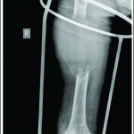
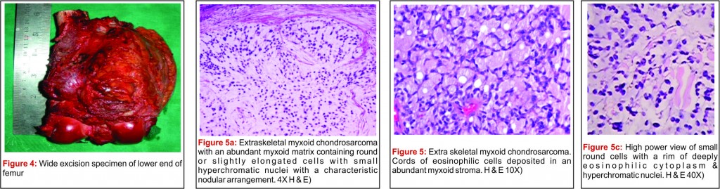
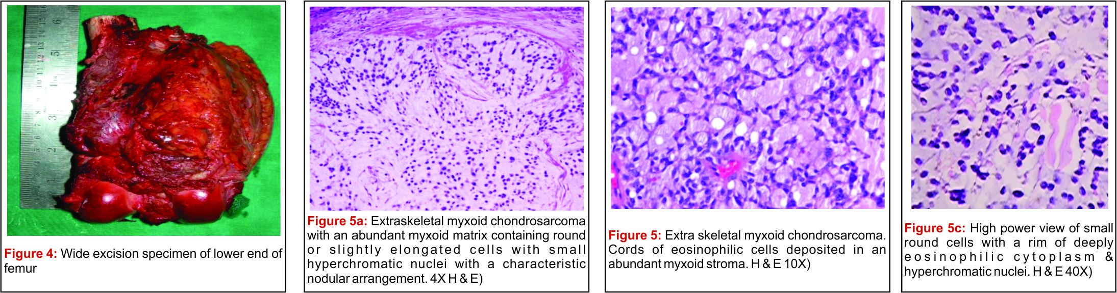
Case 1: 40 year old housewife presented with lower limb swelling and pain just above the knee joint which started since 5 months (Fig 1). Plain radiographs reveal a soft tissue mass abutting the femoral surface with minimum bony involvement in supracondylar region of femur suggestive of a soft tissue neoplasm (Fig. 2). MRI revealed a large extra-osseous soft tissue lesion with minimal intra-osseous extension suggestive of a juxtacortical neoplasm or soft tissue sarcoma (Fig. 3). Open biopsy was done elsewhere and reports were reviewed at authors institute. It showed microscopic features of ESMC. Considering the interosseous involvement and safe oncological margins, a wide resection was planned. The patient underwent limb salvage surgery in form of wide local excision of distal femur along with the mass and reconstruction with a megaprosthesis. Pathological Findings: On gross examination of the wide local excision specimen of distal femur, there was an extra-osseous mass at the lower end of femur on the postero-lateral aspect measuring 9×8 cm in size. It was a soft to firm, ovoid, lobulated mass which on cut section, was gelatinous, mucoid and gray-white. There was no evidence of necrosis or haemorrhage noted (Fig 4). Microscopically, it showed a characteristic multi-nodular pattern. Tumor cells were small round with hyperchromatic nuclei and a narrow rim of cytoplasm (Fig 5a). Cells were arranged in cords and strands separated by abundant myxoid material (Fig 5b,c). No cartilaginous matrix production was seen in the stroma. On Immuno-histochemestry (IHC) the cells showed strong positivity for Vimentin and focal positivity for S-100 protein. These cells were negative for Cytokeratin, EMA, Synaptophysin and Chromogranin. Based on morphological and IHC findings, a diagnosis of Extra-skeletal myxoid chondrosarcoma (ESMC) was reached.
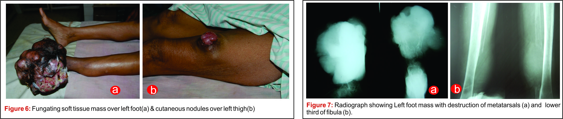
Case 2: 50 year old lady presented with a huge fungating soft tissue mass over left foot associated with similar cutaneous nodules over right arm and left thigh. The foot lesion was progressively increasing over a year but not associated with pain. (Figure 6a, 6b)
X-ray of left foot and lower limb revealed a large soft tissue mass destroying the metatarsals , devoid of any matrix and periosteal reaction (Fig. 7a). Similar lesion was seen destroying the ipsilateral distal fibula (Fig. 7b). Biopsies from the foot mass and the arm nodule revealed histopathological and IHC findings consistent with Extraskeletal Myxoid Chondrosarcoma (ESMC). This patient defaulted for further treatment and follow up.
Discussion:
Extraskeletal myxoid chondrosarcoma (ESMC) is a rare, morphologically distinct soft tissue sarcoma with characteristic nodular architecture & abundant myxoid matrix. In 1972, Enzinger and Shiraki were the first ones who coined ESMC as a distinct entity [1]. In spite of it’s name, Extraskeletal myxoid chondrosarcoma (ESMC) is considered as a “Tumor of uncertain differentiation” because there is no definite evidence of cartilage matrix production in the tumor. Histogenesis of ESMC is still subject of controversy. Incidence of this tumor is only 2.3% of all soft tissue sarcomas as reported by Tsuneyosi et al [2]. Mainly the adult age group (35 years and above) is affected by this tumor, with equal sex predilection [3]. Most common sites are deep soft tissues of the proximal extremities and limb girdles, especially the musculature [4]. However, few uncommon sites are also been described like mediastinum, retroperitoneum, abdomen and the digits [5-7]. In both of our cases, imaging showed that it was a lobulated soft tissue mass without bony periosteal reaction or any radiologically evident matrix production. Histopathologically, both cases showed presence of uniform eosinophilic cells arranged in cords & deposited in an abundant myxoid stroma. There was no evidence of cartilage/osteoid matrix deposition.
The most important clue to the diagnosis of this rare entity is the typical arrangement of cells in cords and columns with a very prominent myxoid background [8]. ESMC appears to exhibit a high tendency of local recurrence & distant metastases, predominantly to the lungs, sometimes years after the initial diagnosis [9]. ESMC should be considered an intermediate grade tumor rather than a low-grade malignant neoplasm as the estimated 5, 10 and 15-year survival rates described by Meis-Kindblom et al were 90%, 70% & 60% respectively [4]. Wide local excision of the tumor is the treatment of choice. If a wide margin can not be obtained, a high rate of local recurrence is observed with poor response to chemotherapy & radiotherapy. Therefore, surgery with appropriate adequate margins continues to be the treatment of choice, for primary as well as recurrent or metastatic tumors.
On follow up part, our first patient is disease free at 6 years from date of surgery with excellent function in the operated limb (MSTS score 97%). The second patient defaulted for further treatment and was lost to follow up. Some adverse pathological prognostic factors reported in literature include Tumor size ≥ 10 cm, high cellularity, anaplasia or rhabdoid features, mitotic activity more than two per 10 high-power fields, and Ki-67 proliferative index of ≥ 10%. These indicate more aggressive behavior, requiring a closer follow-up of the patient [10]. In summary, ESMC is an uncommon but distinct soft tissue sarcoma, clearly different from conventional skeletal chondrosarcoma. Knowledge of this entity and accurate diagnosis is essential because of the difference in its behaviour and prognosis.
References
1. Enzinger FM, Shiraki M. Extraskeletal myxoid chondrosarcoma. An analysis of 34 cases.Hum Pathol 3: 421-35,1972.
2. Tsuneyoshi M, Enjoji M, Iwasaki H and Shirahama N: Extraskeletal myxoid chondrosarcoma: a clinicopathologic and electron microscopic study. Acta Pathol Jpn 31: 201-208, 1981.
3. Antonescu CR, Argani P, Erlandson RA, et al: Skeketal and extraskeletal myxoid chondrosarcoma: a comparative clinicopathologic, ultrastructural, and molecular study. Cancer 83:1504-1521, 1998.
4. Meis-Kindblom JM, Bergh P, Gunterberg B, et al: Extraskeletal myxoid chondrosarcoma: a reappraisal of its morphologic spectrum and prognostic factors based on 117 cases. Am J Surg Pathol 23: 636-650, 1999.
5. Oliveira AM, Sebo TJ, McGrory JE, et al: Extraskeletal myxoid chondrosarcoma; A clinocopathologic, immunohistochemical, and ploidy analysis of 23 cases. Mod Pathol 13: 900-908, 2000.
6. Patel SR, Burgess MA, Papadopculos NE, Linke KA and Benjamin RS: Extraskeletal myxoid chondrosarcoma: long-term experience with chemotherapy. Am J Clin Oncol 18: 161-163,
1995.
7. Okamoto S, Hara K, Sumita S, et al: Extraskeletal myxoid chondrosarcoma arising in the finger. Skeletal Radiol 31: 296-300, 2002.
8. Jakowski JD, Wakely PE Jr. Cytopathology of extra-skeletal myxoid chondrosarcoma: Report of 8 cases. Cancer 2001; 111:298-305
9. Weiss SW and Goldblum JR: Extraskeletal myxoid chondrosarcoma. In: Soft Tissue Tumors 4th Edition. Mosby, St. Louis,pp1368-1379, 2001.
10. Oliveria Am, Sebo TJ, McGrory JE, Gaffey TA, Rock MG, Nascimento AG. Extraskeletal myxoid chondrosarcoma: A clinicaqopthologic, immunohistochemical & ploidy analysis of 23 cases. Mod Pathol 2000; 13:900-8.
| How to Cite this article:Biradar S, Joshi S, Panchwagh Y, Ghanekar V, Kothadiya P. Extraskeletal Myxoid Chondrosarcoma- Rare ‘Non-chondroid’ soft tissue Sarcoma!!!. Journal of Bone and Soft Tissue Tumors Jan-Apr 2016;2(1):36-38 . |