Tag Archive for: extraskeletal

Skeletal Muscle Metastases Arising from Renal Cell Carcinoma in a 58-Year-Old Male: A Case Report

Original Article | Volume 6 | Issue 3 | JBST September – December 2020 | Page 20-25 | Sybill Sue M. Moser, Edwin Joseph R. Guerzon, John Christopher R. Ragasa DOI: 10.13107/jbst.2020.v06i03.035

Author: Sybill Sue M. Moser[1], Edwin Joseph R. Guerzon[1], John Christopher R. Ragasa[1]

[1]Department of Orthopedics, Institute of Orthopedics and Sports Medicine, St. Luke’s Medical Center – Quezon City, Manila, Philippines.

Address of Correspondence:
Dr. Sybill Sue M. Moser
2632 Molave St. United Hills Village,
Parañaque City, Metro Manila, Philippines.
E-mail: sybillsuemoser@gmail.com


Introduction: Renal cell carcinoma (RCC) is the most common malignant kidney tumor, commonly metastasizing to the lung, lymph nodes, bones, and brain. Here, we present a rare case of renal cell skeletal muscle metastases (SMMs), accounting for only <1% of all RCC metastases.
Methods: This is a descriptive report on the clinical course, diagnostic investigations, and surgical treatment of a case of SMM in a patient previously diagnosed with RCC.
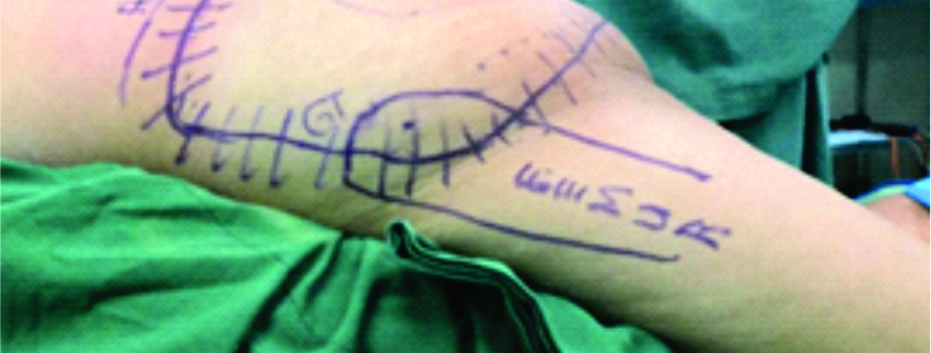
Results: This is a 58-year-old male who previously underwent radical nephrectomy for RCC, presenting with a 5-month history of a rapidly enlarging left gluteal mass. The mass was confirmed to be renal clear cell metastasis through percutaneous biopsy. On magnetic resonance imaging, two heterogeneously enhancing lesions in the left gluteal muscle and right paralumbar muscles at the level of L4 and L5 were noted. Positron emitted tomography scan confirmed no other metastases. He underwent wide excision of the right paraspinal mass and buttockectomy for the left gluteal mass.
Conclusion: SMM in RCC is rare, thus tissue diagnosis and imaging is deemed necessary to rule out any other primary sarcoma. In these cases, patients may benefit from metastasectomy. Regular follow-up and surveillance are recommended for these patients to rule out recurrence.
Keywords: Renal cell carcinoma, metastases, extraskeletal, buttockectomy.


Reference:

1. Graves A, Hessamodini H, Wong G, Lim WH. Metastatic renal cell carcinoma: Update on epidemiology, genetics, and therapeutic modalities. ImmunoTargets Ther 2013;2:73-90.
2. Sakamoto A, Yoshia T, Matsuura S, Tanaka K, Matsuda S, Oda Y, et al. Metastasis to the gluteus maximus muscle from renal cell carcinoma with special emphasis on MRI features. World J Surg Oncol 2007;5:88.
3. Surov A, Hainz M, Holzhausen HJ, Arnold D, Katzer M, Schmidt J, et al. Skeletal muscle metastases: Primary tumours, prevalence, and radiological features. Eur J Radiol 2010;20:649-58.
4. Sountoulides P, Metaxa L, Cindolo L. Atypical presentations and rare metastatic sites of renal cell carcinoma: A review of case reports. J Med Case Rep 2011;5:429.
5. Ramchandani P. Post-treatment surveillance of renal cancer renal cell. In: Patel U, editor. Carcinoma of the Kidney. 1st ed. Cambridge: Cambridge University Press; 2008. p. 185-202.
6. Goger Y, Piskin M, Balasar M, Kilinc M. Unusual Presentation of Renal Cell Carcinoma: Gluteal Metastasis. Case Reports in Urology; 2013.
7. Haygood TM, Sayyouh M, Wong J, Lin J, Matamoros A, Sandler C, et al. Skeletal muscle metastasis from renal cell carcinoma. Sultan Qaboos Univ Med J 2015;15:327-37.
8. Crispen PL, Blute ML. Role of cytoreductive nephrectomy in the era of targeted therapy for renal cell carcinoma. Curr Urol Rep 2012;13:38-46.


How to Cite this article: Goyal AK, Vaish A, Vaishya R, Baweja P| A Rare Recurrent Chondromyxoid Fibroma of the Proximal Tibia in an Adolescent Female | Journal of Bone and Soft Tissue Tumors | September-December 2020; 6(3): 20-25.

[Full Text HTML] [Full Text PDF] [XML]


A Clinicopathological Study of Ewing’s Sarcoma/PNET experience from a Tertiary Cancer Centre in North East India

Original Article | Volume 6 | Issue 2 | JBST May-August 2020 | Page 21-24 | Jagannath Dev Sharma, Argha Baruah, Anupam Sarma, Lopa Mudra Kakoti, Nizara Baishya, Shiraj Ahmed. DOI: 10.13107/jbst.2020.v06i02.27

Author: Jagannath Dev Sharma[1], Argha Baruah[1], Anupam Sarma[1], Lopa Mudra Kakoti[1], Nizara Baishya[1], [2], Shiraj Ahmed[1]

[1]Department of Pathology, Dr.B.Borooah Cancer Institute, Guwahati, Assam, India.
[2]Department of Hospital based Cancer registry, Dr.B. Borooah Cancer Institute, Guwahati, Assam.

Address of Correspondence
Dr. Argha Baruah,
Department of Pathology, Dr.B.Borooah Cancer Institute,Guwahati-781028, Assam, India.
E-mail: argha20@gmail.com


Abstract

Introduction: Ewing sarcoma (ES)/PNET is an aggressive malignant tumor with small round cell morphology affecting mainly children and adolescents. The aim of this study was to study the clinicopathological parameters and immunohistochemical panel of skeletal and extraskeletal ES and to correlate with overall survival.
Case Report: Medical files of 70 patients with ES treated at our center between 2009 and 2015 were retrospectively evaluated. The clinico pathological parameters were extracted and statistically correlated with overall survival(OS). Among 70cases of ES ,41 cases were males and 29 cases were females. Most common age group was 10–20 years. Skeletal involvement was seen in 45 cases(64.2%) and 25cases (35.8%) were extraskeletal. The most common skeletal sites of involvement was lower extremity involving  the Femur (24%) and the most common extraskeletal site involved in our study was sinonasal area(5.7%), followed by chestwall, thigh, orbital, calf, gluteal, kidney, and vulva. Two cases showed involvement of the central nervous system(CNS) involving pineal gland and the ventricle. Two cases showed multiple sites of involvement both including chest wall and thigh. Twenty-nine cases(41.4%) showed metastasized disease. The most common site of metastasis was lung followed by bone and brain. Recurrence was seen in 14 cases(20%). Overall 5-year survival was 24%. There was statistically significant correlation found between tumor size (≥8cm) and 5year survival. Furthermore, significant correlation was found between metastasis and 5-year survival.
Conclusion: ES is an aggressive tumor involving skeletal and extraskeletal sites affecting commonly young people, with a poor prognosis for patients with maximum diameter ≥8cm. Metastasisis common in ES and is also a poor prognostic factor.
Keywords: Ewing’sarcoma, skeletal, extraskeletal, survival, metastasis.


Reference:
1. Paulssen M, Ahrens S, Dunst J, Winkelmann W, Exner GU, Kotz R, et al. Localized Ewing tumor of bone: Final results of the cooperative Ewing’s sarcoma study CESS 86. J Clin Oncol 2001;19:1818-29.
2. Ewing J. Diffuse endothelioma of bone. Proc N Y Path Soc 1921;7:17-24.
3. Angervall L, Enzinger FM. Extraskeletal neoplasm resembling Ewing’s sarcoma. J Cancer 1975;36:240-51.
4. Askin FB, Rosai J, Sibley RK, Dehner LP, McAlister WH. Malignant small cell tumor of the thoracopulmonary region in childhood: A distinctive clinicopathologic entity of uncertain histogenesis. J Cancer 1979;43:2438-51.
5. Bellan DG, Filho RJ, Garcia JG, de Toledo Petrilli M, Viola DC, Schoedl MF, et al. Ewing’s sarcoma: Epidemiology and prognosis for patients treated at the pediatric oncology institute, IOP-GRAACC-UNIFESP. Rev Bras Ortop 2015;47:446-50.
6. Akhavan A, Binesh F, Shamshiri H, Ghanadi F. Survival of patients with Ewing’s sarcoma in Yazd-Iran. Asian Pac J Cancer Prev 2014;15:4861-4.
7. Biswas B, Rastogi S, Khan SA, Mohanti BK, Sharma DN, Sharma MC, et al. Outcomes and prognostic factors for Ewing-family tumors of the extremities. J Bone Joint Surg Am 2014;96:841-9.
8. Lee JA, Kim DH, Lim JS, Koh JS, Kim MS, Kong CB, et al. Soft-tissue Ewing sarcoma in a low-incidence population: Comparison to skeletal Ewing sarcoma for clinical characteristics and treatment outcome. Jpn J Clin Oncol 2010;40:1060-7.
9. Worch J, Matthay KK, Neuhaus J, Goldsby R, DuBois SG. Ethnic and racial differences in patients with Ewing sarcoma. J Cancer 2010;116:983-8.
10. Louati S, Senhaji N, Chbani L, Bennis S. EWSR1 rearrangement and CD99 expression as diagnostic biomarkers for Ewing/PNET sarcomas in a Moroccan population. Dis Markers 2018;2018:7971019.
11. Folpe AL, Hill CE, Parham DM, O’Shea PA, Weiss SW. Immunohistochemical detection of FLI-1 protein expression: A study of 132 round cell tumors with emphasis on CD99-positive mimics of Ewing’s sarcoma/primitive neuroectodermal tumor. Am J Surg Pathol 2000;24:1657-62.
12. Rossi S, Orvieto E, Furlanetto A, Laurino L, Ninfo V, Dei Tos AP. Utility of the immunohistochemical detection of FLI-1 expression in round cell and vascular neoplasm using a monoclonal antibody. Mod Pathol 2004;17:547-52.
13. Ahmed SH, Rahman NA, Meng L. Cytokeratin immunoreactivity in Ewing sarcoma/primitive neuroectodermal tumour. Malays J Pathol 2013;35:139-45.
14. Lucas DR, Bentley G, Dan ME, Tabaczka P, Poulik JM, Mott MP. Ewing sarcoma vs lymphoblastic lymphoma. A comparative immunohistochemical study. Am J Clin Pathol 2001;115:11-7.
15. Machado I, Noguera R, Pellin A, Lopez-Guerrero JA, Piqueras M, Navarro S, et al. Molecular diagnosis of Ewing sarcoma family of tumors: A comparative analysis of 560 cases with FISH and RT-PCR. Diagn Mol Pathol 2009;18:189-99.
16. Tirode F, Surdez D, Ma X, Parker M, Le Deley MC, Bahrami A, et al. Genomic landscape of Ewing sarcoma defines an aggressive subtype with co-association of STAG2 and TP53 mutations. Cancer Discov 2015;4:1342-53.
17. Stahl M, Ranft A, Paulussen M, Bölling T, Vieth V, Bielack S, et al. Risk of recurrence and survival after relapse in patients with Ewing sarcoma. Pediatr Blood Cancer 2011;57:549-53.
18. Obata H, Ueda T, Kawai A, Ishii T, Ozaki T, Abe S, et al. Clinical outcome of patients with Ewing sarcoma family of tumors of bone in Japan: The Japanese musculoskeletal oncology group cooperative study. J Cancer 2007;109:767-75.
19. Oksuz DC, Tural D, Dincbas FO, Dervisoglu S, Turna H, Hiz M, et al. Non-metastatic Ewing’s sarcoma family of tumors of bone in adolescents and adults: Prognostic factors and clinical outcome-single institution results. Tumor J 2014;100:452-8.
20. El Weshi A, Allam A, Ajarim D, Al Dayel F, Pant R, Bazarbashi S, et al. Extraskeletal Ewing’s sarcoma family of tumours in adults: Analysis of 57 patients from a single institution. Clin Oncol 2010;22:374-81.
21. Pradhan A, Grimer RJ, Spooner D, Peake D, Carter SR, Tillman RM, et al. Oncological outcomes of patients with Ewing’s sarcoma: Is there a difference between skeletal and extra-skeletal Ewing’s sarcoma? J Bone Joint Surg Br 2011;93:531-6.
22. Bosma SE, Ayu O, Dijkstra PD, Fiocco M, Gelderblom H. Prognostic factors for survival in Ewing sarcoma: A systematic review. Surg Oncol 2018;27:603-10.


How to Cite this article: Sharma JD, Baruah A, Sarma A, Kakoti LM, Baishya N, Ahmed S | A Clinicopathological Study of Ewing’s Sarcoma/PNET experience from a Tertiary Cancer Centre in North East India | Journal of  Bone and Soft Tissue Tumors | May-August 2020; 6(2): 21-24.

[Full Text HTML] [Full Text PDF] [XML]