JBST- Special IMSOS 2019 Issue

Vol 5 | Issue 1 | Jan-April 2019 | page:1 | Dr. Yogesh Panchwagh, Dr. Ashish Gulia,  Dr. Ashok Shyam.


Author: Yogesh Panchwagh [1], Ashish Gulia [2], Ashok Shyam [1,3].

[1] Orthopaedic Oncology Clinic, Pune, India.
[2] Orthopedic Oncology Services, Department of Surgical Oncology, Tata Memorial Hospital, Mumbai.
[3] Indian Orthopaedic Research Group, Thane, India,
[4] Sancheti Institute for Orthopaedics &Rehabilitation, Pune, India

Address of Correspondence
Dr. Yogesh Panchwagh.
Orthopaedic Oncology Clinic, 101, Vasant plot 29, Bharat Kunj Society -2, Erandwana, Pune – 38, India.
Email: drpanchwagh@gmail.com


JBST- Special IMSOS 2019 Issue

JBST is entering the fifth year of publication . This would not have been possible without the help of the contribution by authors, guest editors, reviewers and the publication house team.

We take this opportunity to thank each and every one of them. Already indexed with Index Copernicus, google scholar and ETH Bibliothek , JBST is very close to pubmed indexation.

Our attempt over the last few issues has been to emphasise on publishing original work than invited or review articles so as to make the journal the chosen one for all those working in the field of bone and soft tissue tumors. We continue to bring to the readers the current concept in management of common bone and soft tissue tumors and tumor like conditions through sections like Students corner which is very useful for residents and consultants alike.

JBST is now affiliated with the IMSOS ( Indian musculo skeletal oncology society) and APMSTS ( Asia Pacific musculo skeletal oncology society) and will be showcasing their activities from time to time. Members of these associations can have the benefit of availing the opportunity of freely publishing in JBST. We are sure that this will add a new dimension and depth to the content of the journal.

We would like to thank the readers for their contribution and would like to hear from you about your experience and feedback so as to make JBST better.

Dr. Yogesh Panchwagh
Dr. Ashish Gulia
Dr. Ashok Shyam


How to Cite this article: Panchwagh Y, Gulia A, Shyam AK. JBST – Special IMSOS 2019 Issue. Journal of Bone and Soft Tissue Tumors Jan-April 2019; 5(1):1.

(Abstract Full Text HTML) (Download PDF)


Giant Ancient Solitary Schwannoma Masquerading as Juxtacortical Osteosarcoma of Femur – A Rare Case Report and Literature Review

Vol 5 | Issue 1 | Jan-April 2019 | page: 28-30 | Julfiqar, Mohd. Aslam, Najmul Huda, Ajay Pant.


Authors: Julfiqar [1], Mohd. Aslam [1], Najmul Huda [1], Ajay Pant [1].

[1] Dept of Orthopaedic Surgery, Faculty of Medicine, J.N.Medical College, Aligarh Muslim University, Aligarh, India
[2] Department of Orthopaedics, Teerthanker Mahaveer Medical College and Research Centre, TMU, Moradabad, India

Address of Correspondence
Dr. Julfiqar:
Assistant professor, Department of Orthopaedic Surgery, Faculty of Medicine, J.N.Medical College, Aligarh Muslim University, Aligarh, India.
Email: ???


Abstract

Introduction: Ancient schwannomas are rare variant of peripheral nerve sheath tumors characterized by the degeneration and hypocellular areas due to long-standing growth. Clinicoradiologically, these tumors can masquerade other tumors arising from the adjacent tissues. Their resemblance to malignant bone tumor has been reported very rarely in the literature. We tend to report a case of benign peripheral nerve schwannoma that greatly mimicked a juxtacortical osteosarcoma of femur.
Case Report: A 23-year-old male presented with a slow-growing painless mass with paresthesias in his right thigh for the last 2½ years. Clinically, it was suspected to be soft tissue tumor with secondary involvement of adjacent neurovascular bundle; however, plain radiograph and magnetic resonance imaging of his right thigh were suggestive of juxtacortical osteosarcoma of the right femur. Surgical exploration of the mass revealed a well-defined encapsulated mass over the anterior aspect of the right thigh, under the quadriceps muscle without infiltration into the surrounding tissue. Histopathological examination confirmed it to be an ancient schwannoma.
Results: The patient was extremely satisfied with outcomes of surgery, and he was symptom-free and there was no clinical evidence of the recurrence on subsequent follow-up.
Conclusion: A correct pre-operative diagnosis of benign peripheral nerve sheath tumors can be difficult at times. However, a slow-growing mass with the absence of other features of a malignant growth and subsequent histopathological examination including immunostaining can settle the diagnosis in almost all the cases.
Keywords: Benign, Peripheral nerve, Ancient schwannoma, Juxtacortical osteosarcoma.


References

1. Dahl I. Ancient neurilemmoma (schwannoma). Acta Pathol Microbiol Scand 1977;85:812-8.
2. Kuriakose S, Vikram S, Salih S, Balasubramanian S, Pareekutty NM, Nayanar S. Unique surgical issues in the management of a giant retroperitoneal schwannoma and brief review of literature. Case Rep Med 2014;2014:781347.
3. Isobe K, Shimizu T, Akahane T, Kato H. Imaging of ancient schwannoma. AJR Am J Roentgenol 2004;183:331-6.
4. Mwaka ES, Senyonjo P, Kakyama M, Nyati M, Orwotho N, Lukande R. Giant, solitary, ancient schwannoma of the cervico-thoracic spine: A case report and review of the literature. OA Case Reports 2013;2:2.
5. Choudry HA, Nikfarjam M, Liang JJ, Kimchi ET, Conter R, Gusani NJ, et al. Diagnosis and management of retroperitoneal ancient schwannomas. World J Surg Oncol 2009;7:12.
6. Jayaraj SM, Levine T, Frosh AC, Almyeda JS. Ancient schwannoma masquerading as parotid pleomorphic adenoma. J Laryngol Otol 1997;111:1088-90.
7. Lee YS, Kim JO, Park SE. Ancient schwannoma of the thigh mimicking a malignant tumour: A report of two cases, with emphasis on MRI findings. Br J Radiol 2010;83:e154-7.
8. Ackerman LV, Taylor FH. Neurogenous tumors within the thorax; A clinicopathological evaluation of forty-eight cases. Cancer 1951;4:669-91.
9. Enzinger FM, Weiss SW. Benign tumours of peripheral nerves. Soft Tissue Tumours. 3rd ed. St Louis: Mosby; 1995. p. 821-88.
10. Takeuchi M, Matsuzaki K, Nishitani H, Uehara H. Ancient schwannoma of the female pelvis. Abdom Imaging 2008;33:247-52.
11. Kransdorf MJ, Murphey MD, editors. Neurogenic tumours. Imaging of Neurogenic Tumours. 2nd ed. Philadelphia, PA: Lippincot Williams &Wilkins; 2006. p. 328-80.
12. Dahlin DC, Coventy MB. Osteogenic sarcoma: A study of six hundred cases. J Bone Joint Surg Am 1967;49:101-10.
13. Wong KT, Haywood T, Dalinka MK, Kneeland B. Chondroblastic, grade 3 periosteal osteosarcoma. Skeletal Radiol 1995;24:69-71.
14. Sorensen DM, Gokden M, El-Naggar A, Byers RM. Quiz case 1. Periosteal osteosarcoma (PO) of the mandible. Arch Otolaryngol Head Neck Surg 2000;126:550, 552.
15. Murphey MD, Jelinek JS, Temple HD, Flemming DJ, Gannon FH. Imaging of periosteal osteosarcoma: Radiologic-pathologic comparison. Radiol 2004;233:129-38.


How to Cite this article: Julfiqar, Aslam M, Huda N, Pant A. Giant Ancient Solitary Schwannoma Masquerading as Juxtacortical Osteosarcoma of Femur-A Rare Case Report and Literature Review. Journal of Bone and Soft Tissue Tumors Jan-April 2019;5(1): 28-30.


                  (Abstract    Full Text HTML)   (Download PDF)


Chondroblastoma of the Naviculum: A Diagnostic Dilemma

Vol 5 | Issue 1 | Jan-April 2019 | page: 25-27 | Monish Malhotra, Sandeep Vijayan, Sourab Shetty.


Authors: Monish Malhotra [1], Sandeep Vijayan [2], Sourab Shetty [2].

[1] Department of Orthopaedics, Pandit Bhagwat Dayal Sharma PGIMS, Rohtak, Haryana, India,
[2] Department of Orthopaedics, Kasturba Medical College, Manipal Academy of Higher Education, Manipal, Karnataka, India.

Address of Correspondence
Dr. Monish Malhotra,
Department of Orthopaedics, Pandit Bhagwat Dayal Sharma PGIMS, Rohtak – 124001, Haryana, India.
E-mail: drmonish.malhotra14@gmail.com


Abstract

Introduction: Chondroblastoma is a benign tumor characteristically seen in the epiphyseal region of long bones. Its occurrence in the tarsal bones is extremely rare.
Case Report: We report the case of a 17-year-old girl with chondroblastoma mimicking an aneurysmal bone cyst in the naviculum. Along with the primary enbloc excision of the naviculum, a tricortical iliac crest bone graft was placed in the gap to prevent collapse of the medial longitudinal arch.
Conclusion: Chondroblastoma in uncommon sites can present with atypical features and cause dilemma in diagnosis and thereby management.
Keywords: Chondroblastoma, Bone grafting, Aneurysmal bone cyst, Tarsal bones, Naviculum.


References

1. Suneja R, Grimer RJ, Belthur M, Jeys L, Carter SR, Tillman RM, et al. Chondroblastoma of bone: Long-term results and functional outcome after intralesional curettage. J Bone Joint Surg Br 2005;87:974-8.
2. Li XN, Peng ZG, Zhao JP, Zhang ZK. Chondroblastoma of the navicular bone. Iran J Radiol 2014;11:e10848.
3. Fang Z, Chen M. Chondroblastoma associated with aneurysmal cyst of the navicular bone: A case report. World J Surg Oncol 2013;11:50.
4. Springfield DS, Capanna R, Gherlinzoni F, Picci P, Campanacci M. Chondroblastoma. A review of seventy cases. J Bone Joint Surg Am 1985;67:748-55.
5. McLeod RA, Beabout JW. The roentgenographic features of chondroblastoma. Am J Roentgenol Radium Ther Nucl Med 1973;118:464-71.
6. Atalar H, Basarir K, Yildiz Y, Erekul S, Saglik Y. Management of chondroblastoma: Retrospective review of 28 patients. J Orthop Sci 2007;12:334-40.
7. Huvos AG, Marcove RC. Chondroblastoma of bone. A critical review. Clin Orthop Relat Res 1973;95:300-12.


How to Cite this article: Malhotra M, Vijayan S, Shetty S. Chondroblastoma of theNaviculum: A Diagnostic Dilemma. Journal of Bone and Soft Tissue Tumors Jan-Apr 2019;5(1): 25-27.


                  (Abstract    Full Text HTML)   (Download PDF)


Parosteal Lipoma of the Lower Limb: A Report of Two Cases

Vol 5 | Issue 1 | Jan-April 2019 | page: 21-24 | Imed Sboui, Hend Riahi, Mouna Chelli-Bouaziz, Mohamed Samir Daghfous, Mohamed Fethi Ladeb, Faten Farah.


Authors: Imed Sboui [1], Hend Riahi [2], Mouna Chelli-Bouaziz[2], Mohamed Samir Daghfous [1], Mohamed Fethi Ladeb [2], Faten Farah [3].

[1] Department of Orthopaedic, Institut Mohamed Kassab d’orthopédie, Manouba, Tunisia,
[2] Department of Radiology, Institut Mohamed Kassab d’orthopédie, Manouba, Tunisia,
[3] Department of Pathology, Institut Mohamed Kassab d’orthopédie, Manouba, Tunisia.

Address of Correspondence
Dr. Hend Riahi,
10 rue Khalifas Rachidites Menzeh 5 Ariana Tunisia.
E-mail: hend.riahi@gmail.com


Abstract

Introduction: Parosteal lipoma is a rare type of lipoma. Usually, asymptomatic and affecting mainly adults aged over 40.
Case Report: We report two cases localized in the lower limb. The first happened in a man presenting a painless swelling of the fibula, represented by a well-defined fatty tissue lesion. The second case concerned women presenting an asymmetry of the calf, represented by a well-defined mass composed of fatty tissue. The lesions were resected from surrounding soft tissues and underlying periosteum. The diagnosis was confirmed by histology.
Conclusion: Parosteal lipoma has the same characteristics of computed tomography (CT) and magnetic resonance imaging (MRI) as subcutaneous fat. The identification of fat is best performed with CT or MRI. Its recognition is mandatory to optimize clinical management including diagnosis, biopsy, and treatment.
Keywords: Deep-seated lipoma, Computed tomography, Magnetic resonance imaging.


References

1. Fleming RJ, Alpert M, Garcia A. Parosteal lipoma. AJR Am J Roentgenol 1962;87:1075-84.
2. Rodriguez-Peralto JL, Lopez-Barea F, Gonzales-Lopes J, Lamas-Lorenzo M. Case report 821. Skeletal Radiol 1994;23:67-9.
3. Asirvatham R, Linjawi T. Ossifying parosteal lipoma with exuberant cortical reaction. A case report. Int Orthop 1994;18:55-6.
4. Amores-Ramírez F, Hierro Martín I, Montañez Heredia E, Garcia-Fortea P, Garcia Salguero AI, Fernandez de Rota Conde A, et al. Painless mass in leg: Diagnosis and discussion. Skeletal Radiol 2009;38:1105-6, 1119-20.
5. Murphey MD, Johnson DL, Bhatia PS, Neff JR, Rosenthal HG, Walker CW, et al. Parosteal lipoma: MR imaging characteristics. AJR Am J Roentgenol 1994;162:105-10.
6. Schajowicz F. Neoplasia Óssea e Lesões Pseudotumorais. 2nd ed. Rio de Janeiro: Revinter; 2000. p. 403-46.
7. John SH, Chad CB, Kathleen SB, Valerie AF, Marcia FB, Joseph B. Parosteal lipoma of the proximal radius. Austin J Musculoskelet Disord 2016;3:1027.
8. Murphey MD, Arcara LK, Fanburg-Smith J. From the archives of the AFIP: Imaging of musculoskeletal liposarcoma with radiologic-pathologic correlation. Radiographics 2005;25:1371-95.
9. Murphey MD, Carroll JF, Flemming DJ, Pope TL, Gannon FH, Kransdorf MJ, et al. From the archives of the AFIP: Benign musculoskeletal lipomatous lesions. Radiographics 2004;24:1433-66.
10. Greco M, Mazzocchi M, Ribuffo D, Dessy LA, Scuderi N. Parosteal lipoma. Report of 15 new cases and a review of the literature. Ann Ital Chir 2013;84:229-35.
11. Rosenberg AE, Bridge JA. Lipoma of bone. In: Fletcher CD, Unni KK, Mertens F, editors. Pathology and Genetics of Tumours of the Soft Tissues and Bones. Lyon: IARC Press; 2002. p. 328-9.
12. Kawashima A, Magid D, Fishman EK, Hruban RH, Ney DR. Parosteal ossifying lipoma: CT and MR findings. J Comput Assist Tomogr 1993;17:147-50.
13. Seki N, Okada K, Miyakoshi N, Shimada Y, Nishida J, Itoi E, et al. Common peroneal nerve palsy caused by parosteal lipoma of the fibula. J Orthop Sci 2006;11:88-91.
14. Resnick D, editor. Tumor and tumor-like diseases. In: Diagnosis of Bone and Joint Disorders. 4th ed. Philadelphia, PA: Saunders; 1995. p. 3745-4128.
15. Dorfman HD. Miscellaneous mesenchymal tumors. In: Dorfman H, Czerniak B, editors. Bone Tumors. St Louis, Mo: Mosby; 1998. p. 913-48.
16. Petit MM, Swarts S, Bridge JA, Van de Ven WJ. Expression of reciprocal fusion transcripts of the HMGIC and LPP genes in parosteal lipoma. Cancer Genet Cytogenet 1998;106:18-23.
17. Kransdorf M, Murphey M. Lipomatous tumors. In: Imaging of Soft Tissue Tumors. Philadelphia, PA: Saunders; 1997. p. 57-101.
18. Obermann EC, Bele S, Brawanski A, Knuechel R, Hofstaedter F. Ossifying lipoma. Virchows Arch 1999;434:181-3.
19. Burt AM, Huang BK. Imaging review of lipomatous musculoskeletal lesions. SICOT J 2017;3:34.
20. Yu JS, Wei L, Becker W. MR imaging of a parosteal lipoma. J Clin Imaging 2000;24:15-8.
21. Balani A, Sankhe A, Dedhia T, Bhuta M, Lakhotia N, Yeshwante J, et al. Lump on back: A rare case of parosteal lipoma of scapula. Case Rep Radiol 2014;2014:169157.
22. Go T, Nakajima N, Yokota N, Yokomise H. Thoracoscopic resection of parosteal lipoma of the rib using orthopedic electric micro drill. Gen Thorac Cardiovasc Surg 2018;66:675-8.


How to Cite this article: Sboui I, Riahi H, Chelli-Bouaziz M, Daghfous MS, Ladeb MF, Farah F. Parosteal Lipoma of the Lower Limb: A Report of Two Cases. Journal of Bone and Soft Tissue Tumors Jan-Apr 2019;5(1): 21-24.


                  (Abstract    Full Text HTML)   (Download PDF)


Health-Related Quality of Life in Patients with Bone Tumor around the Knee after Resection Arthrodesis

Vol 5 | Issue 1 | Jan-April 2019 | page: 17-20 | Wilasinee Sirichativapee, Weerachai Kosuwon, Winai Sirichativapee.


Authors: Wilasinee Sirichativapee [1], Weerachai Kosuwon [1], Winai Sirichativapee [1].

[1] Department of Orthopaedics, Faculty of Medicine, Khon Kaen University, Khon Kaen, Thailand.

Address of Correspondence
Dr. Winai Sirichativapee,
Department of Orthopaedics, Srinagarind Hospital, 123 Khon Kaen University, Nai Mueang Sub-District, Mueang District, Khon Kaen Province – 40002, Thailand.
E-mail: winaisiri@yahoo.com


Abstract

Background: This study aimed to compare the health-related quality of life (HRQoL) of patient with bone tumor around the knee after resection arthrodesis.
Methods: Patients between 15 and 70 years of age who underwent resection arthrodesis in Srinagarind Hospital >1 year were recruited. Patients were interviewed using a short form-36 questionnaire (social functioning-36 [SF-36] Ver2.0 Thai version) regarding their daily life problems.
Results: Eighteen patients with the mean age of 36.6 years (15–63 years) were included (15 females) in the study. Histological diagnoses were giant cell tumor 10 cases, osteosarcoma seven cases, and low-grade chondrosarcoma one case. Site of lesions was distal femur 15 cases and proximal tibia 3 cases. According to HRQoL, patients have good quality of life (score SF-36 >70) in all domains: Mean score: Physical functioning 75.55 ± 21.88, role physical 71.18 ± 22.70, bodily pain 85.41 ± 22.51, vitality 77.43 ± 16.76, general health 74.44 ± 19.16, SF 83.05 ± 26.40, role emotional 80.09 ± 22.89, and mental health 77.77 ± 21.29. Complications post-operative are broken implants (3 cases, 16.7%) and infections (4 cases, 22.2%).
Conclusion: In patients with bone tumor around the knee after resection, arthrodesis has a good quality of life in all domains in SF-36 version 2.0 questionnaire including function, pain, and mentality.
Keywords: Limb salvage, Arthrodesis, Quality of life, social functioning-36 version 2.0, Osteosarcoma, Giant cell tumor.


References

1. Tarnawska-Pierścińska M, Hołody Ł, Braziewicz J, Królicki L. Bone metastases diagnosis possibilities in studies with the use of 18F-NaF and 18F-FDG. Nucl Med Rev Cent East Eur 2011;14:105-8.
2. Sampath SC, Sampath SC, Mosci C, Lutz AM, Willmann JK, Mittra ES, et al. Detection of osseous metastasis by 18F-NaF/18F-FDG PET/CT versus CT alone. Clin Nucl Med 2015;40:e173-7.
3. Harisankar CN, Agrawal K, Bhattacharya A, Mittal BR. F-18 fluoro-deoxy-glucose and F-18 sodium fluoride cocktail PET/CT scan in patients with breast cancer having equivocal bone SPECT/CT. Indian J Nucl Med 2014;29:81-6.
4. Roop MJ, Singh B, Singh H, Watts A, Kohli PS, Mittal BR, et al. Incremental value of cocktail 18F-FDG and 18F-NaF PET/CT over 18F-FDG PET/CT alone for characterization of skeletal metastasesin breast cancer. Clin Nucl Med 2017;42:335-40.
5. Chan HP, Hu C, Yu CC, Huang TC, Peng NJ. Added value of using a cocktail of F-18 sodium fluoride and F-18 fluorodeoxyglucose in positron emission tomography/computed tomography for detecting bony metastasis: A case report. Medicine (Baltimore) 2015;94:e687.
6. Iagaru A, Mittra E, Mosci C, Dick DW, Sathekge M, Prakash V, et al. Combined 18F-fluoride and 18F-FDG PET/CT scanning for evaluation of malignancy: Results of an international multicenter trial. J Nucl Med 2013;54:176-83.
7. Gradishar WJ, Anderson BO, Balassanian R, Blair SL, Burstein HJ, Cyr A, et al. NCCN Clinical Practice Guidelines in Oncology Breast Cancer Version 2; 2016. Available from: https://www.nccn.org/professionals/physician_gls/pdf/breast.pdf. [Last accessed on 2016 Oct 19].
8. Yoon SH, Kim KS, Kang SY, Song HS, Jo KS, Choi BH, et al. Usefulness of (18)F-fluoride PET/CT in breast cancer patients with osteosclerotic bone metastases. Nucl Med Mol Imaging 2013;47:27-35.
9. Israel O, Goldberg A, Nachtigal A, Militianu D, Bar-Shalom R, Keidar Z, et al. FDG-PET and CT patterns of bone metastases and their relationship to previously administered anti-cancer therapy. Eur J Nucl Med Mol Imaging 2006;33:1280-4.
10. Lapa P, Saraiva T, Silva R, Marques M, Costa G, Lima JP. Superiority of 18F-Fna PET/CT for detecting bone metastases in comparison with other diagnostic ımaging modalities. Acta Med Port 2017;30:53-60.
11. Araz M, Aras G, Küçük ÖN. The role of 18F-NaF PET/CT in metastatic bone disease. J Bone Oncol 2015;4:92-7.
12. Schirrmeister H, Glatting G, Hetzel J, Nüssle K, Arslandemir C, Buck AK. Prospective evaluation of the clinical value of planar bone scans, SPECT, and (18)F-labeled NaF PET in newly diagnosed lung cancer. J Nucl Med 2001;42:1800-4.
13. Piccardo A, Puntoni M, Morbelli S, Massollo M, Bongioanni F, Paparo F, et al. 18F-FDG PET/CT is a prognostic biomarker in patients affected by bone metastases from breast cancer in comparison with 18F-naF PET/CT. Nuklearmedizin 2015;54:163-72.
14. Iagaru A, Young P, Mittra E, Dick DW, Herfkens R, Gambhir SS. Pilot prospective evaluation of 99mTc-MDP scintigraphy, 18F NaF PET/CT, 18F FDG PET/CT and whole-body MRI for detection of skeletal metastases. Clin Nucl Med 2013;38:e290-6.
15. Hillner BE, Siegel BA, Hanna L, Duan F, Quinn B, Shields AF. 18F-fluoride PET used for treatment monitoring of systemic cancer therapy: Results from the national oncologic PET registry. J Nucl Med 2015;56:222-8.
16. Iagaru A, Mittra E, Dick DW, Gambhir SS. Prospective evaluation of (99m)Tc MDP scintigraphy, (18)F NaF PET/CT, and (18)F FDG PET/CTfor detection of skeletal metastases. Mol Imaging Biol 2012;14:252-9.


How to Cite this article: Koç Z P, Kara P Ö, Sezer E, Erçolak V.Diagnostic Comparison of F-18 Sodium FluorideNaF, Bone Scintigraphy, and F-18 Fluorodeoxyglucose Positron Emission Tomography/Computed Tomography in the Detection of Bone Metastasis. Journal of Bone and Soft Tissue Tumors Jan-Apr 2019;5(1): 17-20.


  (Abstract    Full Text HTML)   (Download PDF)


Guest Editorial: Message from IMSOS President

Vol 5 | Issue 1 | Jan-April 2019 | page: 2 | Bhavin Jhankaria


Author: Bhavin Jhankaria [1].

[1] Picture This By Jankharia, Bhaveshwar Vihar, 383 Sardar V P Rd, Opposite Vanita Vishram, Mumbai, Maharashtra, 400004, India.

Address of Correspondence
Dr. Bhavin Jhankaria
Picture This By Jankharia, Bhaveshwar Vihar, 383 Sardar V P Rd, Opposite Vanita Vishram, Mumbai, Maharashtra, 400004, India.
Email: bhavin@jankharia.com


Asia Pacific Musculoskeletal Tumor Society Conference 2018

Message from IMSOS President

IMSOS or the Indian Musculoskeletal Oncology Society is truly a multi-disciplinary society comprising of orthopedic surgeons, radiologists, pathologists and medical and radiation oncologists. All of these specialties and their doctors have come together to work towards providing optimal care to patients with bone and soft tissue tumors. IMSOS also encourages non-doctor members…anyone who makes a difference to the care of patients with bone and soft tissue tumors is welcome to be part of the society.
IMSOS’ vision is to “promote scientific, evidence based, comprehensive multidisciplinary management of bone and soft tissue sarcomas and encourage basic and clinical research”. IMSOS’ mission is to do this through meetings, conferences, workshops, white papers and an active website where people engage with each other to learn and teach and to eventually use that knowledge to optimally manage patients and alleviate their pain and suffering.
IMSOS is orthopedic-surgeon-driven given the very nature of the subject. However, radiologists, pathologists, oncologists and the other members are also an integral part of the Society. By electing me President for the next 2 years, IMSOS has truly proven its multi-disciplinary character.
The new IMSOS secretary and I, will ensure that IMSOS’ mission and vision are fulfilled and that we continue the work done by Ajay Puri and Ashish Gulia. We will strive to create a platform that will allow those who come after us to continue and enhance the work done by us.
We are actively soliciting suggestions from IMSOS’ members on various topics and issues to ensure that IMSOS stays and continues to be relevant and make a difference.
Let’s have a great meeting in Kolkata and work towards an equally good or better meeting in Bengaluru in March 2020.

Dr Bhavin Jhankaria
President – Indian Musculo Skeletal Oncology Society


How to Cite this article: Jhankaria B. Message from IMSOS President. Journal of Bone and Soft Tissue Tumors Jan-April 2019; 5(1):2.


(Abstract Full Text HTML) (Download PDF)


Diagnostic Comparison of F-18 Sodium FluorideNaF, Bone Scintigraphy, and F-18 Fluorodeoxyglucose Positron Emission Tomography/Computed Tomography in the Detection of Bone Metastasis

Vol 5 | Issue 1 | Jan-April 2019 | page: 9-12 | Zehra PınarP Koç, Pelin Ö Kara, Emel Sezer, Vehbi Erçolak


Authors: Zehra PınarP Koç [1], Pelin Ö Kara [1], Emel Sezer [2], Vehbi Erçolak [2]

[1] Department of Nuclear Medicine, Mersin University, Mersin/, Turkey.,
[2] Department of Oncology, Mersin University, Mersin, Turkey. Mersin/Turkey.

Address of Correspondence
Dr. Zehra PınarP ınar Koç,
Mersin University Nuclear Medicine Dpt., Mersin – 33343, Turkey.
E-mail: zehrapinarkoc@gmail.com


Abstract

Objective: The aim of this study is to compare the diagnostic efficiency of bone scintigraphy, fluorodeoxyglucose (FDG), and sodium fluoride (NaF) positron emission tomography/computed tomography (PET/CT) in the evaluation of bone metastasis of the several malignant tumors.
Materials and Methods: A total of Thirteen13 patients (9nine Ffemales and, 4four Mmales; mean 62,.3 ± 7,.1 years) with diagnosis of different malignant tumors were included in the study. The comparison of bone scintigraphy, FDG, and NaF PET/CT results were was performed retrospectively.
Results: The NaF PET/CT demonstrated all the metastatic patients in this series; however, FDG PET/CT missed 7/13 and bone scintigrapyhy 1/13 of the patients with bone metastasis. NaF PET/CT showed significantly higher number of metastatic lesions in all the patients.
Conclusion: The lesion- based analysis showed that NaF PET/CT is significantly superior to FDG PET/CT and bone scintigraphy and patient- based analysis lower detection rate for the FDG PET/CT.
Keywords: Bone, scintigraphy, metastasis, sodium fluorideNaF, fluorodeoxyglucosefdg.


References

1. Tarnawska-Pierścińska M, Hołody Ł, Braziewicz J, Królicki L. Bone metastases diagnosis possibilities in studies with the use of 18F-NaF and 18F-FDG. Nucl Med Rev Cent East Eur 2011;14:105-8.
2. Sampath SC, Sampath SC, Mosci C, Lutz AM, Willmann JK, Mittra ES, et al. Detection of osseous metastasis by 18F-NaF/18F-FDG PET/CT versus CT alone. Clin Nucl Med 2015;40:e173-7.
3. Harisankar CN, Agrawal K, Bhattacharya A, Mittal BR. F-18 fluoro-deoxy-glucose and F-18 sodium fluoride cocktail PET/CT scan in patients with breast cancer having equivocal bone SPECT/CT. Indian J Nucl Med 2014;29:81-6.
4. Roop MJ, Singh B, Singh H, Watts A, Kohli PS, Mittal BR, et al. Incremental value of cocktail 18F-FDG and 18F-NaF PET/CT over 18F-FDG PET/CT alone for characterization of skeletal metastasesin breast cancer. Clin Nucl Med 2017;42:335-40.
5. Chan HP, Hu C, Yu CC, Huang TC, Peng NJ. Added value of using a cocktail of F-18 sodium fluoride and F-18 fluorodeoxyglucose in positron emission tomography/computed tomography for detecting bony metastasis: A case report. Medicine (Baltimore) 2015;94:e687.
6. Iagaru A, Mittra E, Mosci C, Dick DW, Sathekge M, Prakash V, et al. Combined 18F-fluoride and 18F-FDG PET/CT scanning for evaluation of malignancy: Results of an international multicenter trial. J Nucl Med 2013;54:176-83.
7. Gradishar WJ, Anderson BO, Balassanian R, Blair SL, Burstein HJ, Cyr A, et al. NCCN Clinical Practice Guidelines in Oncology Breast Cancer Version 2; 2016. Available from: https://www.nccn.org/professionals/physician_gls/pdf/breast.pdf. [Last accessed on 2016 Oct 19].
8. Yoon SH, Kim KS, Kang SY, Song HS, Jo KS, Choi BH, et al. Usefulness of (18)F-fluoride PET/CT in breast cancer patients with osteosclerotic bone metastases. Nucl Med Mol Imaging 2013;47:27-35.
9. Israel O, Goldberg A, Nachtigal A, Militianu D, Bar-Shalom R, Keidar Z, et al. FDG-PET and CT patterns of bone metastases and their relationship to previously administered anti-cancer therapy. Eur J Nucl Med Mol Imaging 2006;33:1280-4.
10. Lapa P, Saraiva T, Silva R, Marques M, Costa G, Lima JP. Superiority of 18F-Fna PET/CT for detecting bone metastases in comparison with other diagnostic ımaging modalities. Acta Med Port 2017;30:53-60.
11. Araz M, Aras G, Küçük ÖN. The role of 18F-NaF PET/CT in metastatic bone disease. J Bone Oncol 2015;4:92-7.
12. Schirrmeister H, Glatting G, Hetzel J, Nüssle K, Arslandemir C, Buck AK. Prospective evaluation of the clinical value of planar bone scans, SPECT, and (18)F-labeled NaF PET in newly diagnosed lung cancer. J Nucl Med 2001;42:1800-4.
13. Piccardo A, Puntoni M, Morbelli S, Massollo M, Bongioanni F, Paparo F, et al. 18F-FDG PET/CT is a prognostic biomarker in patients affected by bone metastases from breast cancer in comparison with 18F-naF PET/CT. Nuklearmedizin 2015;54:163-72.
14. Iagaru A, Young P, Mittra E, Dick DW, Herfkens R, Gambhir SS. Pilot prospective evaluation of 99mTc-MDP scintigraphy, 18F NaF PET/CT, 18F FDG PET/CT and whole-body MRI for detection of skeletal metastases. Clin Nucl Med 2013;38:e290-6.
15. Hillner BE, Siegel BA, Hanna L, Duan F, Quinn B, Shields AF. 18F-fluoride PET used for treatment monitoring of systemic cancer therapy: Results from the national oncologic PET registry. J Nucl Med 2015;56:222-8.
16. Iagaru A, Mittra E, Dick DW, Gambhir SS. Prospective evaluation of (99m)Tc MDP scintigraphy, (18)F NaF PET/CT, and (18)F FDG PET/CTfor detection of skeletal metastases. Mol Imaging Biol 2012;14:252-9.


How to Cite this article: PKoç Z P, Kara P Ö, Sezer E, Erçolak V.Diagnostic Comparison of F-18 Sodium FluorideNaF, Bone Scintigraphy, and F-18 Fluorodeoxyglucose Positron Emission Tomography/Computed Tomography in the Detection of Bone Metastasis. Journal of Bone and Soft Tissue Tumors Jan-Apr 2019;5(1): 9-12.


(Abstract    Full Text HTML)   (Download PDF)


Tumour-like Lesions—- Are We Over Treating Them?

Vol 5 | Issue 1 | Jan-April 2019 | page: 3-8 | Dominic Puthoor, Dijoe Davis


Authors: Dominic Puthoor [1], Dijoe Davis [1].

[1] Department of Orthopedics, Amala Institute of Medical Sciences, Thrissur, Kerala, India.

Address of Correspondence
Dr. Dominic Puthoor,
Orthopedic Oncologist, Amala Institute of Medical Sciences, Thrissur, Kerala, India.
E-mail: dkputhur@gmail.com


Abstract

Introduction: Tumour-like lesions of the bone is area frequently used term but has have not yet been clearly defined. There are no definite guidelines available for their management. The present study was aimed to evaluate the tumour-like lesions and their management.
Case Report: A total of 164 cases of tumour-like lesions managed by the senior author in a Cancer Institute during the past three decades were systematically analyzed. By and large non-aggressive and non-operative treatment was given in all conditions. Outcome of conservative management of tumour-like lesions was very encouraging on long-term follow-up .
Conclusions: Most of the cases with lesser interventions produced better results. They need to be treated only if they are symptomatic or likely to produce a pathological fracture. Even in such situations, one need not take a radical approach.
Keywords: Tumour-like lesions, Cystic lesions, Fibrous lesions.


References

1. Dominic KP, Dijoe D, Manathara LT. Tumour like lesions and their management: A retrospective study. Int J Res Orthop 2018;4:159-65.
2. Puthur DK. Tumour like lesions: Understand the difference. Kerala J Orthopaedics 2013;26:137-41.
3. Mohan H. Text Book of Pathology. 7th ed. New Delhi: Jaypee Brothers Medical Publishers (P) Ltd.; 2015. p. 828.
4. Szendröi M, Sim FL. Color Atlas of Clinical Orthopedics. Berlin Heidelberg: Springer Verlag; 2009. p. 209-29.
5. Motamedi K, Seeger LL. Benign bone tumors. Radiol Clin North Am 2011;49:1115-34, 5.
6. Unni KK, Carrie Y. Inwards Dahlin’s Bone Tumors. Philadelphia, PA: Lippincott Williams and Wilkins; 2010. p. 305-47.
7. Wold LE, Unni KK. Atlas of Orthopedic Pathology. 3rd ed. Pennsylvania, PA: Saunders; 2008. p. 460-2
8. Puri A, Agarwal M. Current Concepts in Bone and Soft Tissue Tumors. ; 2007. p. 93-106.
9. Rastogi S, Varshney MK, Trikha V, Khan SA, Choudhury B, Safaya R, et al. Treatment of aneurysmal bone cysts with percutaneous sclerotherapy using polidocanol. A review of 72 cases with long-term follow-up. J Bone Joint Surg Br 2006;88:1212-6.
10. Varshney MK, Rastogi S, Khan SA, Trikha V. Is sclerotherapy better than intralesional excision for treating aneurysmal bone cysts? Clin Orthop Relat Res 2010;468:1649-59.
11. Balach T, Stacy GS, Peabody TD. The clinical evaluation of bone tumors. Radiol Clin North Am 2011;49:1079-93, 5.
12. Bhardwaj JR, Deb P. Boyd’s Text Book of Pathology.10th ed. Philadelphia, PA: Williams and Wilkins; 2013. p. 1676.
13. Christopher F, Bridge JA, Hogendoorn PC, Fredrik M. WHO Classification of Soft Tissue and Bone. 4th ed. ; 2013. p. 240- 1, 301.
14. Schajowicz F. Histological typing of bone tumours. World Health Organization; International histological classification of tumours. 2nd ed. World Health Organization; 1993. p. 36-42.
15. Cohen J. Etiology of simple bone cyst. J Bone Joint Surg Am 1970;52:1493-7.
16. Resnick D, Kransdorf MJ. Bone and Joint Imaging. 3rd ed. Philadelphia, PA: ???; 2005. p. 2408-593.
17. Vergel De Dios AM, Bond JR, Shives TC, McLeod RA, Unni KK. Aneurysmal bone cyst. A clinicopathologic study of 238 cases. Cancer 1992;69:2921-31.
18. McQueen MM, Chalmers J, Smith GD. Spontaneous healing of aneurysmal bone cysts. A report of two cases. J Bone Joint Surg Br 1985;67:310-2.
19. Donati D, Frisoni T, Dozza B, DeGroot H, Albisinni U, Giannini S, et al. Advance in the treatment of aneurysmal bone cyst of the sacrum. Skeletal Radiol 2011;40:1461-6.
20. Dicprio M, Enneking W. Current concept review of fibrous dyplasia pathophysiology evolution and treatment. J Bone Joint Surg 2005;87:1848-63.
21. Meredith DS, Healey JH. Twenty-year follow-up of monostotic fibrous dysplasia of the second cervical vertebra a case report and review of the literature. Investigation performed at memorial Sloan-Kettering cancer center. Bone Joint Surg Am 2011;93:e74.
22. Kim SH, Smith SE, Mulligan ME. Hematopoietic tumors and metastases involving bone. Radiol Clin North Am 2011;49:1163-83, 6.
23. Available from: http://www.bonetumor.org.
24. Gasbarrini A, Cappuccio M, Donthineni R, Bandiera S. Management of benign tomors of the mobile spine. Orthoped Clin North Am 2009;40:1-178.
25. Han I, Suh ES, Lee SH, Cho HS, Oh JH, Kim HS, et al. Management of eosinophilic granuloma occurring in the appendicular skeleton in children. Clin Orthop Surg 2009;1:63-7.
26. Mirels H. Metastatic disease in long bones. A proposed scoring system for diagnosing impending pathologic fractures. Clin Orthop Relat Res 1989;249:256-64.


How to Cite this article: Puthoor D, Davis D. Tumour-like Lesions—- Are We Over Ttreating Them?. Journal of Bone and Soft Tissue Tumors Jan-Apr 2019;5(1): 3-8.


                  (Abstract    Full Text HTML)   (Download PDF)


Preliminary Results of Curettage and Cementation in the Treatment of Fibrous Dysplasia of the Proximal Radius

Vol 5 | Issue 1 | Jan-April 2019 | page: 13-16 | Ismail Tawfeek Badr, Sanjiv Rampal Ahmed Shahin


Authors: Ismail Tawfeek Badr [1], Sanjiv Rampal [2], Ahmed Shahin [1]

[1] Department of Orthopaedic, Faculty of Medicine, Menoufia University, Faculty of Medicine, Egypt.
[2] Department of Orthopeaedic Department, Faculty of Medicine, Putra University, Faculty of Medicine, Malaysia.

Address of Correspondence

Dr. Ismail Tawfeek Badr,
Department of Orthopaedic, Faculty of Medicine, Menoufia University, Egypt.
E-mail: ismail.tawfeek@yahoo.com


Abstract

Introduction: Fibrous dysplasia (FD)is a benign pathological condition usually observed in the first three decades of life. A single bone may be involved either wholly or partially, or multiple bones may be affected, we aimed to use curettage and cementation as a control method of FDfibrous dysplasia of the proximal radius.
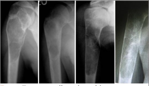
Methods: We describe our finding with the treatment of FDfibrous dysplasia of the proximal radius in five patients(four females and, one male), the mean age of 28.6 years (22 to –39 years). The lesions were in the metaphysis extending to the diaphysis. Persistent pain and pain after pathological fracture were the indications for surgical intervention. We used an extensile approach to expose the lesion then extended curettage using a high-speed burr and filling the cavity with bone cement. Functional outcome and radiological findings were monitored on follow-up visits.
Results: The mean follow-up period was 3.2 years (ranged from 2 years to 5 years).There waswereno recurrences and no patient sustained a fracture at the end of the filling cement. At the time of the last follow-up, all patients have excellent score(mean 27 points) according to the musculoskeletal tumor society scoring system.
Conclusion: Extended curettage and cementation provide a safe and reliable alternative for control of FDfibrous dysplasia of the proximal radius with little morbidity with little risk of recurrence and low incidence of complications.
Keywords: Fibrous dysplasia, Curettage, Cement.


References

1. Lichtenstein L. Fibrous dysplasia of bone. Arch Pathol 1942;33:777-816.
2. Chapurlat RD, Meunier PJ. Fibrous dysplasia of bone. Best Pract Res Clin Rheumatol 2000;14:385-98.
3. Boyce AM, Kelly MH, Brillante BA, Kushner H, Wientroub S, Riminucci M, et al. A randomized, double blind, placebo-controlled trial of alendronate treatment for fibrous dysplasia of bone. J Clin Endocrinol Metab 2014;99:4133-40.
4. Hart ES, Kelly MH, Brillante B, Chen CC, Ziran N, Lee JS, et al. Onset, progression, and plateau of skeletal lesions in fibrous dysplasia and the relationship to functional outcome. J Bone Miner Res 2007;22:1468-74.
5. Kelly MH, Brillante B, Collins MT. Pain in fibrous dysplasia of bone: Age-related changes and the anatomical distribution of skeletal lesions. Osteoporos Int 2008;19:57-63.
6. Smith SE, Kransdorf MJ. Primary musculoskeletal tumors of fibrous origin. Semin Musculoskelet Radiol 2000;4:73-88.
7. Kumar R, Madewell JE, Lindell MM, Swischuk LE. Fibrous lesions of bones. Radiographics 1990;10:237-56.
8. Guille JT, Kumar SJ, MacEwen GD. Fibrous dysplasia of the proximal part of the femur. Long-term results of curettage and bone-grafting and mechanical realignment. J Bone Joint Surg Am 1998;80:648-58.
9. Leet AI, Boyce AM, Ibrahim KA, Wientroub S, Kushner H, Collins MT, et al. Bone-grafting in polyostotic fibrous dysplasia. J Bone Joint Surg Am 2016;98:211-9.
10. Cabral CE, Guedes P, Fonseca T, Rezende JF, Cruz LC Jr., Smith J. Polyostotic fibrous dysplasia associated with intramuscular myxomas: Mazabraud’s syndrome. Skeletal Radiol 1998;27:278-82.
11. Leslie WD, Reinhold C, Rosenthall L, Tau C, Glorieux FH. Panostotic fibrous dysplasia. A new craniotubular dysplasia. Clin Nucl Med 1992;17:556-60.
12. Cutler CM, Lee JS, Butman JA, FitzGibbon EJ, Kelly MH, Brillante BA, et al. Long-term outcome of optic nerve encasement and optic nerve decompression in patients with fibrous dysplasia: Risk factors for blindness and safety of observation. Neurosurgery 2006;59:1011-7.
13. Leet AI, Magur E, Lee JS, Wientroub S, Robey PG, Collins MT, et al. Fibrous dysplasia in the spine: Prevalence of lesions and association with scoliosis. J Bone Joint Surg Am 2004;86-A:531-7.
14. Kumta SM, Leung PC, Griffith JF, Kew J, Chow LT. Vascularised bone grafting for fibrous dysplasia of the upper limb. J Bone Joint Surg Br 2000;82:409-12.
15. Plotkin H, Rauch F, Zeitlin L, Munns C, Travers R, Glorieux FH, et al. Effect of pamidronate treatment in children with polyostotic fibrous dysplasia of bone. J Clin Endocrinol Metab 2003;88:4569-75.
16. Leet AI, Collins MT. Current approach to fibrous dysplasia of bone and mcCune-albright syndrome. J Child Orthop 2007;1:3-17.
17. Adetayo OA, Salcedo SE, Borad V, Richards SS, Workman AD, Ray AO, et al. Fibrous dysplasia: An overview of disease process, indications for surgical management, and a case report. Eplasty 2015;15:e6.
18. Keijser LC, Van Tienen TG, Schreuder HW, Lemmens JA, Pruszczynski M, Veth RP, et al. Fibrous dysplasia of bone: Management and outcome of 20 cases. J Surg Oncol 2001;76:157-66.
19. Stanton RP, Ippolito E, Springfield D, Lindaman L, Wientroub S, Leet A. The surgical management of fibrous dysplasia of bone. Orphanet J Rare Dis 2012;7:S1.
20. Bryant DD 3rd, Grant RE, Tang D. Fibular strut grafting for fibrous dysplasia of the femoral neck. J Natl Med Assoc 1992;84:893-7.
21. Shih HN, Chen YJ, Huang TJ, Hsu KY, Hsu RW. Treatment of fibrous dysplasia involving the proximal femur. Orthopedics 1998;21:1263-6.
22. Weiland AJ, Moore JR, Daniel RK. Vascularized bone autografts. Experience with 41 cases. Clin Orthop Relat Res 1983;174:87-95.
23. Errani C, Ruggieri P, Asenzio MA, Toscano A, Colangeli S, Rimondi E, et al. Giant cell tumor of the extremity: A review of 349 cases from a single institution. Cancer Treat Rev 2010;36:1-7.
24. Gitelis S, Mallin BA, Piasecki P, Turner F. Intralesional excision compared with en bloc resection for giant-cell tumors of bone. J Bone Joint Surg Am 1993;75:1648-55.
25. Keçeci B, Küçük L, Isayev A, Sabah D. Effect of adjuvant therapies on recurrence in aneurysmal bone cysts. Acta Orthop Traumatol Turc 2014;48:500-6.
26. Enneking WF, Dunham W, Gebhardt MC, Malawar M, Pritchard DJ. A system for the functional evaluation of reconstructive procedures after surgical treatment of tumors of the musculoskeletal system. Clin Orthop Relat Res 1993;286:241-6.
27. Stephenson RB, London MD, Hankin FM, Kaufer H. Fibrous dysplasia. An analysis of options for treatment. J Bone Joint Surg Am 1987;69:400-9.
28. Weiland AJ, Phillips TW, Randolph MA. Bone grafts: A radiologic, histologic, and biomechanical model comparing autografts, allografts, and free vascularized bone grafts. Plast Reconstr Surg 1984;74:368-79.
29. Rosario MS, Hayashi K, Yamamoto N, Takeuchi A, Miwa S, Taniguchi Y, et al. Functional and radiological outcomes of a minimally invasive surgical approach to monostotic fibrous dysplasia. World J Surg Oncol 2017;15:1..


How to Cite this article: Badr I T, Rampal S, Shahin A. Preliminary Results of Curettage and Cementation in the Treatment of Fibrous Dysplasia of the Proximal Radius. Journal of Bone and Soft Tissue Tumors Jan-Apr 2019;5(1): 13-16.


(Abstract    Full Text HTML)   (Download PDF)