A Giant Parosteal Lipoma with Exostosis of the Right Proximal Humerus
Case Report | Volume 5 | Issue 2 | JBST May – August 2019 | Page 4-7| Abdaud Rasyid, Mujaddid Idulhaq, Pamudji Utomo, Ambar Mudigdo, Handry Tri Handojo. DOI: 10.13107/jbst.2019.v05i02.424
Author Abdaud Rasyid[1],[2], Mujaddid Idulhaq[1],[2], Pamudji Utomo[1],[2], Ambar Mudigdo[3], Handry Tri Handojo[4]
[1]Department of Orthopaedic and Traumatology, Faculty of Medicine, Sebelas Maret University, Surakarta, Indonesia,
[2]Department of Orthopaedic and Traumatology, Prof. DR. R. Soeharso Orthopaedic Hospital, Surakarta, Indonesia,
[3]Department of Anatomical Pathology, Sebelas Maret University, Surakarta, Indonesia,
[4]Department of Radiology, at Prof. DR. R. Soeharso Orthopaedic, Hospital, Surakarta, Indonesia.
Address of Correspondence
Dr. Abdaud Rasyid,
Jalan Ahmad Yani, Pabelan, Kartasura, Sukoharjo, Jawa Tengah, 57162, Indonesia.
E-mail: abdaudry@gmail.com
Abstract
Introduction: Lipomas are the most frequent benign soft-tissue tumors. Soft -tissue lipomas are categorized by anatomic location as either superficial (subcutaneous) or deep (intermuscular). Deep lipomas can be located in any part of the body, including the superior extremities. Lipomas typically reach a diameter of several centimeters and are localized in a single anatomical region. Parosteal lipoma is a rare subtype of deep lipoma that has a broad attachment to the underlying periosteum that forms an exostoses-like bone prominence. There has been no reliable literature; about two pathological processes occur in one extremity at the same time.
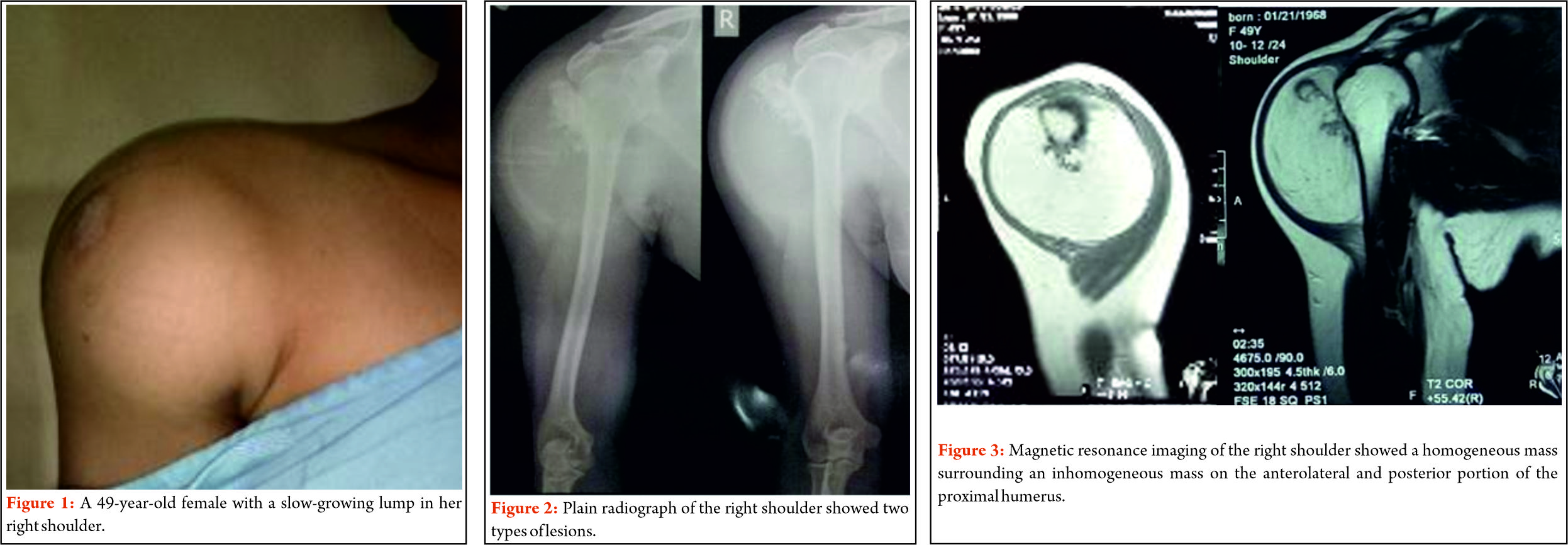
Case Presentation: A 49-years -old female presented at our institution with a painless, slow -growing lump in her right shoulder region since for 2 years ago, with no other symptoms, and no history of trauma. A palpable non-tender mobile mass was present on the right shoulder region. Plain radiographs showed a well-delineated ovoid radiolucent lesion and a radiopaque lesion over the right proximal humerus. The fine -needle biopsy result suggested a liposarcoma. Wide-excision surgery was performed for both the masses. On contrary, the histological examination of the specimen confirmed a giant lipoma with pieces of adult bone tissues.
Conclusion: Deep-seated lipomas are most commonly discovered in men between the ages of thirties 30s and sixties60s. In our patient, the lipoma also accompanied with an exostoses-like cartilaginous mass over the proximal humerus as in parosteal lipoma. Plain radiographs study of parosteal lipoma is associated with a false osteochondroma appearance, which also found in this patient. Histological examination suggested a giant lipoma for this patient, but the possibility of two pathological processes is still in question.
Keywords: Giant lipoma, shoulder, exostosis, surgery.
References
1. Slavchev S, Georgiev PP, Penkov M. Giant lipoma extending between the heads of biceps brachii muscle and the deltoid muscle: Case report. J Curr Surg 2012;2:146-8.
2. Stevenson J, Parry M. Tumours. In: Apley and Solomon’s System of Orthopaedics and Trauma, 10th ed. Vol. 9. Ch. 9. Florida: CRC Press; 2018. p. 223-4.
3. Singh V, Kumar V, Singh AK. Case report: A rare presentation of giant palmar lipoma. Int J Surg Case Rep 2016;26:21-3.
4. Elbardouni A, Kharmaz M, Salah Berrada M, Mahfoud M, Elyaacoubi M. Well-circumscribed deep-seated lipomas of the upper extremity. A report of 13 cases. Orthop Traumatol Surg Res 2011;97:152-8.


| How to Cite this article: Rasyid A, Idulhaq M, Utomo P, Mudigdo A, Handojo H T. A Giant Parosteal Lipoma with Exostosis of the Right Proximal Humerus. Journal of Bone and Soft Tissue Tumors May-August 2019;5(2): 4-7. |
[Full Text HTML] [Full Text PDF] [XML]
[rate_this_page]
Dear Reader, We are very excited about New Features in JOCR. Please do let us know what you think by Clicking on the Sliding “Feedback Form” button on the <<< left of the page or sending a mail to us at editor.jocr@gmail.com